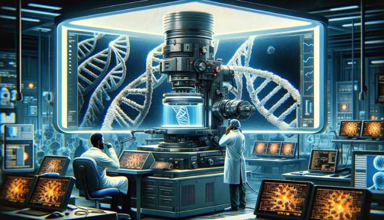
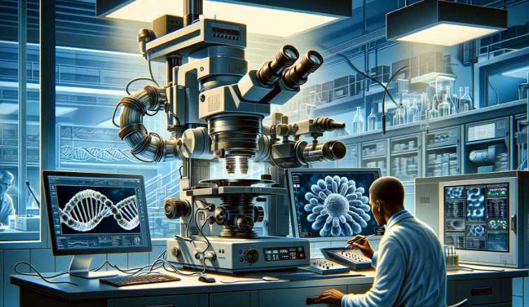
No, Transmission Electron Microscopes (TEM) cannot directly visualize DNA. DNA is not electron-dense, and electron microscopes primarily detect dense materials. However, TEM can be used to study the structures associated with DNA, such as chromatin and nuclear membranes.
Transmission Electron Microscopes use electron beams to create detailed images of thin sections of specimens. The electrons interact with dense regions of a sample, leading to the formation of an image. DNA is not electron-dense, consisting of light atoms like carbon, hydrogen, nitrogen, and oxygen.
In contrast, materials like heavy metals, which can stain or label DNA-associated structures, are electron-dense and can be visualized with TEM. Therefore, DNA is indirectly studied in TEM by examining structures like chromatin, which is composed of DNA and associated proteins.

Here’s a table summarizing the electron density of key components in the context of Transmission Electron Microscopy:
| Component | Electron Density in TEM |
|---|---|
| DNA | Low |
| Heavy Metals (Stains) | High |
| Chromatin | Indirectly visualized |
| Nuclear Membranes | Indirectly visualized |
Understanding Electron Microscopes
Electron microscopes use electrons instead of light to achieve much higher magnification and resolution compared to optical microscopes. There are two main types: transmission electron microscopes (TEM) and scanning electron microscopes (SEM).
- Transmission Electron Microscope (TEM):
- Principle: Transmits electrons through a thin specimen.
- Resolution: High resolution (up to 0.1 nm).
- Magnification: Ultra-high magnification (up to 50 million times).
- Sample: Thin sections (biological tissues, cells, nanoparticles).
- Scanning Electron Microscope (SEM):
- Principle: Scans the specimen with a focused electron beam, detecting emitted electrons.
- Resolution: Lower resolution than TEM (around 1-10 nm).
- Magnification: High magnification (up to 2 million times).
- Sample: Surface imaging (3D topography, morphology).
Electron microscopes employ electrons instead of light for imaging, allowing for much higher magnification and resolution. TEM involves transmitting electrons through a thin specimen, providing detailed internal structures. SEM, on the other hand, scans the specimen surface and detects emitted electrons, offering 3D images of surface morphology.
Table:
| Feature | Transmission Electron Microscope (TEM) | Scanning Electron Microscope (SEM) |
|---|---|---|
| Principle | Transmits electrons through a thin specimen | Scans specimen surface with a focused electron beam |
| Resolution | High resolution (up to 0.1 nm) | Lower resolution than TEM (1-10 nm) |
| Magnification | Ultra-high magnification (up to 50 million times) | High magnification (up to 2 million times) |
| Sample | Thin sections (biological tissues, cells, nanoparticles) | Surface imaging (3D topography, morphology) |
A Transmission Electron Microscope (TEM) is a powerful imaging tool used to study the tiny structures and details of various specimens, including DNA. It works by transmitting a beam of electrons through a thin sample, producing a highly magnified image on a screen or photographic film. Unlike a traditional light microscope, a TEM can magnify objects up to 50 million times, allowing scientists to observe intricate structures at the atomic level.
Challenges Of Imaging DNA
| Challenge | Explanation |
|---|---|
| Resolution Limitations | The nanoscale size of DNA makes achieving high resolution for visualizing individual base pairs challenging. |
| Sample Preparation Complexity | Complex sample preparation techniques impact accuracy and reproducibility in DNA imaging. |
| Dynamic Nature of DNA | Capturing real-time dynamic structural changes and interactions in DNA is a significant technical challenge. |
| Labeling Techniques and Artifacts | Labels used for imaging may introduce artifacts or interfere with the natural behavior of DNA. |
| Speed and Throughput | Balancing high-speed imaging with throughput is crucial for studying dynamic processes in DNA. |
| Single-Molecule Sensitivity | Advanced technologies are needed for detecting and imaging individual DNA molecules with high sensitivity. |
| Environmental Conditions | Maintaining optimal conditions for DNA imaging, especially inside living cells, is challenging. |
| Cost and Accessibility | High costs associated with cutting-edge DNA imaging technologies limit accessibility for some researchers. |
This table summarizes the challenges of imaging DNA, providing a concise overv
Advancements In Tem Technology
Transmission Electron Microscopes (TEM) have undergone significant advancements in technology, enabling enhanced resolution for imaging biological samples, including DNA. The implementation of innovative resolution enhancement techniques has improved the imaging capabilities of TEM, allowing for the visualization of molecular structures with unprecedented clarity. Additionally, improvements in sample preparation techniques have contributed to the advancement of TEM technology, ensuring the preservation and visualization of biological specimens at an unparalleled level of detail. These advancements have revolutionized the study of biological molecules and structures, providing valuable insights into the intricate world of DNA and other biomolecular entities.
Visualizing DNA With TEM
Visualizing DNA with Transmission Electron Microscopy (TEM) is challenging due to the low contrast of DNA molecules. However, it is possible to observe DNA in TEM by using heavy metal stains, such as uranyl acetate or phosphotungstic acid, which bind to the DNA and increase contrast.
Transmission Electron Microscopy (TEM) is a powerful technique for imaging structures at the nanoscale. DNA, being a small and low-contrast biological molecule, is not inherently visible in TEM. To overcome this limitation, heavy metal stains are often employed. These stains bind to the DNA molecules, enhancing their contrast and making them visible under the electron beam.
Here is a table summarizing the key points:
| Challenges in Visualizing DNA with TEM | Solution |
|---|---|
| Low contrast of DNA molecules | Use heavy metal stains (e.g., uranyl acetate, phosphotungstic acid) to increase contrast and make DNA visible in TEM. |
| Small size of DNA | TEM’s high resolution allows imaging of structures at the nanoscale, making it suitable for visualizing DNA. |
Applications Of TEM In Biology
Transmission electron microscopes (TEMs) have revolutionized the field of biology by allowing scientists to investigate molecular structures and cellular processes with unprecedented detail and clarity.
TEMs are invaluable tools for studying the structure of molecules, including DNA. Their high resolution and ability to penetrate samples with electron beams enable researchers to visualize the intricate arrangement of atoms within biomolecules. This information is crucial for understanding the mechanisms of biological processes and designing targeted drugs.
With TEMs, scientists can observe and analyze various cellular processes in real-time. They can track the movement of organelles, observe protein synthesis and degradation, and study cellular signaling pathways. This knowledge helps unravel the complexities of biological systems and sheds light on disease mechanisms.
| Application | Description |
|---|---|
| Cellular Ultrastructure Analysis | Study of cellular components at the ultrastructural level, providing detailed images of organelles, membranes, and other cellular structures. |
| Virus Morphology Studies | Visualization and study of the morphology of viruses, aiding in the understanding of their structure and life cycle. |
| Subcellular Localization of Molecules | Precise localization of molecules within cells, helping researchers identify the exact subcellular compartments where specific activities occur. |
| Nanoparticle Characterization | Characterization of nanoparticles, providing insights into their size, shape, and distribution, valuable for nanomedicine and drug delivery research. |
| Study of Biological Macromolecules | Visualization of biological macromolecules, such as proteins and nucleic acids, providing information about their structure and interactions. |
| Investigation of Cellular Pathology | Study of cellular abnormalities and pathological conditions, aiding in the diagnosis of diseases at the cellular level. |
| Cellular and Tissue Development Research | Investigation of cellular and tissue development, providing insights into morphological changes during processes like embryogenesis and tissue differentiation. |
| Imaging of Membrane Dynamics | Observation and analysis of dynamic changes in cellular membranes, contributing to our understanding of membrane structure and function. |
| Analysis of Intracellular Transport | Study of intracellular transport processes, including vesicle formation, trafficking, and fusion, providing insights into cellular logistics. |
| Molecular Biology Applications | Role in various molecular biology applications, such as the visualization of DNA and RNA structures, aiding in the understanding of genetic processes. |
Ethical Considerations
The use of transmission electron microscopes (TEMs) in DNA analysis raises important ethical considerations, particularly when it comes to privacy concerns. TEMs have the potential to provide unparalleled insights into the genetic makeup of individuals, allowing researchers to view DNA at a molecular level. This level of detail could unlock new discoveries in genetic research and lead to significant advancements in various fields, including medicine and forensics.
However, the highly sensitive nature of genetic information also raises concerns about privacy and data security. Access to an individual’s DNA sequence can reveal not only their predispositions to certain diseases but also their ancestry and other personal information. Safeguarding this data and ensuring its responsible use is vital to protect individuals’ privacy rights and prevent any potential misuse.
Furthermore, the implications of TEMs on genetic research are vast. The ability to observe DNA at such a detailed level opens up possibilities for uncovering new insights and understanding genetic disorders. Researchers can gather a wealth of information on DNA structure, potentially leading to more targeted and effective treatment options for a wide range of conditions.
In conclusion, while TEMs offer exciting opportunities in genetic research, it is essential to address the ethical considerations surrounding privacy concerns and ensure responsible use of this technology to protect individuals’ rights.
Future Prospects And Challenges
The potential for DNA imaging using transmission electron microscopes (TEM) presents promising avenues for future research in the field of molecular biology. Challenges associated with imaging DNA using TEM include the need for advanced sample preparation techniques and imaging technologies that can mitigate radiation damage. Overcoming technical barriers is crucial for realizing the full potential of TEM in DNA imaging applications.
Final Thoughts
While transmission electron microscopes can visualize DNA indirectly, they can’t directly see it due to its small size and the limitations of electron microscopy. Nevertheless, advancements in technology may offer potential solutions in the future, sparking new opportunities for studying DNA at a subcellular level.
Books:
- “Electron Microscopy: Principles and Techniques for Biologists” by John J. Bozzola and Lonnie D. Russell.
- “Transmission Electron Microscopy: A Textbook for Materials Science” by David B. Williams and C. Barry Carter.

Fahim Foysal is a well-known expert in the field of binoculars, with a passion for exploring the great outdoors and observing nature up close. With years of experience in the field, Fahim has honed his skills as a binocular user and has become a go-to resource for those seeking advice on choosing the right binoculars for their needs.
Fahim’s love for the natural world began during his time at The Millennium Stars School and College and BIAM Laboratory School, where he spent much of his free time exploring the outdoors and observing the wildlife around him. This passion for nature led him to pursue a degree in Fine Arts from the University of Dhaka, where he gained a deep understanding of the importance of observation and attention to detail.
Throughout his career, Fahim has used his expertise in binoculars to help others discover the beauty of the natural world. His extensive knowledge of binocular technology and optics has made him a trusted advisor for amateur and professional wildlife observers alike. Whether you’re looking to spot rare birds or observe animals in their natural habitats, Fahim can help you choose the perfect binoculars for your needs. With his guidance, you’ll be able to explore the outdoors with a newfound appreciation for the beauty of the natural world.
Table of Contents


Pingback: Are Electron Microscopes Expensive? Unveiling the Costs
Pingback: 5 Things You Need To Know About Microscopes for 10-Year-Olds on a Scientific Journey