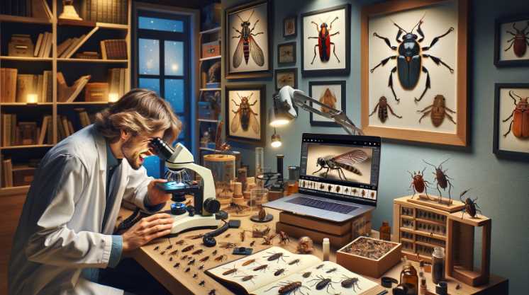
Microscope

Can You See Golgi Apparatus under a Light Microscope

Can You See Golgi Apparatus under a Light Microscope

Yes, you can see the Golgi Apparatus under a light microscope. The Golgi Apparatus, though small, can be observed under a light microscope. However, due to its size and the limitations of light microscopy, it may not appear as detailed as with electron microscopy. Light microscopy provides a lower resolution compared to electron microscopy, so […]

Can You See Golgi Apparatus under a Light Microscope Read More »

Most Powerful Optical Microscope

5 Must-Know Facts About Most Powerful Optical Microscope

The most powerful optical microscope, such as the advanced super-resolution microscopes, can achieve resolutions down to a few nanometers, allowing researchers to visualize structures at the molecular level. These microscopes use techniques like structured illumination, stimulated emission depletion, or stochastic optical reconstruction microscopy to surpass the diffraction limit of conventional light microscopes. They enable unprecedented

5 Must-Know Facts About Most Powerful Optical Microscope Read More »

Are Viruses Visible under a Microscope

Are Viruses Visible under a Microscope: (Video Explained)

Yes, viruses are visible under a microscope. They are too small to be seen without magnification. When viewed under a microscope, viruses appear as tiny particles with unique shapes and structures. Their size and features can vary depending on the type of virus. Scientists and researchers use powerful microscopes to study viruses and understand their

Are Viruses Visible under a Microscope: (Video Explained) Read More »

Are centrioles visible under a light microscope

Are centrioles visible under a light microscope?

No, centrioles are not typically visible under a light microscope. Centrioles are small, cylindrical structures found in animal cells, and they play a crucial role in the organization of microtubules during cell division. However, centrioles are very small and are below the resolution limit of a light microscope, which is approximately 200 nanometers. Therefore, they

Are centrioles visible under a light microscope? Read More »

No, electron microscopes cannot generally be used to observe living cells.

Can we see living cells in an electron microscope?

No, electron microscopes cannot generally be used to observe living cells. The preparation process for electron microscopy involves dehydration and vacuum conditions that are incompatible with maintaining living cells. Electron microscopes use electron beams instead of light to achieve much higher resolution images than light microscopes. However, the sample preparation for electron microscopy involves fixing,

Can we see living cells in an electron microscope? Read More »

What Does Staph Look Like under a Microscope

What Does Staph Look Like Under Microscope: A Close-Up View

Staphylococcus bacteria appear as spherical clusters or grape-like structures under a microscope. Staphylococcus, commonly known as staph, is a genus of bacteria characterized by its round shape and tendency to form clusters. When viewed under a microscope, these bacteria appear as spherical clusters resembling grapes or bunches. The name “staphylococcus” is derived from the Greek

What Does Staph Look Like Under Microscope: A Close-Up View Read More »

Are Electron Microscopes Expensive? Unveiling the Costs

Yes, electron microscopes are expensive. Electron microscopes are sophisticated scientific instruments that use electron beams to achieve extremely high magnification and resolution, allowing researchers to observe objects at the nanoscale. The technology and precision required in electron microscopes contribute to their high cost. These instruments often involve complex components, including electron sources, electromagnetic lenses, detectors,

Are Electron Microscopes Expensive? Unveiling the Costs Read More »

What Does a Kidney Stone Look Like under a Microscope

What Does a Kidney Stone Look Like Under Microscope: A Microscopic Marvel

Under a microscope, a kidney stone typically appears as a crystalline structure with various shapes and sizes, such as jagged edges or smooth surfaces, depending on the composition of the stone. Kidney stones are formed from accumulating substances in the urine, such as calcium, oxalate, and phosphate. These substances can crystallize and aggregate, leading to

What Does a Kidney Stone Look Like Under Microscope: A Microscopic Marvel Read More »

Under a microscope, nail fungus typically appears as hyphae invading and spreading within the nail tissue.

What Does Nail Fungus Look Like under Microscope: Revealing Close-Ups!

Under a microscope, nail fungus (onychomycosis) typically appears as hyphae (thread-like structures) invading and spreading within the nail tissue. Nail fungus is primarily caused by dermatophyte fungi, commonly Trichophyton rubrum, Trichophyton mentagrophytes, or Candida species. When observed under a microscope, these fungi exhibit distinctive characteristics. The hyphae, which are branching filaments, penetrate and invade the

What Does Nail Fungus Look Like under Microscope: Revealing Close-Ups! Read More »