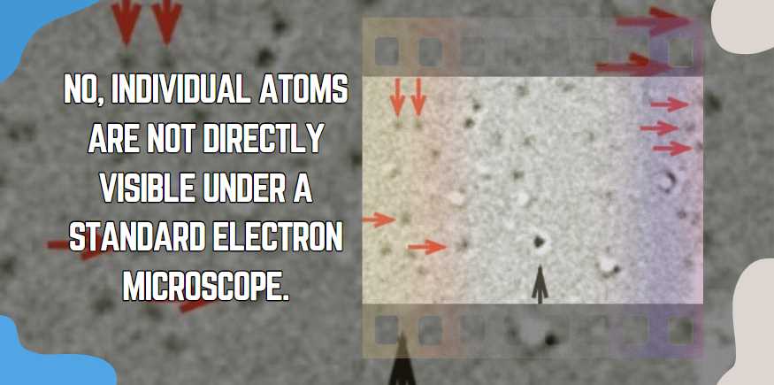
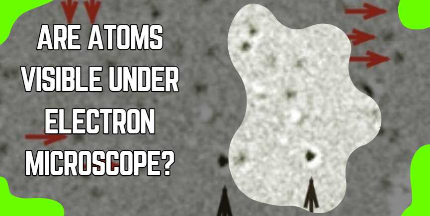
No, individual atoms are not directly visible under a standard electron microscope.
The wavelength of electrons used in electron microscopes is much shorter than that of visible light, allowing for much higher resolution. However, even with this high resolution, the individual atoms are generally not directly visible. Instead, electron microscopes can provide detailed images of the surfaces and structures of objects at the atomic level. Scientists often use techniques such as electron diffraction or imaging specific elements to infer the positions of atoms within a sample indirectly.

| Property | Value |
|---|---|
| Atomic Number | Varies based on the element |
| Atomic Mass | Varies based on the isotope |
| Atomic Radius | Typically in the range of 30-300 pm |
| Electron Configuration | Varies based on the element |
| Ionization Energy | Varies based on the element |
| Electronegativity | Varies based on the element |
| Boiling Point | Varies widely across elements |
| Melting Point | Varies widely across elements |
Understanding Electron Microscopes
An electron microscope is a type of microscope that uses a beam of electrons to create an image of a specimen. They are capable of much higher resolutions than light microscopes, which use photons. There are two main types of electron microscopes:
- Transmission Electron Microscope (TEM): It transmits electrons through a specimen and forms an image from the interaction of the electrons with the specimen.
- Scanning Electron Microscope (SEM): It scans a surface with a focused beam of electrons and creates an image from the signals that are emitted from the interaction of the electrons with the sample’s surface.
These powerful tools have allowed us to visualize the nano-world in ways that were previously thought impossible.
Can Electron Microscopes See Atoms?
Yes, electron microscopes can see atoms.
Electron microscopes use a beam of electrons instead of light to achieve much higher magnification and resolution than traditional light microscopes. Due to the shorter wavelength of electrons compared to photons, electron microscopes can visualize objects at the atomic level. This enables scientists to observe and study the fine details of atomic structures.
In a transmission electron microscope (TEM), electrons pass through a thin specimen, and the resulting image is formed by the interactions of electrons with the specimen. Scanning electron microscopes (SEM) use a focused beam of electrons to scan the surface of a specimen, creating detailed 3D images. Both types of electron microscopes have been instrumental in advancing our understanding of the structure and behavior of atoms.
| Type of Electron Microscope | Visibility of Atoms |
|---|---|
| Transmission Electron Microscope (TEM) | Capable of producing images that resolve individual atoms in a material, especially when combined with powerful computational methods. |
| Scanning Transmission Electron Microscope (STEM) | Allows for detailed imaging and composition analysis at the atomic scale, with some specialized variations capable of identifying single atoms. |
| Scanning Electron Microscope (SEM) | Not typically used for visualizing individual atoms due to lower resolution compared to TEM and STEM but excellent for surveying surfaces and nanostructures. |
SEM generally offers lower resolution than TEM and STEM, but it is still a valuable tool for imaging the surface characteristics of materials and nanostructures.
Can Electron Microscopes Capture Images in Color?
Traditional electron microscopes do not capture images in color. Unlike light microscopes that use visible light to produce color images, electron microscopes use a beam of electrons. The interaction of electrons with the specimen provides contrast in black and white images.
The table below outlines the key differences between the imaging mechanisms of light and electron microscopes:
| Imaging Aspect | Light Microscope | Electron Microscope |
|---|---|---|
| Imaging Source | Visible light | Electrons |
| Image Color | Color images | Black and white images |
| Staining Techniques | Dyes and stains can be used for contrast | Heavy metal stains enhance electron scattering for contrast |
To enhance contrast in electron microscopy, scientists often use staining techniques with heavy metal compounds. These stains selectively scatter electrons, highlighting different structures within the specimen.
In summary, electron microscopes produce black and white images, and contrast is achieved through electron scattering, often enhanced by staining techniques using heavy metal compounds.
How Do Electron Microscopes Work?
Electron microscopes operate on the principles of electron optics, utilizing electron beams instead of light to achieve higher resolution. There are two main types of electron microscopes: transmission electron microscopes (TEM) and scanning electron microscopes (SEM).
Transmission Electron Microscope (TEM):
-
Electron Source:
- The electron gun generates a beam of electrons.
-
Condenser Lens:
- Focuses the electron beam onto the specimen.
-
Specimen:
- Electrons pass through the specimen, interacting with its atoms.
-
Objective Lens:
- Focuses the transmitted electrons to form an image.
-
Projector Lens:
- Further magnifies and projects the image onto a detector.
Scanning Electron Microscope (SEM):
-
Electron Source:
- The electron gun produces a focused beam of electrons.
-
Coil/Lens System:
- Scans the electron beam across the specimen’s surface.
-
Detectors:
- Collect signals such as secondary electrons emitted from the specimen.
-
Image Formation:
- Signals are used to create a 3D surface image of the specimen.
Both types of electron microscopes provide high-resolution images, enabling scientists to study structures at the atomic and nanoscale levels.
What Are the Limitations of Electron Microscopy?
Despite their high resolution, electron microscopes have limitations:
| Limitation | Description |
|---|---|
| Sample Preparation: | Specimens must undergo extensive preparation, including dehydration and coating with conductive materials, which can alter their natural state. |
| Vacuum Requirement: | Electron microscopes operate in a vacuum, limiting the study of biological specimens, which may not survive in a vacuum environment. |
| Artifact Formation: | The preparation process can introduce artifacts, leading to distorted images or inaccurate representation of the specimen’s original structure. |
| Cost and Maintenance: | Electron microscopes are expensive to purchase, maintain, and operate, requiring specialized facilities and trained personnel. |
| Complexity: | Operating electron microscopes requires specialized knowledge and training, making them less accessible to researchers without expertise in electron microscopy. |
Imaging Techniques Used to Visualize Atoms
Technological advancements in microscope design and imaging techniques have significantly improved the ability to visualize atoms. Here are some of the approaches used:
- Cryo-Electron Microscopy: Involves freezing specimens to cryogenic temperatures, reducing damage from the electron beam and improving the visibility of atoms.
- Atomic Force Microscopy: Not a type of electron microscopy but a complementary technique that can also resolve atoms by ‘feeling’ surfaces with a mechanical probe.
- High-Resolution TEM (HRTEM): Capable of visualizing the arrangement of atoms within materials.
- Electron Energy Loss Spectroscopy (EELS): Used alongside TEM to provide information about the composition and electronic structure at the atomic scale.
Through these innovative methods, scientists can create detailed images of atomic structures to gain insights into material properties and biological processes.
Real-World Applications of Atom-Level Imaging
The ability to visualize atoms has profound implications for various fields. Here’s a look at some of the areas benefiting from these technological leaps:
- Material Science: Researchers can design new materials with enhanced properties by understanding atomic arrangements.
- Chemistry: Insight into how atoms bond and interact enables the creation of novel compounds and catalysts.
- Nanotechnology: Atom-level imaging helps manufacture nanoscale devices with unprecedented precision.
- Pharmaceuticals: Detailed atomic maps of proteins and viruses assist in drug discovery and vaccine development.
Final Thoughts
The beauty and complexity of the microscopic world are awe-inspiring. As technology continues to advance, encouraging continued scientific exploration becomes paramount. The ongoing quest to visualize atoms fuels our understanding of the fundamental building blocks of our universe, opening doors to discoveries and innovations.

Fahim Foysal is a well-known expert in the field of binoculars, with a passion for exploring the great outdoors and observing nature up close. With years of experience in the field, Fahim has honed his skills as a binocular user and has become a go-to resource for those seeking advice on choosing the right binoculars for their needs.
Fahim’s love for the natural world began during his time at The Millennium Stars School and College and BIAM Laboratory School, where he spent much of his free time exploring the outdoors and observing the wildlife around him. This passion for nature led him to pursue a degree in Fine Arts from the University of Dhaka, where he gained a deep understanding of the importance of observation and attention to detail.
Throughout his career, Fahim has used his expertise in binoculars to help others discover the beauty of the natural world. His extensive knowledge of binocular technology and optics has made him a trusted advisor for amateur and professional wildlife observers alike. Whether you’re looking to spot rare birds or observe animals in their natural habitats, Fahim can help you choose the perfect binoculars for your needs. With his guidance, you’ll be able to explore the outdoors with a newfound appreciation for the beauty of the natural world.
Table of Contents


Pingback: Can we see living cells in an electron microscope?