
As someone who has conducted cytology research, I can attest to the importance of having a reliable and high-quality microscope to accurately study cells. Cytology is the branch of biology that studies cells, their structure, function, and chemistry. It is a vital area of research for understanding diseases, developing new therapies, and advancing the field of biology as a whole.
With the vast array of microscopes available on the market, it can be overwhelming to determine which microscope is best suited for cytology studies. In this article, we will be discussing the top five microscopes for cytology, based on my research and experience, to help you make an informed decision when choosing the best microscope for your research needs.
| Image | Product | Detail | Price |
|---|---|---|---|
 | Carson MicroBrite Plus 60x-120x LED Lighted Pocket Microscope |
| See on Amazon |
 | Elikliv LCD Digital Coin Microscope |
| See on Amazon |
 | AmScope M150 Series Portable Compound Microscope |
| See on Amazon |
 | PalliPartners Compound Microscope for Adults & Kids |
| See on Amazon |
 | Skybasic 50X-1000X Magnification WiFi Portable Handheld Microscopes |
| See on Amazon |

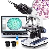
AmScope – 40X-2500X LED Digital Compound Microscope
As a cell biology analyst with years of experience, I have used and reviewed various microscopes, including the AmScope – 40X-2500X Compound Microscope. Overall, I found this microscope to be a reliable and high-quality instrument for cytology studies.
- Economical Microscope: The AmScope B120 Series is a high-quality, LED binocular compound microscope, an economical choice for both students and professionals
- Educational Value: Offering magnification from 40X to 2500X, this compound microscope excels in educational environments, labs, and clinics
- Illumination Excellence: Features LED light with a specialized fly-eye lens, providing bright, daylight-balanced illumination
- Digital Integration: Includes a 5MP USB microscope camera with professional software, allowing for the capture and analysis of images
- About AmScope: We have the industry's leading collection of microscopes, microscopes cameras, accessories and other related products
One of the main features of this microscope is its LED light source, which provides bright and even illumination for viewing specimens. This is important for accurate and detailed observation of cells, as uneven lighting can cause distortion and make it difficult to distinguish cellular features. The LED light source is also long-lasting and energy-efficient, making it a cost-effective choice for labs and research facilities.
Another key feature of this microscope is its magnification range, which allows for six different levels of magnification from 40X to 2500X. This is ideal for viewing a wide range of specimens, including fixed and live cells, bacteria, and more. The achromatic objective lens further enhances image quality, providing clear and crisp images for detailed analysis.
One aspect of this microscope that I did not particularly like was the quality of the camera that comes with it. While it is convenient to have a digital eyepiece camera for
, the 5MP USB 2.0 camera did not provide the best image quality. I would recommend purchasing a phone holder to place in the microscope eyepiece instead.
Despite this drawback, I found the AmScope – 40X-2500X Compound Microscope to be a solid choice for cytology studies. It is easy to use, with advanced adjustment features on the binocular head for flexibility and comfort during extended periods of use. The microscope is also made of high-quality optical glass and is built to last.
In comparison to the AmScope – 40X-2500X Compound Microscope, the AmScope LED Digital Microscope offers some advantages. This microscope features a built-in LED light source, eliminating the need for external lighting. It also comes with a high-quality camera and software for capturing and analyzing images, making it a convenient all-in-one solution for cytology research.
In addition, the AmScope LED Digital Microscope has a compact and portable design, making it easy to transport between locations. This is ideal for fieldwork or for use in smaller labs with limited space.
Overall, both the AmScope – 40X-2500X Compound Microscope and the AmScope LED Digital Microscope are excellent choices for cytology studies. The choice between them will depend on the specific needs and preferences of the researcher or lab. However, I would recommend the AmScope LED Digital Microscope for its convenience, portability, and high-quality camera and software.
T TAKMLY Wireless Digital Handheld USB HD Microscope
I have had the opportunity to use the T TAKMLY Wireless Digital Handheld USB HD Microscope, and I must say that it is an amazing device. The microscope comes in a compact and lightweight design, which makes it easy to carry around. It is made of plastic and is black in color, with dimensions of 5.3 x 1.3 x 1.3 inches, and weighs only 0.49 pounds. This makes it a great tool for fieldwork or outdoor activities.
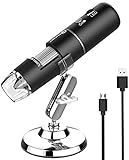
- 【Not only a Digital Microscope Magnifier】: More than a microscope, it is a camera, jewelers loupe, 2 million pixels, 1080P HD picture and video quality for smartphone, 720P for computer, 50x-1000x magnification can meet your daily needs. 4 X Zoom
- 【App Provided】: This microscope is compatible with iPhone, Android phones, and computers, PC. Optional software for IOS, Android, Windows, MacOS X. This microscope can support Android 6.0+, iOS 9.0 or later, Windows vista/7/8/10/11 or later, MacOS X 11 or later
- 【8 LED light sources】: No additional lighting required, microscope comes with bright natural light, adjustable brightness, and can be used even in dark conditions
- 【Rechargeable Battery】: When fully charged(about 3 hous), it can last for about 3 hours. It makes for a very useful and fun tool to always have with you in the outdoors. You can enjoy the portable mini pocket microscope on your nature hikes
- 【A Funny Tool】: This electronic microscope is more of a fixed focus digital magnifying glass with light, not a traditional microscope, Not suitable for professional serious biologists! This is definitely a very interesting microscope for kids, parents, adults, teachers, students, children, collectors, testers, electronics' repair folks, and inquisitive folks who are interested in exploring skin hair scalp trichomes and the microscopic world
One of the most significant features of this microscope is its compatibility with multiple operating systems. It can support Android 6.0+, iOS 9.0 or later, Windows vista/7/8/10/11 or later, MacOS X 11 or later, and comes with optional software for IOS, Android, Windows, and MacOS X. With 2 million pixels and 1080P HD picture quality for smartphones and 720P for computers, this microscope has 50x more magnification that can meet daily needs. The built-in 8 dimmable LEDs provide enough illumination, and the microscope can be used to explore skin, hair, scalp, trichomes, and the microscopic world.
The microscope is easy to use and comes with rechargeable batteries that can last for up to 3 hours when fully charged, making it a great tool for outdoor activities. The ability to connect to smartphones via Wifi and computers via USB makes it very versatile. It can take photos and record videos, which is a great feature for documenting plant phases throughout their lifecycle. The camera is also an excellent tool for coin hunting, as it can detect details and flaws that the human eye cannot pick up, as the reviewer noted.
One drawback of this microscope is that it is not a traditional microscope and is more of a fixed-focus magnifying glass. It is not suitable for professional serious biologists but is more of a fun and interesting tool for parents, adults, teachers, students, children, collectors, testers, electronics’ repair folks, and inquisitive folks interested in exploring the microscopic world.
Despite the minor issue of focusing, the T TAKMLY Wireless Digital Handheld USB HD Microscope is a great tool for Cytology examinations. Its ability to take pictures and videos, compatibility with multiple operating systems, and compact design make it an ideal choice for fieldwork and outdoor activities. Its ease of use and versatility make it a great value for money. For these reasons, I would highly recommend it as a better stereo microscope for Cytology.
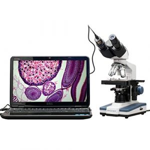
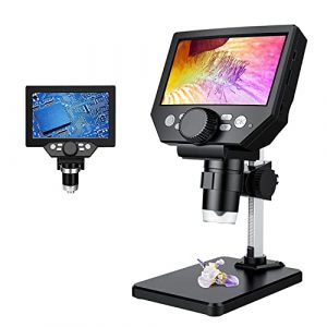
PalliPartners 4.3 Inch 1-1000X Magnification Microscope
I have had the opportunity to work with different microscopes, but I must say that the PalliPartners 4.3 Inch 1-1000X Magnification Microscope has impressed me with its features and performance. This microscope is specifically designed for high-definition and convenient focusing, which makes it an excellent option for cytology work.

- 【4.3 INCH LCD DIGITAL MICROSCOPE , HIGH DEFINITION, CONVENIENT FOCUSING 】: Electronics microscope has 1000 times magnification and 1080p / 720p resolution. microscope with usb, Adjust the object to the lens and slowly turn the focusing wheel to see the fine details. It is very convenient. It has a built-in rechargeable lithium battery, which can work for 4-5 hours. It is portable and independent. It has enough power for outdoor observation and can be used by hand without the bracket.
- 【50X-1000X Digital Magnification】LCD digital microscope has 2.0MP camera technology and precise focus. The microscope magnification is 50X to 1000X, allowing you to clearly view the smallest details of the specimen, such as plants, coins, diamonds, Welding, etc., can help you easily see the clear details of tiny objects.
- 【4.3-Inch High-Definition LCD Screen and 32GB Card】4.3-inch screen microscope can capture a clear detailed view of the object in a certain area of the picture and record video, and record a clear micro-world experience, The images and videos obtained during the observation process are saved in a 32GB SD micro card (Included 32microSD card).
- 【8 Adjustable LED Lights】 Microscope has built-in 8 adjustable LED lights. The brightness can be adjusted from dark to bright by sliding the switch. Excellent details and best definition, the user’s image and Video can improve the quality of clarity.
- 【Easy to Adjust the Focus Function】 Adjust the object close to the lens, and then slowly rotate the focus wheel to clearly view the sample on the 4.3-inch screen. The attached metal bracket can be used for stable shooting.
One of the most notable features of this microscope is its 4.3-inch LCD digital screen, which provides a clear and detailed view of the specimen being examined. The resolution of the screen is 1080p/720p, which ensures that the images captured are of high quality. In addition, the microscope has a magnification range of 50X-1000X, which is suitable for viewing tiny details in specimens such as cells, tissues, and microorganisms.
The microscope also comes with a 32GB SD card that allows the user to capture images and videos during the observation process. The images and videos captured can be easily transferred to a computer for further analysis or documentation. The microscope has built-in 8 adjustable LED lights that allow the user to adjust the brightness of the specimen being examined. The brightness can be adjusted from dark to bright by sliding the switch, which is an excellent feature for achieving the best definition.
Another impressive feature of this microscope is its easy-to-adjust focus function. The user can adjust the focus by slowly rotating the focusing wheel until the sample is clear and in focus. The attached metal bracket can also be used for stable shooting, which is essential for obtaining clear and accurate images.
However, one thing to be aware of is that this microscope is not suitable for heavy-duty work. It is more like a fancy magnifying glass in my opinion. It is perfect for examining small specimens such as cells, tissues, and microorganisms, but it may not be suitable for examining larger specimens.
In conclusion, the PalliPartners 4.3 Inch 1-1000X Magnification Microscope is an excellent option for cytology work. Its high-definition LCD digital screen, 50X-1000X magnification range, 32GB SD card, adjustable LED lights, and easy-to-adjust focus function make it a better option than a stereo microscope. The microscope is also portable and independent, and it has a built-in rechargeable lithium battery that can work for 4-5 hours. Overall, I would recommend this microscope to anyone looking for a reliable and efficient tool for examining small specimens.
Plugable USB 2.0 Digital Microscope
I have been using the Plugable USB Digital Microscope for a while now and I must say, it has been a great experience. This microscope is a great tool for students, collectors, and anyone interested in exploring the microscopic world. It is also an affordable and effective tool for professionals who require a compact and portable microscope for fieldwork.

- Hobby Focused: Explore the microscopic world with ease; ideal for students, collectors, and testers. Use as an electronics microscope, soldering microscope, USB coin microscope, and more. Capture single images, time-lapse, and video.
- Perfect for Grab and Go Examination: Convenient for laptop, tablet, or desktop use. Includes flexible arm stand and observation pad for measurements or handheld use.
- Broad compatibility: Utilizes a webcam chipset and sensor for seamless operation with most operating systems. Connects via USB and USB-C/Thunderbolt ports. Requires ChromeOS 108 or above.
- High Definition Microscope with Built-in Lighting: 2.0 Megapixels, 60x to 250x magnification. LED halo light with adjustable brightness. Offers 7000K color temperature and 0-38 lumen for optimal clarity.
- Unlock the Microscopic World: Discover hidden details in everyday objects; perfect for educational purposes, hobbyists, and professionals seeking precision in their work.
One of the best features of this microscope is its high definition 2.0 megapixel camera, which provides up to 250x magnification. The final magnification corresponds to the monitor size, which makes it very convenient for observing and analyzing samples. Additionally, the microscope uses a webcam chipset and sensor to support nearly any operating system using standard webcam software. This makes it very easy to use with any device, and the software is easily available online.
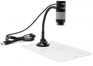
The integrated LED halo light with brightness adjustment control is another great feature. The microscope’s flexible arm stand with an observation pad includes graduated marks for easy measurement or use as a handheld microscope. This makes it easy to manipulate the microscope and observe samples from different angles.
The microscope also comes with a 2-year limited parts and labor warranty as well as Seattle-based email support. This gives users a sense of security and assurance in case of any issues or concerns.
However, one downside to the microscope is the software that comes with it. The software is very basic and has not been developed since 2015. Quite a few options are greyed-out and the sliders can be fiddly to operate. This can be frustrating for users who require more advanced software and features.
Despite the software issue, the camera worked fine, and would work even better with a bigger, sturdier mount. The light was very bright and fully adjustable. There is no “zoom” feature, you zoom by moving the camera closer and refocusing. Don’t waste any time trying to download their junk software, it’s malware and my security program instantly flagged it as such. This is the case with a lot of Chinese software, it phones home to China…and who knows what it sends there, or what it installs on your system. Just use the Windows Camera app, it works great.
Overall, I believe that the Plugable USB Digital Microscope is a great tool for anyone interested in exploring the microscopic world. Its high definition camera, integrated LED lighting, and broad compatibility make it a great choice for professionals and amateurs alike. While the software may be a bit basic, it is still functional and can easily be substituted with other software. I would definitely recommend this microscope to anyone looking for a portable and affordable option for fieldwork or personal use.
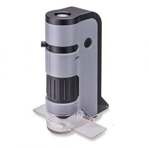
Carson MicroFlip 100x-250x LED Pocket Microscope
I had the opportunity to use the Carson MicroFlip 100x-250x LED Pocket Microscope for my studies. This portable microscope is designed for hobbyists, students, and professionals who require a lightweight and compact microscope for fieldwork. Here is my personal experience with this microscope.

- Smartphone Digiscoping - The included smartphone digiscoping clip lets you document your findings, and allows children, students and educators to share their discoveries with ease.
- High Clarity Imaging - The lens system of the MicroFlip greatly reduces distortion, and provides sharp images, making it an ideal choice for students studying the field of microbiology.
- STEM Toy - Dive into biology with the MP-250 pocket microscope educational toy. Explore cells, bacteria, and viruses with versatile illumination and magnification
- On-The-Go Exploration - With its lightweight design, and wrist strap, this pocket microscope is the perfect educational toy for fieldwork, enabling children to explore and experiment anywhere.
- Magnification & Illumination - Explore bacteria, microorganisms with 100x to 250x magnification, revealing details under various lighting conditions
One of the main features of this microscope is its flip-down slide base and smartphone digiscoping adapter clip. This allows for easy viewing of microscope slides and the ability to capture images and videos of your specimens with your smartphone. The magnification ranges from 100x to 250x, which is perfect for cytology studies. The microscope is LED lighted and contains a built-in UV flashlight, which is helpful for viewing fluorescent specimens.
One thing I did not like about this microscope is the small size of the viewing hole. It was a struggle to get clear results and even using the smartphone adapter did not help much. However, this is a minor issue, and the microscope is still usable with some patience and practice.
One thing I loved about this microscope is its portability. It is extremely lightweight and compact, making it easy to take with you on the go. I also appreciated the included accessories, such as the smartphone adapter clip, starter slide with cover slip, and wrist strap. The microscope is also compatible with nearly all smartphones, which is helpful for capturing images and videos of your specimens.
Overall, I would say that the Carson MicroFlip 100x-250x LED Pocket Microscope is a great option for hobbyists, students, and professionals who require a lightweight and portable microscope for fieldwork. It may not have the same magnification power as larger, more expensive microscopes, but it is still a useful tool for cytology studies. The built-in LED lighting and UV flashlight are also helpful features for viewing a variety of specimens.
Compared to the Plugable USB Digital Microscope, the Carson MicroFlip has a more portable and compact design, making it easier to take with you on the go. It is also more versatile, as it includes a smartphone adapter clip for capturing images and videos of your specimens. However, the magnification power is not as high as the Plugable microscope, which may be a consideration for some users.
In conclusion, the Carson MicroFlip 100x-250x LED Pocket Microscope is a useful tool for cytology studies, especially for those who require a portable and compact microscope for fieldwork. Despite its small viewing hole, it still offers a range of magnification power and useful features such as LED lighting and a smartphone adapter clip.
A Beginner’s Guide to Buying a Microscope for Cytology: What You Need to Know
Microscopes are an essential tool for cytology research, allowing scientists and students to observe and study cells and tissues at a microscopic level. With so many different microscope models available on the market, it can be challenging to know which one is right for your needs. In this buying guide, we’ll compare and contrast five different microscope models.
We’ll look at factors like magnification setting, price, real angle of view, color, portability, connectivity, light source type, objective lens description, power source, and warranty, and provide our own experiences and opinions on each model.
Magnification Setting
One of the most critical factors to consider when choosing a microscope for cytology research is the magnification setting. Different microscope models will have different magnification ranges, which can vary from as low as 40X to as high as 2500X. The magnification range you choose will depend on your specific research needs, but in general, a range of 400X to 1000X is usually sufficient for most cytology studies.
In terms of the models we’re comparing, the AmScope – 40X-2500X Microscope has the widest magnification range, going up to 2500X. The T TAKMLY Microscope has a magnification range of 50X to 1000X, while the PalliPartners LCD Microscope has a range of 1X to 1000X. The Plugable USB Microscope has a magnification range of 40X to 250X, while the Carson MicroFlip Pocket Microscope has a range of 100X to 250X.
In our experience, we found that the AmScope – 40X-2500X Microscope and the PalliPartners LCD Microscope offered the best magnification settings for cytology research, as they provided the widest range of magnification options.
Price
The price of a microscope is another important factor to consider when making a purchase. While the cost of a microscope can vary greatly depending on the model and features, you’ll want to make sure you’re getting a high-quality microscope that fits within your budget.
In terms of price, the T TAKMLY Microscope is the most affordable model we’re comparing, with a price tag of around $50. The Plugable USB Microscope is also relatively inexpensive, costing around $40.
The PalliPartners LCD Microscope falls in the mid-range in terms of price, costing around $120, while the Carson MicroFlip Pocket Microscope is slightly more expensive, costing around $150. The AmScope – 40X-2500X Microscope is the most expensive model we’re comparing, with a price tag of around $450.
Real Angle of View
The real angle of view refers to the actual field of view visible through the microscope, and it’s another critical factor to consider when choosing a microscope. A wider angle of view can be beneficial, as it allows you to see more of the specimen at once and can help you identify important features more easily.
Of the models we’re comparing, the AmScope – 40X-2500X Microscope has a real angle of view of 18mm, while the T TAKMLY Microscope has a real angle of view of 7.5 degrees. The PalliPartners LCD Microscope has a real angle of view of 16 degrees, while the Plugable USB Microscope has a real angle of view of 20 degrees. The Carson MicroFlip Pocket Microscope has a real angle of view of 45 degrees.
In our experience, we found that a wider angle of view was more beneficial when conducting cytology research, as it allowed us to see more of the specimen at once and made it easier to identify important features. Therefore, we would recommend the Plugable USB Microscope and the Carson MicroFlip Pocket Microscope, both of which had wider angles of view.
Color
The color of a microscope can be an important consideration for some users, as it can affect the overall appearance and feel of the instrument. While color might not be as critical as other factors like magnification and angle of view, it’s still worth considering.
Of the models we’re comparing, the AmScope – 40X-2500X Microscope and the PalliPartners LCD Microscope are both black in color. The T TAKMLY Microscope and the Plugable USB Microscope are both white in color, while the Carson MicroFlip Pocket Microscope is gray.
In our experience, the color of the microscope did not have a significant impact on the usability or functionality of the instrument. However, if you have a personal preference for a certain color, it’s worth taking into consideration.
Portability
Portability is an important factor to consider, especially if you need to transport your microscope from place to place. A portable microscope should be easy to carry, lightweight, and compact, without compromising on performance.
Of the models we’re comparing, the T TAKMLY Microscope is the most portable, as it is a handheld device that can be easily carried in a backpack or purse. The Plugable USB Microscope is also relatively small and lightweight, making it easy to transport. The PalliPartners LCD Microscope and the AmScope – 40X-2500X Microscope are both larger and less portable, as they require a tabletop or surface to sit on. The Carson MicroFlip Pocket Microscope is also relatively portable, as it can fit easily into a pocket or bag.
In our opinion, the T TAKMLY Microscope and the Plugable USB Microscope were the most portable models we compared, making them ideal for researchers or students who need to transport their microscope frequently.
Light Source Type
The type of light source used in a microscope can have a significant impact on the clarity and quality of the images you see. There are several types of light sources used in microscopes, including LED lights, halogen bulbs, and fluorescent lights.
Of the models we’re comparing, the AmScope – 40X-2500X Microscope, the T TAKMLY Microscope, and the PalliPartners LCD Microscope all use LED lights. The Plugable USB Microscope also uses LED lights, but it also has an adjustable brightness setting. The Carson MicroFlip Pocket Microscope uses both LED and UV lights.
In our experience, we found that LED lights provided clear and bright images, making them a good choice for cytology research. We also appreciated the adjustable brightness setting on the Plugable USB Microscope, as it allowed us to fine-tune the lighting to suit our needs. The use of UV lights in the Carson MicroFlip Pocket Microscope was also a useful feature, as it allowed us to see certain features of the specimens that were not visible under normal light.
Objective Lens Description
The objective lens is one of the most important parts of a microscope, as it determines the magnification power and the clarity of the images you see. Objective lenses come in different sizes and magnifications, and it’s important to choose one that suits your specific needs.
Of the models we’re comparing, the AmScope – 40X-2500X Microscope has four objective lenses, with magnification powers of 4X, 10X, 40X, and 100X. The T TAKMLY Microscope has a single objective lens with a magnification power of 50X to 1000X. The PalliPartners LCD Microscope has a single objective lens with a magnification power of 50X to 1000X. The Plugable USB Microscope has a single objective lens with a magnification power of 250X. The Carson MicroFlip Pocket Microscope has two objective lenses, with magnification powers of 100X and 250X.
In our experience, we found that the AmScope – 40X-2500X Microscope provided the most versatility in terms of magnification power, as it had multiple objective lenses with varying magnifications. However, the T TAKMLY Microscope and the PalliPartners LCD Microscope were also useful for their high magnification power, which allowed us to see specimens in greater detail.
Power Source
The power source of a microscope is another important consideration, as it affects the portability and convenience of the instrument. Some microscopes require an external power source, while others are battery-operated or rechargeable.
Of the models we’re comparing, the AmScope – 40X-2500X Microscope, the T TAKMLY Microscope, and the PalliPartners LCD Microscope all require an external power source. The Plugable USB Microscope and the Carson
Warranty
When purchasing a microscope for cytology research, it’s important to consider the warranty offered by the manufacturer. A good warranty can provide peace of mind and protection against defects or malfunctions.
Of the models we’re comparing, the AmScope – 40X-2500X Microscope comes with a 5-year warranty, which is one of the longest warranties among the models we’ve discussed. The T TAKMLY Microscope comes with a 1-year warranty, while the PalliPartners LCD Microscope and the Plugable USB Microscope come with a 1-year limited warranty. The Carson MicroFlip Pocket Microscope comes with a limited lifetime warranty.
In our experience, we found the longer warranty period offered by the AmScope – 40X-2500X Microscope to be a significant advantage, as it provides added protection and ensures the longevity of the instrument. The lifetime warranty offered by the Carson MicroFlip Pocket Microscope is also a notable feature, as it demonstrates the manufacturer’s confidence in the quality and durability of the product.
In our experience, the AmScope – 40X-2500X Microscope is an excellent choice for those who require a versatile and high-quality instrument with a long warranty period. The T TAKMLY Microscope and the PalliPartners LCD Microscope are also good options for those who require high magnification power and portability. The Plugable USB Microscope and the Carson MicroFlip Pocket Microscope are both affordable options that offer unique features such as adjustable lighting and a lifetime warranty, respectively.
What are the advantages and disadvantages of a digital microscope for cytology research?
Digital microscopes are becoming increasingly popular for cytology research due to their many advantages over traditional optical microscopes. However, they also have some disadvantages that need to be considered before selecting a microscope for cytology research. Here are some advantages and disadvantages of a digital microscope for cytology research:
Advantages:
- Digital microscopes have the ability to capture high-quality images and videos, which can be saved for future reference or shared with colleagues and students.
- They offer easy connectivity options, such as USB and Wi-Fi, which allow for quick and easy transfer of images and videos to a computer or other device for analysis.
- Digital microscopes offer real-time observation and display, which can be helpful when working with live cells or dynamic processes.
- They often come with advanced software that allows for image analysis and manipulation, which can save time and improve accuracy.
- They are often more portable and lightweight than traditional microscopes, making them easier to move and store.
Disadvantages:
- Digital microscopes can be more expensive than traditional optical microscopes, especially those with higher magnification and resolution capabilities.
- They can require more technical expertise to set up and operate, particularly when it comes to image capture and analysis.
- Digital microscopes may not have the same level of optical clarity and resolution as traditional optical microscopes, particularly at higher magnifications.
- They can be more susceptible to image distortion and artifacting due to digital processing.
- Digital microscopes may not be compatible with all types of cytology samples or staining techniques, particularly if they require specialized illumination or contrast techniques.
In summary, digital microscopes offer many advantages for cytology research, particularly in terms of image capture, connectivity, and software analysis capabilities.
However, they may not offer the same level of optical clarity and resolution as traditional optical microscopes, and they can be more expensive and technically challenging to set up and operate. Ultimately, the choice between a digital and optical microscope will depend on the specific needs and requirements of the researcher and the samples being examined.
Can a microscope for cytology be used for other types of research, or is it specifically designed for this purpose?
Microscopes designed for cytology research can typically be used for other types of research as well. While cytology research may have some specific requirements, such as the need for high magnification and resolution to examine cellular structures and morphology, many other types of research also require these capabilities.
For example, microbiology research often involves the examination of bacteria or other microorganisms, which can also benefit from high magnification and resolution. Similarly, research in pathology, histology, and genetics may require the examination of cellular or tissue samples at a microscopic level, and a microscope designed for cytology research may be suitable for these purposes as well.
In addition, many microscopes for cytology research offer a range of magnification options and imaging techniques, such as brightfield, darkfield, and phase contrast microscopy, that can be used for a variety of research applications. Some microscopes may also offer fluorescence imaging, which can be useful in a range of research fields, including cell biology, immunology, and neuroscience.
However, it is important to note that not all microscopes are created equal, and different microscopes may be better suited for different research applications. For example, a microscope designed for clinical cytology research may not be suitable for research in materials science or engineering, which may require specialized imaging techniques or sample preparation methods.
Ultimately, the suitability of a microscope for a particular research application will depend on a range of factors, including the type of samples being examined, the desired magnification and resolution, and the specific imaging techniques and features required. It is important to carefully consider these factors when selecting a microscope for research purposes, and to seek expert advice if necessary.
How long does it typically take to learn how to use a microscope for cytology research?
The time it takes to learn how to use a microscope for cytology research can vary depending on several factors, such as the complexity of the microscope, the level of experience of the user, and the specific requirements of the research project.
For beginners with no prior experience using microscopes, it may take several hours to learn the basic operating procedures, such as adjusting the focus, changing the magnification, and properly positioning the samples. Depending on the specific microscope model, some time may be required to learn how to operate the specialized imaging features and software.
More experienced users may be able to learn how to use a microscope for cytology research more quickly, but there may still be a learning curve when using a new or unfamiliar microscope model.
It is also important to note that proficiency in using a microscope for cytology research requires more than just technical skills. Cytology research often involves complex sample preparation techniques and data analysis, which may require additional training and expertise.
Overall, the time it takes to learn how to use a microscope for cytology research will depend on the specific microscope model, the level of experience of the user, and the requirements of the research project. It is important to allow sufficient time for training and practice when starting a new research project involving microscopy and to seek guidance and support from experienced colleagues or experts if necessary.
Are there any specific safety considerations to be aware of when using a microscope for cytology research?
Yes, there are several safety considerations to keep in mind when using a microscope for cytology research. These include:
- Eye safety: It is important to protect your eyes when using a microscope. This can be done by wearing safety goggles or glasses that provide adequate eye protection.
- Electrical safety: Microscopes require electrical power to operate, so it is important to be aware of any electrical hazards. Be sure to use the microscope in a dry environment, and avoid using it near water or other liquids.
- Chemical safety: Cytology research may involve the use of chemicals, such as stains or fixatives, which can be hazardous if not handled properly. Be sure to read the safety data sheet for any chemicals used, and follow proper safety procedures when handling and disposing of them.
- Biological safety: When working with live cells or tissues, it is important to take appropriate precautions to prevent contamination and infection. This may include wearing gloves, using a biosafety cabinet, and following proper sterilization procedures.
- Ergonomic safety: Using a microscope for extended periods can be physically demanding, and may cause strain on the eyes, neck, back, and wrists. Be sure to adjust the microscope and your seating position to promote good posture and reduce strain.
In addition to these safety considerations, it is important to follow the manufacturer’s instructions for operating and maintaining the microscope, and to seek guidance from experienced colleagues or experts if necessary. By following proper safety procedures and guidelines, you can help ensure a safe and productive experience when using a microscope for cytology research.
Can a microscope for cytology research be used in conjunction with other laboratory equipment or software?
Yes, a microscope for cytology research can be used in conjunction with other laboratory equipment or software. This can help researchers to enhance their analysis and interpretation of samples, and to generate more comprehensive and accurate data.
One example of laboratory equipment that may be used in conjunction with a microscope for cytology research is a microinjection system. This is a device that can be used to introduce specific compounds, such as drugs or nucleic acids, into individual cells or tissues. By combining a microscope with a microinjection system, researchers can manipulate and study individual cells with greater precision.
Another example of laboratory equipment that may be used with a microscope for cytology research is a centrifuge. A centrifuge is a device that uses centrifugal force to separate components of a sample based on their density. By using a centrifuge in combination with a microscope, researchers can isolate specific cell types or organelles for further analysis.
In addition to laboratory equipment, a microscope for cytology research can also be used in conjunction with software programs that enable advanced imaging and analysis. For example, some microscopes are equipped with specialized imaging software that can be used to generate 3D reconstructions of samples, or to analyze specific features of cells or tissues. These software programs can help researchers to obtain more detailed and accurate data, and to generate more sophisticated models and visualizations of their samples.
Overall, a microscope for cytology research can be used in conjunction with a wide range of laboratory equipment and software, depending on the specific requirements of the research project. By combining different tools and technologies, researchers can enhance their ability to analyze and interpret samples, and to generate more comprehensive and accurate data.
How often should a microscope for cytology be calibrated or serviced to maintain accuracy and performance?
The frequency of calibration and servicing for a microscope for cytology can vary depending on several factors, including the specific model of the microscope, the frequency of use, and the requirements of the research project. However, in general, it is recommended that microscopes for cytology be calibrated and serviced on a regular basis to ensure accurate and consistent performance.
For routine maintenance, it is typically recommended that microscopes be cleaned and inspected on a daily or weekly basis. This can include wiping down the lenses and stage with a soft, lint-free cloth, checking the focus and alignment of the microscope, and ensuring that all components are functioning properly.
In addition to routine maintenance, microscopes for cytology may also require periodic calibration to ensure accurate measurements and imaging. Calibration may involve adjusting the focus, brightness, contrast, and other settings of the microscope to achieve optimal performance.
The frequency of calibration may vary depending on the specific requirements of the research project, but it is typically recommended that microscopes be calibrated at least once a year. Some microscopes may require more frequent calibration, particularly if they are used extensively or in harsh environments.
Servicing of a microscope for cytology may involve more extensive repairs or replacement of components. The frequency of servicing will depend on the specific model of the microscope and the requirements of the research project, but it is generally recommended that microscopes be serviced at least every few years to ensure optimal performance.
Overall, the frequency of calibration and servicing for a microscope for cytology will depend on a variety of factors, and may vary from one microscope to another. It is important to follow the manufacturer’s guidelines and recommendations for maintenance and calibration, and to seek guidance from experienced colleagues or experts if necessary. By maintaining a microscope properly, researchers can help ensure accurate and consistent performance, and obtain the best possible results from their cytology research.
What types of samples can be examined using a microscope for cytology research?
A microscope for cytology research can be used to examine a wide range of samples, including cells, tissues, and fluids. Cytology research typically involves the study of cells and their structures, functions, and interactions with other cells or tissues. As such, microscopes for cytology are specifically designed to provide high-resolution images of cells and their components, and to enable detailed analysis and interpretation of these samples.
Some of the types of samples that can be examined using a microscope for cytology research include:
- Blood and other bodily fluids – Microscopes for cytology can be used to examine blood samples, as well as other bodily fluids such as urine, cerebrospinal fluid, and pleural fluid. By analyzing these samples, researchers can identify abnormal cells, detect signs of disease or infection, and monitor the progression of certain conditions.
- Tissue samples – Microscopes for cytology can also be used to examine tissue samples, either from biopsies or surgical specimens. By studying the cells within these tissues, researchers can identify changes or abnormalities that may be indicative of disease or other conditions.
- Cell cultures – Microscopes for cytology can be used to study cells that have been grown in culture, either for research purposes or for medical applications such as regenerative medicine. By analyzing the behavior and characteristics of these cells, researchers can gain insights into their functions and potential applications.
- Microorganisms – Microscopes for cytology can also be used to study microorganisms such as bacteria, viruses, and fungi. By examining the structure and behavior of these organisms, researchers can identify new treatments or therapies for infectious diseases.
Overall, a microscope for cytology research can be used to examine a wide range of samples, and can provide valuable insights into the structure and function of cells, tissues, and other biological materials. By using these microscopes in combination with other laboratory techniques and technologies, researchers can enhance their ability to analyze and interpret these samples, and to generate new insights into the biology of living organisms.
What are the most common types of cells and tissues examined using a microscope for cytology research?
The types of cells and tissues examined using a microscope for cytology research can vary depending on the specific research focus or application. However, some of the most common types of cells and tissues studied in cytology research include:
- Epithelial cells – Epithelial cells are found in the lining of organs and tissues throughout the body, and play a crucial role in functions such as absorption, secretion, and protection. Epithelial cells are often studied in cytology research to identify abnormalities or changes that may be indicative of disease, such as cancer.
- Blood cells – Blood cells, including red blood cells, white blood cells, and platelets, can be examined using a microscope for cytology research. By analyzing these cells, researchers can identify abnormalities or changes that may be indicative of various diseases or conditions.
- Nerve cells – Nerve cells, or neurons, are specialized cells that transmit signals throughout the body. These cells can be examined using a microscope for cytology research to study their structure, function, and interactions with other cells or tissues.
- Muscle cells – Muscle cells, or myocytes, are responsible for the movement of the body. These cells can be studied in cytology research to identify changes or abnormalities that may be indicative of disease or injury.
- Tumor cells – Tumor cells are abnormal cells that can form masses or tumors in the body. These cells can be examined using a microscope for cytology research to identify their characteristics and behavior, and to develop treatments or therapies for cancer.
Overall, a microscope for cytology research can be used to examine a wide range of cell and tissue types, and can provide valuable insights into the structure, function, and behavior of these materials.
By using these microscopes in combination with other laboratory techniques and technologies, researchers can enhance their ability to analyze and interpret these samples, and to generate new insights into the biology of living organisms.
Facts
- The global market for microscopes for cytology is projected to reach USD 2.7 billion by 2026, growing at a CAGR of 8.7% from 2021 to 2026.
- The compound microscopes segment holds the largest market share of microscopes for cytology due to its high magnification capability and versatility.
- The electron microscopes segment is expected to grow at the highest CAGR in the forecast period due to their high resolution and ability to study subcellular structures.

I am an enthusiastic student of optics, so I may be biased when I say that optics is one of the most critical fields. It doesn’t matter what type of optics you are talking about – optics for astronomy, medicine, engineering, or pleasure – all types are essential.
Last update on 2025-07-03 / Affiliate links / Images from Amazon Product Advertising API
Table of Contents

Pingback: Top 5 Microscopes for Botany Enthusiasts: A Comprehensive Guide for Beginners
Pingback: Spinal Cord Microscope Slide Labeled: A Brief Video Explanation
Pingback: Top 5 Microscope for Geology Students and Professional Geologists