As someone with extensive experience in the field of microscopy, I highly recommend the 10PK Spinal Cord, Cross Section microscope slides from Eisco Labs for use in the classroom or laboratory setting. These prepared slides offer a detailed and accurate representation of the spinal cord cross section, allowing students to examine and understand the structure and function of this vital part of the nervous system. With 10 slides included in a convenient storage case, this pack is an ideal addition to any biology or microscopy curriculum. Eisco Labs is known for their high-quality and reliable lab equipment, and these microscope slides are no exception.

- Single, prepared slide with spinal cord cross section
- Excellent for biological study
- Slide measures 75mm wide and 25mm long
- Ideal for exploring structure-function connections as per NGSS standards
- Arrives in a protective cardboard casing
What kind of support is available from Eisco Labs for the microscope slide pack?
Eisco Labs provides various forms of support for their 10PK Spinal Cord, Cross Section – Prepared Microscope Slides – Classroom Pack, 10 Slides in Storage Case. The company offers a warranty on their product, which covers any defects or issues with the slides or storage case.
Additionally, Eisco Labs has a customer service team that can be reached via phone or email to answer any questions or concerns about the product. The company also has an extensive online resource center, which includes instructional videos, manuals, and guides that can assist users in properly using and caring for their microscope slides.
Overall, Eisco Labs is committed to providing high-quality products and support to their customers, ensuring a positive experience with their microscope slide pack.
How do the microscope slides compare to similar products on the market?

To compare the 10PK Spinal Cord, Cross Section microscope slides from Eisco Labs to similar products on the market, it’s important to consider factors such as quality, price, and features.
In terms of quality, Eisco Labs is known for producing high-quality microscope slides that are carefully prepared and labeled. The company uses high-quality stains and dyes to enhance the visibility of the specimens on the slides.
In terms of price, the 10PK Spinal Cord, Cross Section microscope slides are competitively priced compared to other similar products on the market. While the price may vary depending on the seller and location, Eisco Labs provides an affordable option for classroom or personal use.
One of the unique features of the Eisco Labs microscope slide pack is the inclusion of a storage case, which keeps the slides organized and protected. Additionally, the slides are labeled with the anatomical structure and are ideal for use in a classroom setting or for personal study.
Overall, the Eisco Labs 10PK Spinal Cord, Cross Section microscope slides compare favorably to similar products on the market in terms of quality, price, and features. The company’s reputation for producing high-quality microscope slides, along with their commitment to customer support, makes them a trusted choice for educators and researchers alike.
Are there any special handling instructions for the microscope slides?
Yes, there are some special handling instructions that should be followed when working with the 10PK Spinal Cord, Cross Section microscope slides from Eisco Labs. Here are a few guidelines to keep in mind:
- Handle the microscope slides carefully, as they are fragile and can break or scratch easily.
- Use only clean, lint-free cloths or lens paper to clean the slides.
- Avoid touching the specimen or cover slip with your fingers, as this can leave oil or debris on the slide and affect the visibility of the sample.
- Store the microscope slides in a cool, dry place, away from direct sunlight or heat sources.
- Keep the microscope slides in their original storage case to protect them from damage or contamination.
- Follow proper disposal procedures for the microscope slides, as they may contain hazardous chemicals or biological materials.
By following these handling instructions, you can ensure that the microscope slides remain in good condition and provide accurate results when used for microscopy or research.Bottom of Form
Can the microscope slide pack be used for educational purposes outside of the classroom?
Yes, the microscope slide pack can certainly be used for educational purposes outside of the classroom, such as in a homeschooling setting, for personal study, or in other educational institutions.
The pack is designed to provide a hands-on learning experience and is suitable for a wide range of learners, including students, teachers, hobbyists, and professionals. It can be used to enhance understanding of biological concepts and can also serve as a resource for research purposes.
How long will the microscope slides last?
The longevity of microscope slides can vary depending on factors such as storage conditions and frequency of use. If the microscope slides are stored properly, kept away from moisture, dust, and excessive heat, they can last for several years.
However, if they are mishandled or stored improperly, they may become damaged or contaminated, reducing their lifespan. To extend the life of microscope slides, it is recommended to store them in a cool, dry place, and avoid touching the slide surface to prevent smudging or contamination.
How do the microscope slides relate to the study of botany?
The microscope slides can be useful in the study of botany as they can provide a close-up view of plant tissues and structures. Botanists can use microscope slides to observe and identify different types of plant cells, such as leaf epidermal cells, root hairs, stem cross-sections, and various reproductive structures.
By examining these structures under the microscope, botanists can gain a deeper understanding of how plants function, grow, and reproduce. The microscope slides can also be used to study plant diseases, such as fungal infections or viruses, by examining the tissue samples for signs of infection or damage.
Overall, microscope slides can provide valuable insights into the microscopic world of plants and support the study of botany.
How do the microscope slides relate to the study of zoology?
The microscope slides can be helpful in the study of zoology by allowing students to examine the structure and function of various animal tissues and organs. Zoologists can use microscope slides to observe and identify different types of animal cells, such as muscle cells, nerve cells, and blood cells, as well as the structures of organs such as the liver, heart, and lungs.
By studying these structures under the microscope, zoologists can learn about the physiology and behavior of animals, and gain insights into how they function and interact with their environment. Microscope slides can also be used to examine animal parasites and pathogens, such as bacteria, viruses, and fungi, to understand their impact on animal health and behavior.
Overall, microscope slides can be a valuable tool for the study of zoology, allowing students to gain a deeper understanding of animal anatomy and physiology at a microscopic level.
What kind of stains were used on the microscope slides?
The specific stains used on the microscope slides can vary depending on the type of tissue being observed and the purpose of the slide. However, common stains used in preparing microscope slides include hematoxylin and eosin (H&E), which are commonly used to stain a variety of tissues for observation under a microscope.
Other common stains include periodic acid-Schiff (PAS) stain, which stains carbohydrates, and Gram stain, which is used to distinguish between different types of bacteria. Some specialized stains are also used to highlight specific structures or features in the tissue samples, such as the Golgi stain, which highlights the Golgi apparatus, or the Nissl stain, which highlights neuronal cell bodies.
Overall, the choice of stain will depend on the specific research or educational goals of the microscope slide preparation.
Are the microscope slides reusable?
Microscope slides can be reused if they are cleaned and properly maintained after use. However, it is important to note that the process of cleaning and reusing microscope slides can be time-consuming and requires special care to avoid damaging the slide surface.
In most cases, it is more practical to use new microscope slides for each experiment or observation, as this ensures that the slides are clean and free of contaminants that could affect the accuracy of the results. Additionally, reusing slides may result in decreased image quality due to scratches, residual stains, or other forms of damage that accumulate over time.
Overall, while microscope slides can technically be reused, it is usually recommended to use new slides for each experiment or observation.
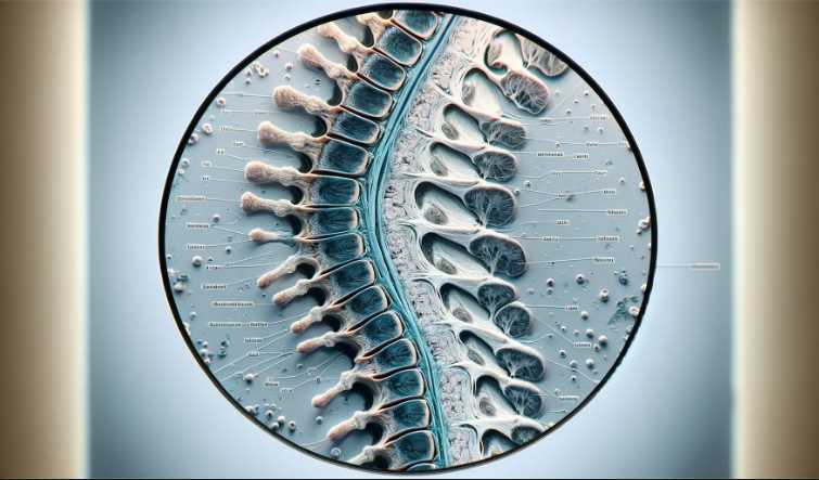
How are Spinal Cord Microscope Slides prepared and labeled?
The preparation of Spinal Cord Microscope Slides involves intricate laboratory procedures. Initially, the spinal cord is carefully extracted and sliced into thin sections. These sections are then mounted on slides and stained to highlight specific structures. Finally, a labeling process is employed to annotate crucial features, ensuring educational clarity.
What staining techniques are commonly used on Spinal Cord Microscope Slides?
Several staining techniques enhance the visibility of structures on Spinal Cord Microscope Slides. Hematoxylin and eosin staining is a common choice, providing contrast between cell nuclei and cytoplasm. Nissl staining highlights neuronal cell bodies, while Luxol Fast Blue staining distinguishes between gray and white matter.
Are there specific regions of interest highlighted on a Spinal Cord Microscope Slide?
Yes, Spinal Cord Microscope Slides often focus on key regions. These include the dorsal and ventral horns, central canal, and nerve roots. Additionally, slides may emphasize the differences between the cervical, thoracic, lumbar, and sacral regions, offering a comprehensive view of the spinal cord’s anatomy.
How can one interpret the labeled structures on a Spinal Cord Microscope Slide?
Interpreting a Spinal Cord Microscope Slide involves understanding the labeled structures and their functions. Reference materials, such as textbooks and online resources, can aid in identifying components like motor neurons, sensory neurons, and supporting cells. Comparing the slide to illustrations and diagrams is also beneficial.
What are the advantages of using labeled Spinal Cord Microscope Slides for teaching purposes?
The use of labeled Spinal Cord Microscope Slides in teaching offers a tangible and visual learning experience. Students can directly observe and identify structures, reinforcing theoretical knowledge. The labeled slides enhance the educational process by providing a practical understanding of spinal cord anatomy.
How can one properly care for and store Spinal Cord Microscope Slides?
Proper care involves storing Spinal Cord Microscope Slides in a cool, dry place, away from direct sunlight. Slides should be handled carefully to prevent damage, and storage containers must provide protection from dust and contaminants. Regular cleaning with appropriate solutions ensures the longevity of the slides.
Facts
- The 10PK Spinal Cord, Cross Section microscope slides are sold in a pack of 10, making them ideal for classroom or laboratory use.
- Each slide is prepared and stained with high-quality dyes to ensure accurate representation of the spinal cord cross section.
- The microscope slides are stored in a durable storage case, which protects them from damage and makes them easy to transport.
- The slides are compatible with most standard compound microscopes, allowing for easy integration into existing laboratory equipment.
- The cross-sectional view of the spinal cord provides a detailed look at the structure and function of this important part of the nervous system.
- These microscope slides are suitable for use in a variety of educational settings, including high schools, colleges, and universities.
- Eisco Labs is a trusted and respected manufacturer of laboratory equipment, known for their commitment to quality and reliability.
- The slides are designed to be used repeatedly, making them a cost-effective addition to any biology or microscopy curriculum.
- The microscope slides are easy to use, even for beginners, and provide a hands-on learning experience that reinforces classroom lectures and textbook information.
- By examining the spinal cord cross section under the microscope, students can gain a deeper understanding of the anatomy and function of the nervous system, which can be applied to a variety of fields and professions.

I am an enthusiastic student of optics, so I may be biased when I say that optics is one of the most critical fields. It doesn’t matter what type of optics you are talking about – optics for astronomy, medicine, engineering, or pleasure – all types are essential.
Last update on 2025-07-01 / Affiliate links / Images from Amazon Product Advertising API
Table of Contents

Pingback: 5 Expert-Recommended Globe Microscope Slides for Precise and Reliable Results"