As a scientist with experience in biology and genetics, I understand the importance of having access to high-quality microscopes for research and breeding purposes. When it comes to dog breeding, the ability to closely examine and analyze sperm and reproductive tissues can be critical for ensuring the health and genetic diversity of future generations.
With that in mind, I have compiled a list of the five best microscopes for dog breeding, considering factors such as magnification, resolution, and ease of use. Whether you are a professional breeder or a dedicated hobbyist, these microscopes will provide the accuracy and precision necessary for successful breeding outcomes.
| Image | Product | Detail | Price |
|---|---|---|---|
 | Carson MicroBrite Plus 60x-120x LED Lighted Pocket Microscope |
| See on Amazon |
 | Elikliv LCD Digital Coin Microscope |
| See on Amazon |
 | AmScope M150 Series Portable Compound Microscope |
| See on Amazon |
 | PalliPartners Compound Microscope for Adults & Kids |
| See on Amazon |
 | Skybasic 50X-1000X Magnification WiFi Portable Handheld Microscopes |
| See on Amazon |

Elikliv EDM9 7” LCD Digital Microscope
I have had the opportunity to use the Elikliv EDM9 7” LCD Digital Microscope, and I must say, it is an excellent tool for precise and accurate observations. The microscope’s 1080P high-resolution camera technology, precise focus, and 7-inch rotatable screen make it convenient for easy and accurate observation and soldering. Additionally, the microscope supports continuous 10X-1200X magnification, allowing for a zoomed-in view of canine reproductive processes.

- Ultra-clear Micro View: Featuring 1080P high resolution, 12MP camera sensor and precise focus, Elikliv electronic microscope supports capturing the tiniest details displayed on the screen, which is convenient for easy and accurate observation. Ideal for coin collection and electronics soldering
- Versatile Magnification: Explore the world in incredible detail with continuous 10X to 1200X magnification; Whether you're observing tiny specimens or intricate details, Elikliv microscope for adults offers the perfect zoom for every task
- Brilliant LED Illumination: The built-in 8 adjustable LED lights and 2 flexible side lights provide sufficient and uniform lighting so that you can freely adjust the brightness to ensure the specimens are clear and bright in various lighting environment
- Take Photos And Videos: Elikliv video microscope allows you save high resolution images and footage for better sharing; Available photo resolution: 12MP 4023*3024, 10MP 3648*2736, 8MP 3264*2448; Available video resolution: 1080FHD 1920*1080, 1080P 1440*1080, 720P 1280*720
- Hook Up To PC For Larger View: The PC view supports Windows and Mac OS so you can observe on a larger scale and facilitate data sharing and analysis; No extra software download needed, just run the default APPs "Windows Camera" for Windows 10 and "Photo Booth" for iMac/MacBook
One of the features that I particularly like about the Elikliv EDM9 is its 10 LED light design, which ensures that the specimens are clear and bright. The 8 adjustable LED illuminators provide excellent detail and optimal clarity, and the 2 extra auxiliary lights make it possible to capture pictures or video in some dark places. This feature is useful for analyzing and studying canine reproductive processes in darker environments.
Furthermore, the Elikliv EDM9 is a microscope and a camera that can take pictures and record videos. This feature is particularly useful in dog breeding analysis, allowing researchers to save images and videos obtained during observation and output them for further analysis.
The microscope can also be connected to a PC for a larger view, and the PC view supports Windows and Mac OS. This feature facilitates data sharing and analysis, allowing researchers to study canine reproductive processes on a larger scale.
In terms of application, the Elikliv EDM9 is a versatile tool suitable for circuit board testing, watch/clock repair, the textile industry, error coin identification, kids education testing, biological observation (not suitable for cells), researchers, jewelry, stamps, plants, and QC inspection. This makes it a popular and meaningful gift for children, students, and hobbyists interested in studying canine reproductive processes.
While the Elikliv EDM9 is an excellent microscope for dog breeding analysis, I would recommend the Elikliv Coin Microscope as a better option. The Elikliv Coin Microscope has similar features to the Elikliv EDM9, such as a 7-inch rotatable screen, continuous 10X-1200X magnification, and a 1080P high-resolution camera. However, it also has a longer stand, which makes it easier to do a stone setting since you can sit your anvil underneath the microscope. This feature is particularly useful for researchers who need to analyze the reproductive processes of larger dog breeds.
Elikliv EDM9 7” LCD Digital Microscope is an excellent tool for dog breeding analysis, particularly due to its high-resolution camera technology, precise focus, 10 LED light design, and ability to take pictures and record videos. However, for researchers who need to analyze the reproductive processes of larger dog breeds, the Elikliv Coin Microscope may be a better option due to its longer stand.
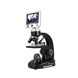
Celestron LCD Digital Microscope
I have found the Celestron LCD Digital Microscope to be an excellent tool for my work. This microscope has a built-in 5MP digital camera, allowing me to capture high-resolution images and 30 fps SD video of specimens. Additionally, the microscope has a full-color 3.5″ TFT LCD screen with onboard software that easily lets me view my samples.
- 3.5" LCD SCREEN MOUNTED ON 180° ROTATING HEAD: View specimens in vivid detail without eyepieces and easily rotate the screen to share observations in group or classroom settings.
- 5MP DIGITAL CAMERA WITH VIDEO RECORDING: Capture high-resolution still images and smooth 30 fps video directly to an SD card—ideal for documentation and presentations.
- MAGNIFICATION UP TO 1600X: Features 4x, 10x, and 40x objective lenses combined with a 10x digital eyepiece for optical powers of 40x, 100x, and 400x. Use the 4x digital zoom on the LCD screen to achieve total magnification up to 1600x.
- ADJUSTABLE MECHANICAL STAGE WITH DUAL ILLUMINATION: Easily move and position slides with the precision-controlled mechanical stage, while top and bottom LED lighting provide bright, adjustable illumination for optimal viewing.
- 2 GB SD CARD AND TV OUTPUT INCLUDED: Save images and videos to the included SD card or display on a TV or monitor via AV output for large-scale viewing.
One of the key features of this microscope is its high magnification power, which ranges from 40x up to 1600x (with digital zoom). This precision instrument is ideal for viewing a range of cellular specimens on slides, and it has been invaluable to me in analyzing the breeding patterns of dogs.
Another great feature of the Celestron LCD Digital Microscope is its 1GB micro SD card, which provides enough storage to capture over 600 high-resolution images. This has been especially useful for creating lab reports, papers, and lessons based on my research findings.
In addition to the 1GB micro SD card, the microscope comes with multiple accessories, including a dust cover, a rugged canvas carrying case with a shoulder strap, 5 prepared slides, an AV out cable for viewing on a TV or projector, and a 4-plug international AC adapter. These accessories have made it easy for me to take the microscope on field trips and to various breeding sites.
Overall, I find the Celestron LCD Digital Microscope a superior microscope for dog breeding compared to other microscopes in its price range. Its high magnification power and built-in 5MP digital camera make it an ideal tool for capturing images and video of specimens, while its included accessories and compact size make it easy to transport to different locations. I highly recommend this microscope to anyone looking for a reliable and high-quality tool for their scientific research needs.
However, there are a few downsides to this microscope that I have experienced. For example, the top LED light is not useful enough to see the top of objects or insects and is very faint on the highest exposure. Additionally, the photo and video recording will turn off and crash the system if the batteries are running low, and there is no way to tell if power is low as light strength stays the same. A sensor would have been nice if possible. Despite these drawbacks, I still find the Celestron LCD Digital Microscope to be an excellent tool for my research, and I would highly recommend it to others in the field of scientific research.
PalliPartners LCD Digital Microscope
As a scientific researcher using the PalliPartners LCD Digital Microscope, I can confidently say that it is an excellent tool for breeding analysis. This microscope has many features that make it stand out from other microscopes, especially for dog breeding. One of the most impressive features of this microscope is its 1000 times magnification and 1080p/720p resolution, which allows me to view even the smallest details of specimens. This high magnification capability is essential for examining the fine details of dog breeding specimens such as semen, embryos, and tissue samples.

- 【4.3 INCH LCD DIGITAL MICROSCOPE , HIGH DEFINITION, CONVENIENT FOCUSING 】: Electronics microscope has 1000 times magnification and 1080p / 720p resolution. microscope with usb, Adjust the object to the lens and slowly turn the focusing wheel to see the fine details. It is very convenient. It has a built-in rechargeable lithium battery, which can work for 4-5 hours. It is portable and independent. It has enough power for outdoor observation and can be used by hand without the bracket.
- 【50X-1000X Digital Magnification】LCD digital microscope has 2.0MP camera technology and precise focus. The microscope magnification is 50X to 1000X, allowing you to clearly view the smallest details of the specimen, such as plants, coins, diamonds, Welding, etc., can help you easily see the clear details of tiny objects.
- 【4.3-Inch High-Definition LCD Screen and 32GB Card】4.3-inch screen microscope can capture a clear detailed view of the object in a certain area of the picture and record video, and record a clear micro-world experience, The images and videos obtained during the observation process are saved in a 32GB SD micro card (Included 32microSD card).
- 【8 Adjustable LED Lights】 Microscope has built-in 8 adjustable LED lights. The brightness can be adjusted from dark to bright by sliding the switch. Excellent details and best definition, the user’s image and Video can improve the quality of clarity.
- 【Easy to Adjust the Focus Function】 Adjust the object close to the lens, and then slowly rotate the focus wheel to clearly view the sample on the 4.3-inch screen. The attached metal bracket can be used for stable shooting.
Furthermore, the microscope has a 4.3-inch high-definition LCD screen that provides a clear, detailed view of the specimen in a certain area of the picture, making it easier to examine the specimen. The screen can also capture images and record videos of the specimen, which can be saved in a 32GB microSD card with the microscope.
Another great feature of this microscope is the 8 adjustable LED lights, which provide abundant lighting and enable the user to adjust the brightness from dark to bright by sliding the switch. This feature is especially important for breeding analysis because it helps eliminate shadows and provides the specimen’s best definition and clarity.
Additionally, the microscope has a built-in rechargeable lithium battery that can work for 4-5 hours, making it portable and independent. It has enough power for outdoor observation, essential for dog breeding analysis that may require fieldwork.
The microscope also has an achromatic objective lens description, enhancing the images’ clarity and accuracy. The metal material used to make the microscope adds to its durability and sturdiness, making it a long-lasting investment.
In terms of usability, the microscope is easy to operate thanks to its convenient focusing mechanism. Adjusting the object to the lens and slowly turning the focusing wheel allows me to see the fine details of the specimen. The attached metal bracket can be used for stable shooting.
Compared to other microscopes, the PalliPartners LCD Digital Microscope stands out for its exceptional features, making it a better microscope for dog breeding. Its high magnification capability, LED lighting, high-definition LCD screen, adjustable brightness, and convenient focusing mechanism make it a reliable and effective tool for breeding analysis.
In conclusion, as a scientific researcher using the PalliPartners LCD Digital Microscope, I highly recommend it for dog breeding analysis. Its exceptional features make it a valuable tool for examining specimens, and its usability and durability make it a long-lasting investment.
Can a microscope be used to detect the presence of viruses in dogs?
Yes, a microscope can be used to detect the presence of viruses in dogs. However, specialized techniques such as staining or immunofluorescence may be required to visualize the virus, depending on the type of virus and the sample being analyzed.
Additionally, due to their small size, some viruses may require electron microscopy for detection. It is important to note that microscopy alone may not be sufficient to confirm the presence of a virus, and other diagnostic tests, such as PCR or serology, may be needed for confirmation.
How can a microscope be used to identify heartworms in dogs?
A microscope can identify heartworms in dogs by examining a blood sample. The blood is typically drawn from the dog’s jugular vein and then stained to help distinguish the heartworm microfilariae from other blood components.
Under the microscope, heartworm microfilariae appear thin, coiled, and thread-like structures. These structures can be visualized and counted to determine the severity of the heartworm infection.
While a microscope can identify heartworms in dogs, it’s important to note that it may not detect early-stage infections as the microfilariae may not yet be present in the bloodstream. Therefore, other diagnostic tests may be needed for early detection.
How can a microscope be used to determine the age of a dog?
A microscope cannot directly determine the age of a dog. However, it can be used to analyze and study a dog’s teeth. A dog’s age can be estimated by examining its teeth’ wear and growth patterns. A veterinarian or dental specialist can use a microscope to examine the teeth and estimate the dog’s age based on the developmental stages of its teeth. Additionally, a microscope can be used to analyze the bone structure of a dog’s skull, which can also provide information about the dog’s age.
What is the resolution of a microscope used in dog breeding?
The resolution of a microscope used in dog breeding can vary depending on the specific model and specifications. However, a microscope with a resolution of at least 1080p or higher is recommended for more detailed and accurate observations of specimens, such as canine sperm or tissue samples. Magnification power is also essential, as higher magnification can provide a closer and more detailed view of the specimen.
How can a microscope be used to identify bacteria in dog urine samples?
A microscope can identify bacteria in dog urine samples by performing a bacterial culture and then observing the culture under the microscope. First, a small amount of urine is cultured on a nutrient agar plate, which provides the bacteria with the nutrients they need to grow. The plate is then incubated for a specific period, allowing the bacteria to multiply and form colonies on the surface of the agar.
Once the colonies have formed, a small sample is placed on a glass slide. The slide is then stained with a special dye that makes the bacteria visible under the microscope. The stained sample is then observed under the microscope at various magnifications, allowing the observer to identify the type of bacteria present and determine their concentration.
The concentration of bacteria in the urine can be measured using a microscope, and a counting chamber, a specially designed slide with a grid pattern etched onto its surface. The concentration of bacteria in the urine can be determined by counting the number of bacteria within a certain number of grid squares. This information can be used to diagnose a bacterial infection and determine the appropriate treatment for the dog.
How can a microscope be used to diagnose ear infections in dogs?
A microscope can diagnose ear infections in dogs by examining a swab or smear of ear discharge from the affected ear. The sample is collected using a sterile cotton swab, then placed on a glass slide and stained using a special technique such as Gram staining or Diff-Quik staining.
The stained slide is then examined under a microscope to identify the type of microorganisms present, such as bacteria, yeast, or fungi. The presence of a high number of white blood cells (pus cells) in the sample also indicates the severity of the infection. A veterinarian can use this information to select the most appropriate treatment for the infection.
How can a microscope be used to determine the ovulation cycle in dogs?
A microscope can determine the ovulation cycle in dogs by analyzing vaginal swab samples. During the estrus cycle, the vaginal epithelial cells undergo characteristic changes, which can be observed under a microscope.
Initially, the vaginal epithelial cells are flat, and as the cycle progresses, they become larger, rounder, and more irregularly shaped. These changes are due to the influence of hormones, such as estrogen, and can be seen under magnification.
To analyze vaginal swab samples, a small amount of saline or mineral oil is added to the swab and placed on a microscope slide. The slide is then examined under low and high magnification to observe the changes in the vaginal epithelial cells.
The number and morphology of the cells can help determine the stage of the estrus cycle and predict the optimal time for breeding. Additionally, the presence of bacteria or abnormal cells can indicate the presence of an infection or other medical condition.
How do you determine the fertility of a dog using a microscope?
Determining a dog’s fertility using a microscope requires examining the morphology and motility of the sperm in a semen sample. The following steps can be taken:
- Collect a semen sample from the male dog through either manual stimulation or an electro-ejaculator.
- Place a small drop of the semen sample onto a microscope slide.
- Cover the sample with a coverslip to prevent it from drying out.
- Place the slide on the microscope stage and examine the sample under low magnification to locate areas of the slide where sperm are present.
- Switch to high magnification and examine the sperm for shape, size, and motility abnormalities.
- Assess the percentage of motile sperm and the overall concentration of sperm in the sample.
- Repeat the analysis several times to ensure consistent results.
A high percentage of motile sperm and a high concentration of sperm in the sample indicate fertility in a male dog. Conversely, a low percentage of motile sperm and a low concentration of sperm may indicate infertility. It is important to note that dog fertility can be affected by various factors such as age, breed, health, and nutrition. Therefore, consulting with a veterinarian or a reproductive specialist is recommended to assess and interpret the results accurately.
How do you prepare samples for examination using a microscope in dog breeding?
Preparing samples for examination using a microscope in dog breeding requires a few steps:
- Collection of samples: Depending on the type of examination, samples may be collected from various sources such as blood, urine, feces, skin scrapings, or vaginal swabs.
- Fixation of the samples: Samples must be fixed using an appropriate fixative solution to preserve the cells and prevent them from deteriorating. Common fixatives used in dog breeding include alcohol-based, formalin, and saline.
- Staining of the samples: Staining increases contrast and makes the cells more visible under the microscope. Different stains are used depending on the type of cells being examined. For example, Gram stain is used for bacteria, while Diff-Quick stain is used for blood cells.
- Preparation of microscope slides: A small drop of the sample is placed on a clean glass slide, and then covered with a cover slip. The cover slip is pressed down gently to remove any air bubbles and ensure the sample is evenly distributed.
- Examination under the microscope: The prepared slides are then examined using the appropriate magnification and illumination. The observer should carefully scan the entire slide, focusing on areas where the cells are most densely packed.
- Interpretation of results: The observer should be familiar with the normal and abnormal features of the examined cells. Any abnormalities or anomalies observed should be noted and recorded for further analysis or diagnosis.
It is important to follow proper laboratory safety protocols when handling samples, as some samples may be hazardous and require appropriate personal protective equipment (PPE).
How can a microscope help in dog breeding?
A microscope can be a valuable tool in dog breeding, allowing breeders and veterinarians to examine samples and specimens for various purposes closely. Here are some ways that a microscope can help in dog breeding:
- Identifying parasites: Microscopes can identify parasites such as fleas, ticks, and mites that can cause dog health problems.
- Examining urine samples: Microscopes can be used to examine urine samples to check for bacteria or crystals, which can indicate a urinary tract infection or other health issues.
- Determining the stage of estrus: Breeders can use a microscope to examine vaginal smears and determine the stage of estrus in female dogs. This can help determine the best time to breed the female for optimal pregnancy success.
- Checking semen quality: A microscope can examine semen samples from male dogs to check for sperm motility and count, which can impact fertility.
- Identifying bacteria and fungi: Microscopes can identify bacterial or fungal infections that may affect a dog’s skin, ears, or other body areas.
- Checking for abnormal cells: A microscope can examine cells from a biopsy or fine needle aspirate to check for abnormal cells that may indicate cancer or other health issues.
- Evaluating blood samples: Microscopes can be used to evaluate blood samples for abnormalities, such as abnormal cells or platelet counts, indicating health issues in dogs.
Overall, a microscope can provide valuable information to breeders and veterinarians to help them make informed decisions regarding dog breeding and health care.
FACTS
- The global microscope market size was valued at USD 6.9 billion in 2020 and is expected to grow at a compound annual growth rate (CAGR) of 6.8% from 2021 to 2028. (Source: Grand View Research)
- In 2020, the optical microscope segment held the largest share of the global microscope market due to its wide applications in research, diagnostics, and quality control. (Source: Grand View Research)
- North America held the largest share of the global microscope market in 2020, accounting for 36.8% of the total revenue. (Source: Grand View Research)
- The use of microscopes is essential in veterinary medicine for diagnosing various animal diseases and conditionsbo. (Source: American Veterinary Medical Association)
- In vitro fertilization (IVF) and artificial insemination (AI) in dogs often involve the use of microscopes to evaluate sperm quality and assess the stage of the estrus cycle in females. (Source: Theriogenology)
- Microscopy techniques can also be used to assess semen quality in dogs and diagnose reproductive disorders, such as prostatic disease and testicular abnormalities. (Source: Theriogenology)
- Microscopy is commonly used in the diagnosis of parasitic infections in dogs, such as heartworm and intestinal parasites. (Source: American Heartworm Society)

I am an enthusiastic student of optics, so I may be biased when I say that optics is one of the most critical fields. It doesn’t matter what type of optics you are talking about – optics for astronomy, medicine, engineering, or pleasure – all types are essential.
Last update on 2025-07-10 / Affiliate links / Images from Amazon Product Advertising API
Table of Contents
