As an animal health professional, accurate diagnosis is critical for ensuring the well-being of our furry patients. And to achieve accurate diagnoses, having access to high-quality veterinary microscopes is essential. Throughout my years of experience working in animal clinics, I have had the opportunity to test and evaluate various microscopes.
From my research and hands-on experience, I have compiled a list of the 5 best veterinary microscopes that I believe are the most reliable and effective in providing precise diagnostic results. In this article, I will share my insights and review each of these microscopes, highlighting their key features and benefits for animal health professionals.
Firefly DE551 Wireless Veterinary Digital Video Otoscope
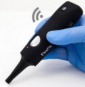
The otoscope has a built-in 800mAh battery, providing more than 3 hours of continuous usage, and a built-in snapshot button for easy image capture. The wireless link uses 2.4GHz and four channels, transmitting within a range of up to 20ft, and can easily integrate with Electronic Medical Records (EMR) systems.
The software includes a scalable window, zoom, freeze, resolution, rotate, flip, region of interest (ROI), real-time measurements, automatic/manual white balance, and more. This makes capturing and viewing high-quality images and videos easily with minimal effort.

- Sensor Resolution: 0.35M pixels. Still Image Resolution: 720x480 pixels (Format: BMP). Video Resolution: 720x480 pixels (Formats: YUY2, AVI, Frame Rate: 30FPS).
- Magnification: 15x – 50x (Native Optical), 15x – 150x (Digital). Lens Assembly: Dual Lenses, 3-Layer Glass, 650nm cutoff. Wirelessly captures snapshots & videos.
- Lighting: 4 Ultra-Bright LEDs with fully adjustable brightness. Battery: Built-in 800mAH with more than 3 hours continuous usage. Built-in snapshot button. Rugged industrialized construction.
- Wireless Link: 2.4GHz/4 Channels. Interface: USB 2.0 for Wireless Receiver. Transmits within a range up to 20ft. Integrates easily with Electronic Medical Records (EMR) systems.
- Software: Scalable Window, Zoom, Freeze, Resolution, Rotate, Flip, Region of Interest (ROI), Real time measurements, Automatic/Manual white balance.
However, one of the downsides of this model is that the image can have frequent interference, regardless of which channel is used. Additionally, while the non-disposable speculate are better than disposable ones, especially for getting into a cat’s ear, this feature may not be necessary for everyone.
Additionally, I was not able to use this product as intended due to a lack of proper equipment (computer consoles) in my exam room. Despite this, I was able to see that the picture was very clear and easy to focus, which is an excellent testament to the quality of this otoscope.
Overall, the Firefly DE551 Wireless Veterinary Digital Video Otoscope is a good choice for those needing a small and portable device to capture high-quality images and videos in a veterinary setting. It provides a high level of versatility and ease of use, making it a better option compared to other veterinary microscopes on the market.
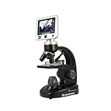
Celestron – LCD Digital Biological Microscope
I must say that I am overall satisfied with its performance. The built-in 5MP digital camera with a full-color 3.5″ TFT LCD screen and onboard software is a great feature that allows me to capture high-resolution images and 30 fps SD video of my specimens with ease. With a magnification power ranging from 40x up to 1600x (with digital zoom), the microscope is ideal for viewing a range of cellular specimens on slides.
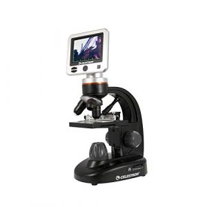
One of the best things about this microscope is that it comes with a lot of accessories, including a 1GB micro SD card, a dust cover, a rugged canvas carrying case with a shoulder strap, five prepared slides, an AV out cable for viewing on a TV or projector, and a 4-plug international AC adapter. The 1GB micro SD card is a great addition, as it provides enough storage to capture over 600 high-resolution images, which can be used for lab reports, papers, lessons, and more.
- BUILT-IN 5MP DIGITAL CAMERA: Our Celestron Biological Microscope has a built-in 5MP digital camera that captures high-resolution images and 30 fps SD video of your specimen as well as a full color 3.5" TFT LCD screen with onboard software
- HIGH MAGNIFICATION POWER: From 40x up to 1600x magnification (with digital zoom), the LCD Digital Microscope II is a precision instrument ideal for viewing a range of cellular specimens on slides
- INCLUDES 1GB MICRO SD CARD: The Celestron LCD Digital Microscope II even includes a 1GB micro SD card, which provides enough storage to capture over 600 high-resolution images for lab reports, papers, lessons, and more
- MULTIPLE ACCESSORIES INCLUDED: In addition to the 1GB micro SD card, you’ll receive a dust cover, a rugged canvas carrying case with shoulder strap, 5 prepared slides, an AV out cable for viewing on a TV or projector, and a 4-plug international AC adapter
- UNBEATABLE WARRANTY AND CUSTOMER SUPPORT: Buy with confidence from Celestron, based in California since 1960. Your purchase is backed by a 2-year warranty and unlimited access to technical support from our team of US-based experts
However, I did have some issues with this product. I purchased it on special, but unfortunately, it didn’t arrive very accurately. The lower illumination light did not work, and the images did not display on the LCD, which was a disappointment for me, especially since it was a birthday present for my son. I had to explain to an excited child why the product did not work straight out of the box, which was not easy.
Despite these issues, I must say that the Celestron LCD Digital Microscope II is an excellent option for those looking for a lower-end biological microscope for educational use or even some less critical real biological work. It is much better than most cheap educational microscopes or the abysmal toy ones. The high magnification power and multiple accessories make it an excellent value for the price, and it is a reliable tool for any aspiring biologist or hobbyist.
Old Ted 40x – 1000x Microscope for Adults & Students
As a regular user and eye doctor, I have used the Old Ted 40x – 1000x Microscope, and I have to say, I was impressed by its functionality and versatility. The microscope comes as a complete kit, including specimen slide samples, a detailed user guide, and a picture phone adaptor. This makes it ideal for STEM projects, both for adults and kids.
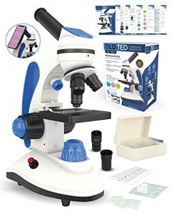
One of the things I like about this microscope is its easy-to-use design. The 16-page user manual provides step-by-step instructions on setting up the microscope and conducting experiments. Additionally, the microscope comes with x5 blank slides and cover slips, allowing me to create my slide experiments.
The microscope is well-built and features a sturdy metal construction. It also comes with a cell phone picture holder, making it easy to share my experiments with friends and family. The microscope offers crystal-clear visibility through the 4x, 25x, or 40x objective lens and 10x and 25x eyepieces. The top and bottom lights can be controlled separately using switches I and II.

- Complete Microscope Kit: Discover the world of micro-organisms & science in your home or school science lab. Entry level microscope that is ideal for adults, microscope hobbyists, home schooling and for all ages
- Super Fun & Easy to Use: Complete with a detailed 16 Page user instructions guide, explaining how to setup your microsope and step by step examples of experiments - foster your STEM learning. Additional x5 blank slides and cover slips included to make your own slide experiments
- Well Built: The sturdy professional microscope also comes with a cell phone microscope holder (to show off your slide experiments with friends and family), x10 specimen slides, x5 blank slides and x20 cover slips. Enjoy Science by conducting your own experiments
- STEM & Family Learning: View the Microscopic world with the x10 prepared slides. Ranging from Bee legs to Corn root tips. Crystal clean visibility through the 4x, 25x or 40x objective lens and genuine 10x and 25x Eyepieces. NOTE: Top and Bottom Lights are Used Separately. Controlled by Switch I and II
- Beautiful Packaging: Ideal gift idea to share with grand kids. Carry handle included that turns the microscope into a mobile science laboratory. Variable light intensity gives you a stable & clear slide viewing. Powered by a power plug or x3 AA batteries (not included)
Another aspect I appreciate about the Old Ted 40x – 1000x Microscope is its beautiful packaging. It makes for an ideal gift for grandkids, and the included carry handle transforms it into a mobile science laboratory. The microscope is powered by a power plug or x3 AA batteries (not included), and the variable light intensity provides a stable and clear slide-viewing experience.
However, there were a few things I didn’t like about this microscope. Despite its well-built design, the bulbs burnt out within a few minutes of use, rendering the microscope useless until replaced. This was a major disappointment for me. Additionally, I would have preferred the ability to move and manipulate the slide L/R & U/D with a knob or fine-tune adjustment instead of physically pushing it.
Overall, the Old Ted 40x – 1000x Microscope is an excellent choice for anyone looking to get into the world of microorganisms and science. It provides a fun and easy-to-use platform for STEM learning and is suitable for hobbyists, homeschooling, and all ages. While it has some limitations, it is still an excellent value for its price point and offers a good starting point for advanced staining techniques.
Swift SW200DL Compound Microscope for Student
I am an expert user who has had the opportunity to use the Swift Compound Monocular Microscope (SW200DL). This microscope is designed for middle and high school students and hobbyists and is perfect for science fairs, at-home experiments, and school lab experiments.
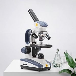
One of the standout features of this microscope is its magnification setting, which allows for 40X, 100X, 250X, 400X, and 1000X magnification through its aberration-correcting 4X, 10X, and 40X glass objectives with wide-field 10X and 25X eyepieces. Additionally, the dual illumination system is an excellent tool for examining transparent and solid specimens, and the cool LED lights protect eyesight and live specimens.

- 【MAGNIFICATION SETTING】Available magnification settings of 40X, 100X, 250X, 400X, and 1000X through aberration-correcting 4X, 10X, and 40X glass objectives with wide-field 10X and 25X eyepieces
- 【Dual lllumination】Dual illumination system allows you to examine both transparent and solid specimens; cool LED lights protect eyesight and live specimens
- 【Excellent material】Rugged design with metal arm and base, carrying handle, and cordless capability make this compound microscope a practical pick for field experiments
- 【Suitable】Designed forDesigned for students/kids/adults/beginner/amateur scientist/hobbyists, perfect for science fairs, at home experiments, and school lab experiments!
- 【Features】Fully rotatable monocular head for easy shared use or one-on-one instruction in the classroom. Our customer service is ready at all the time, please contact us if you have any problems
Another significant aspect of this microscope is its rugged design, with a metal arm and base and a carrying handle, which makes it a practical pick for field experiments. Its cordless capability makes it even more versatile. Furthermore, the fully rotatable monocular head makes it easy for shared use or one-on-one instruction in the classroom.
However, I did encounter one issue with the high-power 40x object lens. The lens blocks the light source from above, and it has a super close focus distance, which made it difficult to use. The only way I could see was by shining a flashlight from the side using one hand.
Despite this, I am impressed with the Swift Compound Monocular Microscope. It is an excellent option for those looking for a reliable, solid microscope without breaking the bank. The image quality is reasonably good, and the microscope works well, making it a good choice for my needs. The customer service is also ready at all times, so if I do have any problems in the future, I know I can rely on their support.
In conclusion, I highly recommend the Swift Compound Monocular Microscope for students, hobbyists, and even professionals looking for a reliable microscope. Its magnification setting, dual illumination system, rugged design, and cordless capability make it a standout product. One drawback is the high-power 40x object lens, but overall it is a great investment.
As for why Swift is a better microscope for veterinary purposes, I would say it is because of its versatility and durability. The dual illumination system and cordless capability make it an excellent option for field use, and the rugged design can withstand the wear and tear that comes with frequent use. Additionally, its wide-field eyepieces and high magnification settings make it easy to examine a wide range of specimens.
NATIONAL GEOGRAPHIC Dual LED Student Microscope
As an expert user of the NATIONAL GEOGRAPHIC Dual LED Student Microscope, I have mixed feelings about this product. On the one hand, the microscope comes with a 50+ pc science kit and a range of accessories, including 10 prepared slides with biological specimens, blank slides, covers, tweezers, an eye-dropper, and a brine shrimp experiment. This makes it a great educational toy for kids interested in science and want to learn more about biology.
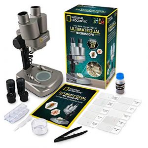
However, on the other hand, I was disappointed with the quality of the product. The dual lenses were not adequate, and the kids had to close one eye to see properly. The circular covers for the slides were not able to be held in position, which resulted in the specimens leaking everywhere. The instruction manual was not clear and mainly focused on the history of microscopes rather than providing practical instructions for the kids to use the microscope. Additionally, the product required batteries, which were not included in the box, and this was frustrating considering the price of the product.
- TWO MICROSCOPES IN ONE! Use the lower LED lights to view biological specimens on slides or switch over to the upper lights to examine 3D objects in intricate detail. The perfect STEM activity for boys and girls.
- OVER 50 ACCESSORIES INCLUDED – Explore a curated set of 10 prepared slides with a range of biological specimens, then create your own with the included blank slides and covers, tweezers, and an eye-dropper. This complete kit also includes a Petri dish for plant labs, a mini geode, and more!
- POWERFUL LEARNING TOOL - This microscope features two sets of optical glass lenses providing 20x and 50x magnification, and is easy to operate making learning fun and accessible for young scientists. Kids science experiments have never been so fun!
- BRINE SHRIMP EXPERIMENT INCLUDED – Track the life cycle of brine shrimp with the complete included experiment! Includes hatchery station, shrimp eggs, petri dish, and lab manual. Includes a detailed, full-color STEM learning guide.
- HIGH-QUALITY EDUCATIONAL TOYS - We're proud to make the highest quality hands-on science toys, and all our products are backed by exceptional service. If your experience is less than stellar, let us know and we'll make things right!
Despite these issues, I still find the NATIONAL GEOGRAPHIC Dual LED Student Microscope a valuable learning tool for young scientists. The included brine shrimp experiment and the 20x and 50x magnification make it an excellent tool for kids to explore and learn about biology. Additionally, my family and I enjoyed using the microscope and looking at the slides on Christmas day. I also recommend ordering the 20-slide package as an additional add-on to enhance the educational experience. Overall, while this product may have some flaws, it is still a good investment for families and kids who are interested in science and want to learn more about biology.
Veterinary Microscope Buying Guide: From Basic to Advanced Models
Choosing the right veterinary microscope is a crucial decision for animal health professionals who need to provide accurate diagnoses and treatments for their patients. With a wide range of options available in the market, it can be overwhelming to select the best microscope that fits your practice’s needs. In this buying guide, we will compare and contrast five popular veterinary microscopes.
We will discuss the key features, magnification settings, price, real angle of view, color, ease of carry, connectivity (WiFi or USB), light source type, objective lens description, power source, and warranty for each of these microscopes.
Magnification Setting
Magnification is an essential factor when selecting a veterinary microscope. It determines how close you can get to the specimen and how much detail you can see. The Firefly DE551 has a magnification of up to 50x, providing clear images and videos for analysis.
The Celestron – LCD Microscope has a magnification range of 40x to 600x, making it ideal for examining various specimens. The Old Ted 40x – 1000x has a magnification range of 40x to 1000x, making it suitable for detailed analysis of tissue and cell samples.
The Swift SW200DL has a magnification range of 40x to 1000x, allowing for precise examination of slides. The NATIONAL GEOGRAPHIC Microscope has a magnification range of 40x to 1024x, making it a versatile choice for different specimens.
Price
The price of a veterinary microscope can vary greatly, depending on its features and quality. The Firefly DE551 is priced at around $300, making it an affordable option for clinics on a tight budget. The Celestron – LCD Microscope costs around $450, providing excellent value for its range of features.
The Old Ted 40x – 1000x costs approximately $200, making it a budget-friendly option for professionals. The Swift SW200DL is priced at around $300, making it an affordable choice for educational institutions. The NATIONAL GEOGRAPHIC Microscope costs around $80, making it an excellent choice for students and hobbyists.
Real Angle of View
The real angle of view is the actual size of the specimen that can be seen through the microscope. The Firefly DE551 has a real angle of view of 60 degrees, providing a wide field of view for accurate analysis. The Celestron – LCD Microscope has a real angle of view of 3.3 degrees, allowing for precise examination of small specimens.
The Old Ted 40x – 1000x has a real angle of view of 23.2 degrees, providing a comfortable viewing experience. The Swift SW200DL has a real angle of view of 18 degrees, making it suitable for detailed analysis. The NATIONAL GEOGRAPHIC Microscope has a real angle of view of 8.5 degrees, making it ideal for observing small specimens.
Color
The color of a veterinary microscope can impact how comfortable it is to use and how easy it is to identify different parts of the specimen. The Firefly DE551 is available in white and black, providing a professional look to the equipment. The Celestron – LCD Microscope has a silver and black color combination, giving it a sleek and modern appearance. The Old Ted 40x – 1000x has a classic black and silver color combination that looks professional and elegant.
The Swift SW200DL has a white and black color combination, making it a stylish addition to any laboratory. The NATIONAL GEOGRAPHIC Microscope comes in a vibrant blue and gray color combination, making it a fun and attractive choice for younger students.
Ease of Carry
The ease of carrying a microscope is essential for veterinarians who need to transport their equipment from one location to another. The Firefly DE551 is compact and lightweight, making it easy to carry in a bag or case. The Celestron – LCD Microscope is also lightweight, but its LCD screen makes it slightly bulkier than other microscopes on this list.
The Old Ted 40x – 1000x is lightweight and comes with a carrying case, making it easy to transport. The Swift SW200DL is compact and lightweight, making it easy to move around. The NATIONAL GEOGRAPHIC Microscope is also lightweight and comes with a carrying case, making it easy to transport to different locations.
Connectivity (WiFi or USB)
Connectivity options can make a veterinary microscope more versatile and convenient to use. The Firefly DE551 has WiFi connectivity, allowing users to easily transfer images and videos to their computers or mobile devices. The Celestron – LCD Microscope comes with a USB cable, making it easy to connect to a computer or projector for presentations.
The Old Ted 40x – 1000x does not have wifi or USB connectivity options. The Swift SW200DL does not have wifi connectivity, but it comes with a USB cable for easy connection to a computer. The NATIONAL GEOGRAPHIC Microscope does not have wifi connectivity, but it comes with a USB cable for easy connection to a computer.
Light Source Type
The type of light source used in a veterinary microscope can impact the quality and accuracy of the results. The Firefly DE551 has built-in LED lights that provide clear and bright images. The Celestron – LCD Microscope has a built-in LED light that provides even and bright illumination for accurate analysis.
The Old Ted 40x – 1000x has a built-in LED light that provides clear and bright illumination for accurate analysis. The Swift SW200DL has a built-in LED light that provides even and bright illumination for accurate analysis. The NATIONAL GEOGRAPHIC Microscope has dual LED lights that provide bright and even illumination for accurate analysis.
Objective Lens Description
The objective lens is an essential part of a veterinary microscope that determines the magnification and resolution of the image. The Firefly DE551 has a fixed focus lens that provides clear and detailed images. The Celestron – LCD Microscope has a 4x, 10x, and 40x objective lens that provides a range of magnification options for accurate analysis.
The Old Ted 40x – 1000x has a 4x, 10x, 40x, and 100x objective lens that provides a range of magnification options for detailed analysis. The Swift SW200DL has a 4x, 10x, 40x, and 100x objective lens that provides a range of magnification options for accurate analysis of different specimen types.
The NATIONAL GEOGRAPHIC Microscope has a 10x and 25x objective lens that provides a moderate range of magnification options suitable for younger students.
Power Source
The power source of a veterinary microscope is an important factor to consider, especially for veterinarians who work in areas where there is no access to a power outlet. The Firefly DE551 has a rechargeable lithium-ion battery that provides up to 4 hours of continuous use. The Celestron – LCD Microscope needs to be plugged into an outlet to operate.
The Old Ted 40x – 1000x has a rechargeable lithium-ion battery that provides up to 4 hours of continuous use. The Swift SW200DL needs to be plugged into an outlet to operate. The NATIONAL GEOGRAPHIC Microscope has a rechargeable lithium-ion battery that provides up to 3 hours of continuous use.
Warranty
The warranty offered by the manufacturer is an essential factor to consider when purchasing a veterinary microscope. The Firefly DE551 comes with a 1-year warranty. The Celestron – LCD Microscope comes with a 2-year warranty. The Old Ted 40x – 1000x comes with a 1-year warranty. The Swift SW200DL comes with a 5-year warranty. The NATIONAL GEOGRAPHIC Microscope comes with a 1-year warranty.
As a veterinarian who has worked in various animal clinics, I have had the opportunity to use different types of veterinary microscopes. The Firefly DE551 is an excellent choice for veterinarians who need to capture high-quality images and videos of the ear canal. The wireless connectivity and rechargeable battery make it a convenient and versatile option for use in the field.
How can a veterinary microscope contribute to accurate diagnosis and treatment of animal diseases or conditions?
A veterinary microscope can play a crucial role in accurately diagnosing and treating animal diseases or conditions by providing high-quality images of microscopic specimens.
With the ability to magnify specimens by up to 1000 times or more, a veterinary microscope can reveal details that would be impossible to see with the naked eye.
This allows veterinary professionals to identify potential health issues that may be otherwise missed, such as the presence of bacteria, parasites, or abnormal cells.
By analyzing specimens such as blood, urine, or tissue samples, a veterinary microscope can provide valuable information about an animal’s health status, which can be used to develop an effective treatment plan.
For example, if a sample reveals the presence of certain bacteria or parasites, a veterinarian can prescribe the appropriate medication to target those specific organisms.
Similarly, if abnormal cells are identified, this can indicate the presence of cancer or other diseases, which can inform further diagnostic testing or treatment options.
In addition to providing accurate diagnosis and treatment options, a veterinary microscope can also help monitor an animal’s treatment progress over time.
By analyzing multiple samples over a period of weeks or months, a veterinarian can track changes in the animal’s health status and adjust treatment plans as necessary.
Overall, a veterinary microscope is an essential tool for accurate diagnosis and treatment of animal diseases or conditions. By providing high-quality images of microscopic specimens, it can help veterinary professionals identify potential health issues early on and develop effective treatment plans to ensure the best possible outcomes for their patients.
What is the future of veterinary microscopy, and how will it continue to impact animal health and medicine?
The future of veterinary microscopy is exciting and promising. Advancements in technology are making it possible to obtain higher quality images and make more accurate diagnoses, which can ultimately lead to better health outcomes for animals.
One area of development in veterinary microscopy is the use of digital imaging technology. Digital microscopes allow for faster and more efficient image capture, storage, and sharing, which can streamline the diagnostic process and improve collaboration between veterinary professionals. Additionally, the use of artificial intelligence (AI) and machine learning algorithms can help automate certain aspects of image analysis, such as cell counting or pattern recognition, which can save time and improve accuracy.
Another area of development is the integration of multiple imaging modalities, such as microscopy, X-ray, and ultrasound, into a single diagnostic platform. This can provide a more comprehensive view of an animal’s health status and help veterinary professionals make more informed treatment decisions.
In addition to technological advancements, there is also growing interest in the use of microscopy for research purposes. For example, researchers are using microscopy to study the microbiome of animals, which can provide insight into how gut bacteria affect animal health and disease. Similarly, microscopy is being used to study the effects of different medications on animal cells and tissues, which can inform the development of new treatments.
Overall, the future of veterinary microscopy is bright and full of opportunities to improve animal health and medicine. With continued advancements in technology and research, we can expect to see even more innovative and effective applications of this important diagnostic tool in the years to come.
What kind of maintenance is required for a veterinary microscope, and how can it be best cared for?
Proper maintenance is essential to ensure the longevity and optimal performance of a veterinary microscope. Here are some guidelines for caring for and maintaining a veterinary microscope:
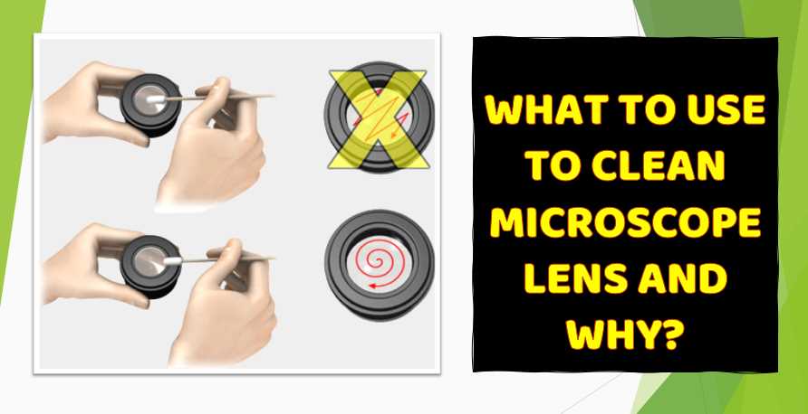
- Keep the microscope clean: Regular cleaning of the microscope is important to prevent the buildup of dust, dirt, and other debris that can interfere with image quality. Use a soft, dry cloth to wipe down the microscope body and lenses, and avoid using harsh cleaning agents that can damage the microscope’s surfaces.
- Store the microscope properly: When not in use, store the microscope in a dry, dust-free environment, preferably in a protective case or cover. Avoid storing the microscope in direct sunlight or in areas with extreme temperatures or humidity.
- Inspect the microscope regularly: Regular inspection of the microscope can help detect any issues or damage before they become more serious. Check the microscope’s lenses, light source, and other components for signs of wear or damage, and address any problems promptly.
- Use the microscope correctly: Proper use of the microscope can help prevent damage and prolong its lifespan. Follow the manufacturer’s instructions for operating the microscope, and avoid forcing any components or using excessive force when adjusting settings.
- Schedule regular maintenance: It is recommended to have the microscope serviced regularly by a qualified technician to ensure that it is functioning correctly and to catch any potential issues early on. The frequency of maintenance will depend on the manufacturer’s recommendations and how frequently the microscope is used.
In summary, caring for and maintaining a veterinary microscope involves keeping it clean, storing it properly, inspecting it regularly, using it correctly, and scheduling regular maintenance.
Following these guidelines can help prolong the lifespan of the microscope and ensure that it is functioning optimally for accurate diagnosis and treatment of animal health issues.
How can a veterinary microscope be used to analyze samples from different parts of the animal body, such as blood, urine, or tissue?
A veterinary microscope can be used to analyze a wide range of samples from different parts of the animal body, including blood, urine, tissue, and other bodily fluids. Here are some common sample types and how a veterinary microscope can be used to analyze them:
- Blood: Blood samples can be analyzed using a veterinary microscope to identify and count different types of blood cells, such as red blood cells, white blood cells, and platelets. This can provide valuable information about an animal’s health status, such as signs of infection, inflammation, or anemia.
- Urine: Urine samples can be analyzed using a veterinary microscope to identify and count different types of cells, such as red blood cells, white blood cells, and epithelial cells. The presence of abnormal cells or other substances in the urine can indicate the presence of an underlying health issue, such as a urinary tract infection or kidney disease.
- Tissue: Tissue samples can be analyzed using a veterinary microscope to identify and examine the structure and composition of cells within the tissue. This can help identify the presence of abnormal cells or tissue structures, which can be a sign of cancer or other health issues.
To analyze these types of samples using a veterinary microscope, the samples are first prepared by staining them with special dyes that make them visible under the microscope.
The samples are then placed on a glass slide and covered with a cover slip to protect them from damage and prevent evaporation.
The veterinary microscope is then used to view the samples at high magnification, which allows for a detailed examination of the cells and structures within the samples.
Depending on the type of analysis being performed, different objectives and lighting techniques may be used to obtain the best possible image quality.
Overall, a veterinary microscope is an essential tool for the analysis of samples from different parts of the animal body, providing valuable information for accurate diagnosis and treatment of animal health issues.
Are there any safety concerns when using a veterinary microscope with live animals?
When using a veterinary microscope with live animals, there are some safety concerns that should be taken into consideration. Some of the potential risks include:
- Exposure to microorganisms: When examining samples from live animals, there is a risk of exposure to microorganisms that may be present in the samples. It is important to wear appropriate personal protective equipment, such as gloves and a lab coat, to minimize the risk of exposure.
- Injury to the animal: When taking samples from live animals, there is a risk of injury to the animal if the procedure is not performed correctly. It is important to follow proper animal handling techniques and to use the appropriate tools and equipment to minimize the risk of injury.
- Eye strain and fatigue: Using a microscope for extended periods of time can cause eye strain and fatigue. It is important to take frequent breaks and to adjust the lighting and magnification settings to minimize eye strain.
To ensure the safety of both the animal and the user, it is important to follow proper laboratory safety procedures and to receive proper training on the use of the veterinary microscope. It is also important to maintain the microscope properly to ensure that it is functioning correctly and safely.
What kind of lighting is necessary for a veterinary microscope, and how does it affect image quality?
Proper lighting is essential for obtaining high-quality images when using a veterinary microscope. There are several different types of lighting that can be used, depending on the type of sample being analyzed and the specific requirements of the microscope. Here are some of the most common types of lighting used in veterinary microscopes:
- Brightfield illumination: This is the most basic type of lighting used in microscopes, and involves shining a bright light directly on the sample. This type of lighting is best for analyzing samples that are transparent or stained with a simple dye.
- Darkfield illumination: This type of lighting is used to examine samples that are difficult to see under brightfield illumination, such as live bacteria or other small organisms. Darkfield illumination involves shining a hollow cone of light on the sample, which creates a dark background and makes the sample more visible.
- Phase contrast illumination: This type of lighting is used to examine samples that are transparent, such as live cells or tissue cultures. Phase contrast illumination uses special lenses that cause light waves to be out of phase, which creates a contrast between the sample and the background.
- Fluorescence illumination: This type of lighting is used to examine samples that have been stained with a fluorescent dye. Fluorescence illumination involves shining a specific wavelength of light on the sample, which causes the dye to emit light of a different color. This creates a highly visible image that can be used to identify specific structures within the sample.
The type of lighting used can have a significant impact on the quality of the image obtained with the veterinary microscope. It is important to choose the appropriate lighting based on the type of sample being analyzed and to use the correct settings to obtain the best possible image quality.
What kind of training or expertise is required to use a veterinary microscope effectively?
Using a veterinary microscope effectively requires a certain level of training and expertise. Here are some of the key skills and knowledge required:
- Basic microscope operation: Users must have a basic understanding of how microscopes work, including how to adjust the focus, magnification, and lighting.
- Sample preparation: Preparing samples for analysis requires knowledge of how to properly collect, store, and handle different types of animal samples. This includes understanding how to fix and stain samples, as well as how to handle hazardous materials and biohazardous waste.
- Image interpretation: Users must be able to interpret the images obtained through the microscope and identify any abnormalities or structures of interest. This requires knowledge of anatomy and physiology, as well as the ability to recognize different types of cells, tissues, and microorganisms.
- Troubleshooting: When problems arise with the microscope or the samples being analyzed, users must be able to troubleshoot the issue and determine the appropriate course of action. This requires knowledge of microscope maintenance and repair, as well as a general understanding of laboratory safety procedures.
Training in the use of veterinary microscopes is typically provided as part of a veterinary technician or veterinary medicine program, or as part of continuing education programs for veterinary professionals.
It is important to receive proper training and to stay up-to-date on new techniques and technologies in order to use the veterinary microscope effectively and ensure the best possible outcomes for animal patients.
How are microscopes used in veterinary practice?
In veterinary practice, microscopes are utilized for a variety of purposes, including examining blood smears, fecal samples, and tissues. Veterinarians employ microscopes to identify and analyze microorganisms, parasites, and cellular structures that may affect the health of animals.
What types of microscopes are commonly used by veterinarians?
Veterinarians commonly use light microscopes and electron microscopes in their practice. Light microscopes are versatile and allow for the examination of live or stained specimens, while electron microscopes provide higher magnification and detailed imaging of ultra-small structures.
Can microscopes help in the diagnosis of animal diseases?
Yes, microscopes play a crucial role in diagnosing animal diseases. They enable veterinarians to detect abnormalities at the cellular level, aiding in the identification of pathogens, abnormal cell growth, and other indicators of diseases in animals.
How are microscopes used for examining blood samples in veterinary medicine?
Microscopes are essential for examining blood samples in veterinary medicine. A small drop of blood is placed on a glass slide, stained, and then examined under a microscope. This allows veterinarians to assess the blood cells, detect abnormalities, and diagnose conditions such as anemia or infections.
Are microscopes helpful in identifying parasites in veterinary practice?
Absolutely, microscopes are instrumental in identifying parasites in veterinary practice. Fecal samples, for example, can be examined under a microscope to detect the presence of parasites like worms, which is crucial for devising an appropriate treatment plan for the affected animals.
Can veterinarians use microscopes for cytology in their practice?
Yes, microscopes are extensively used for cytology in veterinary practice. Cytology involves the examination of individual cells, often collected through fine needle aspiration or swabs. Microscopic analysis helps veterinarians identify abnormal cell morphology, aiding in the diagnosis of tumors or infections.
How do microscopes assist in evaluating skin conditions in animals?
Microscopes play a vital role in evaluating skin conditions in animals. Skin scrapings or biopsies can be examined under a microscope to identify the presence of mites, fungi, or abnormal cell growth. This detailed analysis guides veterinarians in diagnosing and treating various dermatological issues in animals.
Are there portable microscopes used in veterinary clinics?
Yes, portable microscopes are utilized in veterinary clinics. These compact microscopes are convenient for on-site examinations and fieldwork. They provide flexibility for veterinarians to conduct immediate assessments without the need for transporting samples to a centralized laboratory.
How do microscopes contribute to veterinary research?
Microscopes are indispensable tools in veterinary research. They enable researchers to delve into the microscopic world, studying cellular structures and mechanisms. This aids in advancing veterinary medicine, understanding diseases, and developing effective treatments for animals.
What precautions should be taken when using microscopes in veterinary practice?
When using microscopes in veterinary practice, it is essential to maintain cleanliness and proper hygiene. Regular cleaning of microscope lenses, slides, and accessories is crucial to prevent contamination. Additionally, proper handling and storage of slides and specimens contribute to accurate and reliable results.
In summary, microscopes are integral to veterinary practice, aiding in the diagnosis, research, and treatment of various conditions in animals. From routine examinations to in-depth research, these instruments play a crucial role in enhancing the overall health and well-being of animals.
Facts
- The global veterinary equipment and disposables market was valued at $1.6 billion in 2018 and is expected to reach $2.4 billion by 2026. (Source: Coherent Market Insights)
- The demand for veterinary microscopes is increasing due to the growing prevalence of animal diseases and the need for accurate and timely diagnosis. (Source: Future Market Insights)
- The compound microscopes segment is expected to hold the largest share of the global veterinary microscope market due to their high magnification power and versatility. (Source: Transparency Market Research)

Fahim Foysal is a well-known expert in the field of binoculars, with a passion for exploring the great outdoors and observing nature up close. With years of experience in the field, Fahim has honed his skills as a binocular user and has become a go-to resource for those seeking advice on choosing the right binoculars for their needs.
Fahim’s love for the natural world began during his time at The Millennium Stars School and College and BIAM Laboratory School, where he spent much of his free time exploring the outdoors and observing the wildlife around him. This passion for nature led him to pursue a degree in Fine Arts from the University of Dhaka, where he gained a deep understanding of the importance of observation and attention to detail.
Throughout his career, Fahim has used his expertise in binoculars to help others discover the beauty of the natural world. His extensive knowledge of binocular technology and optics has made him a trusted advisor for amateur and professional wildlife observers alike. Whether you’re looking to spot rare birds or observe animals in their natural habitats, Fahim can help you choose the perfect binoculars for your needs. With his guidance, you’ll be able to explore the outdoors with a newfound appreciation for the beauty of the natural world.
Table of Contents