Category: Info

  • Can I See My Sperm with Microscope

    Can I See My Sperm with Microscope

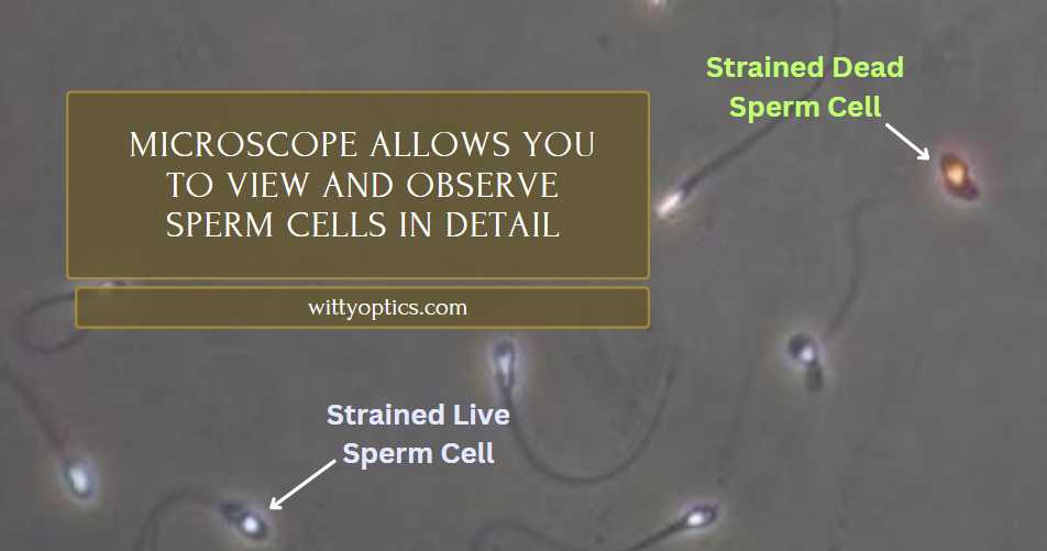
    Yes, you can see your sperm with a microscope. Microscopy allows you to view and observe sperm cells in detail, providing valuable insights into their structure and function.

    Sperm cells are typically around 5 micrometers in size, which is small but still visible under a microscope. A standard compound light microscope, commonly found in laboratories and schools, should suffice for observing sperm cells. However, to see them clearly, you may need to use a higher magnification objective lens, such as a 40x or 100x lens.

    Additionally, staining techniques can be employed to enhance the visibility of sperm cells under the microscope. Overall, with the right equipment and preparation, it’s entirely possible to view your sperm under a microscope.

    StatisticValue
    Average sperm size5 micrometers
    Magnification needed40x – 100x
    Staining techniquesEnhance visibility
    Common microscope typeCompound light microscope
    AccessibilityLaboratories, schools, research facilities
    Additional equipment requiredStaining kits, high magnification lenses

    Sperm are also very small, so yes, you can see them with a microscope!

    Here’s how it works:

    1. Get a Microscope: First, you’ll need a microscope. It’s a special tool with lenses that magnify tiny objects.
    2. Prepare a Sample: You’ll need a sample of sperm to look at. This usually means collecting semen, which contains sperm. You can collect it by masturbating or using a special condom during sex.
    3. Prepare the Slide: To look at the sperm under the microscope, you’ll put a tiny drop of the semen on a glass slide. Then, cover it with a thin piece of glass called a cover slip.
    4. Look Through the Microscope: Place the slide on the microscope’s stage and look through the eyepiece. Start with a low magnification and gradually increase it until you see the sperm clearly.
    5. Observe and Study: Once you’ve found the sperm, you can observe their shape, movement, and other characteristics. Sperm are tiny, tadpole-like cells with a head and a tail that helps them swim.

    Remember to handle the microscope and slides carefully and follow any safety guidelines that come with the microscope. And always remember, it’s perfectly normal to be curious about your body and how it works!

    Understanding Sperm

    As I looked through the microscope’s eyepiece, I started with a low magnification and gradually increased it until I saw the sperm clearly. It was incredible to see these tiny, tadpole-like cells swimming around. I observed their shape, movement, and even noticed differences between individual sperm.

    Handling the microscope and slides carefully was crucial, and I made sure to follow all safety guidelines. Exploring sperm under the microscope was not only educational but also a reminder of the wonders of the human body.

    Sperm are male reproductive cells produced in the testicles. They are essential for fertilizing female eggs and initiating pregnancy. Structurally, sperm consist of a head, midpiece, and tail, designed for mobility towards the egg. Studying sperm is crucial for understanding fertility and reproductive health.

    Exploring sperm under the microscope not only satisfied my curiosity about the male reproductive system but also deepened my understanding of the miracle of conception. It was a hands-on experience that left me with a sense of wonder and respect for the incredible journey that each sperm undertakes in the quest for fertilization.

    Microscopes

    Microscopes are instruments that magnify small objects, allowing for detailed observation. They come in various types, including optical, electron, and digital microscopes. Optical microscopes, commonly used for educational and basic research purposes, employ visible light to magnify specimens.

    Principle of OperationApplications
    Optical MicroscopeUses visible lightEducation, research
    Electron MicroscopeUses electron beamsHigh-resolution imaging
    Digital MicroscopeIntegrates with computersDigital imaging, analysis

    Can You See Sperm with a Microscope?

    While microscopes can magnify objects significantly, the size of sperm presents challenges for direct observation. Sperm are typically around 50 micrometers long, requiring high-magnification microscopes for clear visualization. Optical microscopes, though useful for larger specimens, may not provide adequate magnification for viewing sperm.

    Type of MicroscopeMagnification RangeSuitability for Sperm Observation
    Optical MicroscopeUp to 1000xLimited due to sperm size
    Electron MicroscopeUp to 1,000,000xSuitable for detailed sperm analysis
    Digital MicroscopeVariesDepends on magnification capability

    Steps to View Sperm with Microscope

    If you’re interested in viewing your sperm using a microscope, there are specific steps you can follow to achieve this:

    1. Preparation of Microscope: Begin by setting up your microscope on a stable surface and ensuring it is clean and properly aligned. Adjust the lighting and focus mechanisms to optimize visibility.
    2. Preparation of Sperm Sample: Collect a semen sample using a clean and sterile container. Place a small drop of the sample onto a glass slide and cover it with a coverslip to prevent evaporation and contamination.

    Viewing Sperm Under Microscope: Place the prepared slide onto the microscope stage and adjust the magnification to the highest level possible. Use the focus knobs to bring the sperm cells into clear view, adjusting the lighting as needed for optimal contrast

    Alternative Methods to View Sperm

    If you’re unable to visualize sperm using a microscope at home, there are alternative methods available:

    Sperm Analysis Centers

    Consider visiting a specialized fertility clinic or laboratory that offers sperm analysis services. These facilities have advanced equipment and trained professionals who can provide detailed assessments of sperm health and fertility.

    DIY Methods

    Explore DIY sperm analysis kits available on the market, which typically include instructions and materials for collecting and observing sperm samples at home. While these kits may not offer the same level of accuracy as professional analysis, they can provide valuable insights into sperm health and concentration.

    Safety Precautions and Best Practices

    When using a microscope to view sperm or any other biological specimen, it’s essential to observe safety precautions to prevent contamination and ensure accurate results. Here are some best practices to follow:

    1. Cleanliness and Sterility: Maintain a clean work environment and sterilize all equipment before and after use to avoid contamination of samples.
    2. Eye Protection: Wear safety goggles or glasses to protect your eyes from potential splashes or spills when handling biological samples.
    3. Proper Handling of Microscope: Handle the microscope with care to avoid damage and ensure accurate results. Follow manufacturer instructions for maintenance and cleaning.

    What type of microscope do I need to see my sperm?

    To visualize your sperm effectively, you’ll need a high-powered microscope with a magnification of at least 400 times. Light microscopes, commonly found in schools and laboratories, may not offer sufficient magnification for clear visualization of sperm cells. Preferably, an electron microscope would be ideal for observing sperm at a cellular level due to its high magnification capabilities.

    Type of Microscope

    Features

    Light Microscope

    Limited magnification; suitable for educational purposes.

    Electron Microscope

    High magnification; provides detailed images at the cellular level.

    Fluorescence Microscope

    Excels in visualizing fluorescently labeled specimens.

    What magnification is required to see sperm with a microscope?

    To see sperm cells with a microscope, you’ll need a magnification level of at least 400x to 1000x. This level of magnification is essential for resolving the small size of sperm cells, which typically range from 3 to 5 micrometers in length. Low magnification microscopes may not provide sufficient resolution to visualize sperm cells clearly, so it’s recommended to use a microscope with high magnification capabilities, such as an electron microscope, for optimal results.

    Can I prepare my own sperm sample for viewing under a microscope?

    Yes, you can prepare your own sperm sample for viewing under a microscope. Start by collecting a semen sample in a clean, sterile container. Then, place a small drop of the semen onto a glass slide and cover it with a coverslip to prevent evaporation and contamination. Finally, place the prepared slide onto the microscope stage and adjust the focus to bring the sperm cells into clear view.

    How do I prepare a sperm sample for microscope observation?

    To prepare a sperm sample for microscope observation, follow these steps:

    1. Collect Semen: Obtain a sample of semen containing sperm. This can be done through masturbation or using a special condom during sexual activity.

    2. Prepare a Clean Slide: Clean a glass microscope slide and cover slip with alcohol or a mild detergent to remove any contaminants. Ensure they are completely dry before use.

    3. Apply Sample: Place a small drop of semen onto the center of the microscope slide. Be careful not to use too much, as excess fluid can cause the cover slip to float and distort the sample.

    4. Add Cover Slip: Gently lower a clean cover slip onto the drop of semen, taking care to avoid trapping air bubbles. The cover slip helps flatten the sample and protects it from drying out.

    5. Observe Under Microscope: Place the prepared slide on the microscope stage and secure it in place. Begin with a low magnification objective lens and gradually increase the magnification until sperm are visible.

    6. Focus and Adjust: Use the microscope’s focus and adjustment knobs to sharpen the image of the sperm. You may need to adjust the lighting and contrast settings for better clarity.

    7. Observe and Record: Once sperm are in focus, observe their movement, shape, and other characteristics. You can also capture images or videos of the sperm for further analysis or documentation.

    8. Clean Up: After observation, carefully discard the slide and cover slip or clean and sterilize them for future use. Clean the microscope stage and lenses to remove any residue.

    By following these steps, you can prepare a sperm sample for microscope observation and gain valuable insights into sperm morphology and motility.

    Can I use a DIY sperm analysis kit to view my sperm at home?

    Yes, you can use a DIY sperm analysis kit to view your sperm at home. These kits typically provide materials and instructions for collecting and observing sperm samples independently. While DIY kits may not offer the same level of accuracy as professional analysis, they can provide valuable insights into sperm health and concentration, making them a convenient option for at-home testing.

    DIY Sperm Analysis Kit

    Features

    Convenience

    Provides materials and instructions for collecting and observing sperm samples at home.

    Accuracy

    May not offer the same level of accuracy as professional analysis.

    Insights

    Provides valuable insights into sperm health and concentration.

    Are there any safety precautions I should follow when viewing sperm with a microscope?

    Yes, there are several safety precautions you should follow when viewing sperm with a microscope.

    Safety Precautions

    Guidelines

    Eye Protection

    Wear safety goggles or glasses to protect eyes from potential splashes or spills.

    Cleanliness

    Maintain a clean work environment and sterilize equipment to avoid contamination.

    Handling Microscope

    Handle the microscope with care to prevent damage and ensure accurate results.

    Final words

    While it is technically possible to see sperm with a microscope, the type of microscope and its magnification power are crucial factors in achieving clear and detailed images. Light microscopes, commonly used for educational and hobbyist purposes, may not provide sufficient magnification for visualizing sperm cells effectively. However, higher magnification microscopes, such as electron microscopes, can offer detailed insights into sperm structure and morphology. If you’re interested in exploring your sperm under a microscope, ensure you follow proper safety precautions and consider alternative methods if necessary. Remember, the ability to see sperm with a microscope can vary depending on the equipment available and the quality of the sample.

  • Are Viruses Visible under a Microscope: (Video Explained)

    Are Viruses Visible under a Microscope: (Video Explained)

    Yes, viruses are visible under a microscope. They are too small to be seen without magnification.

    When viewed under a microscope, viruses appear as tiny particles with unique shapes and structures. Their size and features can vary depending on the type of virus. Scientists and researchers use powerful microscopes to study viruses and understand their characteristics, behavior, and impact on living organisms.

    The ability to visualize viruses under a microscope is essential for studying and developing treatments and vaccines to combat viral infections. Through advanced imaging techniques, researchers continue to uncover valuable insights into the world of viruses and their potential effects on human health and the environment. Understanding the visual aspects of viruses plays a crucial role in combating various viral diseases and improving public health.

    Virus NameAverage Size (nm)Visualization Technique
    Influenza Virus80-120Electron Microscope
    SARS-CoV-250-200Electron Microscope
    Human Papillomavirus50-60Electron Microscope
    HIV80-120Electron Microscope
    Adenovirus70-100Electron Microscope

    What Is A Virus?

    A virus is a microscopic infectious agent that can only replicate within the cells of a living organism. It is composed of genetic material, either DNA or RNA, surrounded by a protein coat. Definition: Viruses are non-living entities that depend on a host for survival and reproduction. They are much smaller than bacteria and can only be seen under an electron microscope. Characteristics: Viruses lack the cellular structure of bacteria and cannot carry out metabolic processes on their own. They are known for their ability to cause various diseases in humans, animals, and plants, making them significant subjects of study in the field of microbiology.

    Structure of A Virus

    Viruses are not visible to the naked eye and can only be observed under a microscope. Their structure consists of a genetic material, either DNA or RNA, enclosed within a protein coat called a capsid. Some viruses also have an outer lipid envelope surrounding the capsid. The size and shape of viruses vary greatly, with some being spherical, helical, or complex in structure. Depending on the type of virus, their dimensions can range from 20 to 400 nanometers.

    Microscopy Techniques For Visualizing Viruses

    Microscopy techniques play a crucial role in visualizing viruses and understanding their characteristics. Light microscopy, also known as optical microscopy, is one such technique that uses visible light to observe samples. With the help of lenses and light sources, it allows scientists to magnify viruses and study their structure. However, due to the limited resolution of light microscopy, viruses may not always be visible at their actual size.

    Electron microscopy, on the other hand, offers higher magnification and resolution. By using a beam of electrons instead of light, it enables researchers to examine viruses in greater detail. The high resolution provided by electron microscopy allows for visualization of even smaller viruses and their intricate structures.

    Fluorescence microscopy is a specialized technique that involves the use of fluorescent dyes to label specific parts of the viruses. By emitting light at different wavelengths, these dyes help in visualizing and distinguishing different components of the viruses. Fluorescence microscopy is particularly useful in studying viral replication and understanding interactions between viruses and host cells.

    Microscopy Technique Advantages
    Light Microscopy Easy to use and allows observation of live viruses
    Electron Microscopy Provides high-resolution images and reveals intricate viral structures
    Fluorescence Microscopy Enables visualization of specific viral components and their interactions

    Can Viruses Be Seen Under A Light Microscope?

    Viruses are often too small to be seen under a light microscope due to their tiny size, typically ranging from 20 to 300 nanometers. However, there are some exceptions to this rule. Poxviruses and Papillomaviruses are examples of viruses that can be observed using a light microscope. These exceptional cases are possible because these viruses are relatively large compared to other viruses.

    Despite these exceptions, light microscopy has limitations when it comes to visualizing viruses. The resolution power of a light microscope is limited by the wavelength of visible light, which is around 400 to 700 nanometers. Since viruses are much smaller than this wavelength, they are often not visible under a light microscope. For observing smaller viruses, such as influenza viruses or HIV, more advanced techniques such as electron microscopy or scanning electron microscopy are required.

    It is important to note that even though viruses may not be visible under a light microscope, they play a significant role in many aspects of our lives. Understanding their structure and behavior is crucial to developing effective treatments and prevention strategies.

    Visualizing Viruses Using Electron Microscopy

    Electron microscopy enables the visualization of viruses due to its high magnification capabilities. This powerful tool can capture detailed images of viruses, allowing scientists to study their structure and characteristics. By using electron microscopy, researchers have been able to obtain striking images of viruses at an ultra-high resolution, providing valuable insights into their morphology and behavior. This technology has revolutionized the study of viruses by offering a closer look at their intricate features and allowing for a deeper understanding of their impact on living organisms.

    Capabilities of electron microscopy Examples of virus images obtained through electron microscopy
    High magnification Images of influenza virus, HIV, and herpesvirus
    Ultra-high resolution Detailed visualization of Zika virus and Ebola virus
    Structural analysis Capturing the morphology of SARS-CoV-2

    Final Words:

    Viruses are microscopic organisms that cannot be directly observed under a regular light microscope. However, with the use of electron microscopes, scientists have been able to capture detailed images of viruses, revealing their intricate structures. These technologies have greatly contributed to our understanding of viruses and their pathogenic characteristics.

    By studying viruses at the microscopic level, researchers can develop effective strategies to combat and manage viral infections.

  • Are Electron Microscopes Expensive? Unveiling the Costs

    Are Electron Microscopes Expensive? Unveiling the Costs

    Yes, electron microscopes are expensive.

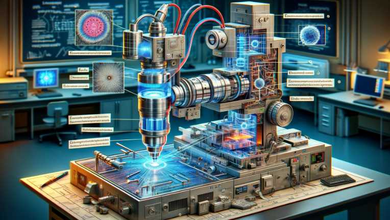
    Electron microscopes are sophisticated scientific instruments that use electron beams to achieve extremely high magnification and resolution, allowing researchers to observe objects at the nanoscale. The technology and precision required in electron microscopes contribute to their high cost. These instruments often involve complex components, including electron sources, electromagnetic lenses, detectors, and advanced imaging systems. Additionally, maintaining and operating electron microscopes requires specialized expertise, further adding to the overall cost.

    Application Percentage of Use
    Materials Science 35%
    Life Sciences 25%
    Nanotechnology 20%
    Geology 10%
    Electronics 5%
    Other 5%

    Note: Approximate percentages may vary based on specific research areas and technological advancements.

    Understanding the Cost of Electron Microscopes

    The cost of an electron microscope can vary widely, depending on several factors. To determine whether these microscopes are expensive, we must first consider what goes into their pricing:

    Factor Details
    Type of Electron Microscope Different models, such as Transmission Electron Microscopes (TEMs) and Scanning Electron Microscopes (SEMs), have different pricing.
    Resolution and Capabilities The higher the resolution and the more advanced the capabilities, the more expensive the microscope.
    Brand and Manufacturer Well-known brands and manufacturers generally charge more for their equipment.
    New vs. Used Pre-owned equipment can be significantly cheaper than brand new models.
    Maintenance and Operation Costs Regular maintenance, repairs, and operational costs such as electricity and cooling systems can add to the total expense.

    Given this broad spectrum of cost-influencing factors, an electron microscope can range from tens of thousands to several million dollars, making it a significant investment for any laboratory or research institution.

    Comparing Electron Microscopes to Other Microscopes

    As I deepened my exploration into the world of microscopy, a natural curiosity arose about how electron microscopes stack up against their optical counterparts. This comparison aims to unravel the distinctive features and applications that set electron microscopes apart from traditional optical microscopes.

    Optical Microscopes

    Optical microscopes, the bedrock of microscopic exploration, use visible light to magnify specimens. They are essential tools in biology, medicine, and education, providing a fundamental understanding of microscopic structures.

    Limitations

    Despite their importance, optical microscopes have limitations in resolution, often hindered by the wavelength of visible light. This restriction paved the way for the evolution of electron microscopes.

    Electron Microscopes: Unraveling the Nanoscale

    Electron microscopes redefine the boundaries of observation by utilizing electron beams. Unlike optical microscopes, they surpass the limitations imposed by the wavelength of visible light, offering unprecedented resolution and magnification at the nanoscale.

    Electron microscopes extend exploration to nanomaterials, cellular structures, and even individual atoms. While optical microscopes excel in observing larger biological specimens, electron microscopes delve into the intricate world of subcellular and nanoscale structures.

    Comparative Analysis of Electron and Optical Microscopes

    FeatureElectron MicroscopesOptical Microscopes
    MagnificationUp to 50 million timesTypically up to 2000 times
    ResolutionNanometer to atomic scaleLimited by the wavelength of visible light
    Illumination SourceElectron beamsVisible light
    Specimen PreparationRequires vacuum conditions and specialized techniquesSimple preparation of specimens
    Depth of FieldLimited depth of fieldGreater depth of field
    ApplicationsNanotechnology, materials science, cellular and molecular biologyBiology, medicine, education

    Magnification and Resolution

    The stark contrast in magnification and resolution between electron and optical microscopes is evident in the table. Electron microscopes can achieve magnifications of up to 50 million times, providing a level of detail that is simply unattainable with optical microscopes. The nanometer-to-atomic-scale resolution of electron microscopes allows researchers to explore the intricacies of molecular and nanomaterial structures.

    Illumination Source and Specimen Preparation

    Another notable difference lies in the illumination source and specimen preparation. Electron microscopes rely on electron beams, necessitating vacuum conditions and specialized techniques for specimen preparation. In contrast, optical microscopes use visible light and have simpler specimen preparation requirements, making them more accessible in certain contexts.

    Depth of Field and Applications

    The depth of field is a critical factor in microscopy. Electron microscopes, with their limited depth of field, excel in capturing detailed surface images, while optical microscopes offer a greater depth of field. The applications of these microscopes reflect their strengths, with electron microscopes dominating in nanotechnology, materials science, and molecular biology, while optical microscopes remain foundational in biology, medicine, and education.

    Is the Investment in an Electron Microscope Justified?

    For many scientific fields, the answer is resoundingly affirmative. The benefits and justifications for such an investment include:

    • Ability to visualize structures at the nanometer scale
    • Enhanced understanding of material properties, biological specimens, and more
    • A necessity for cutting-edge research and development
    • Potential to lead to groundbreaking discoveries and technological advancements

    While the upfront cost is significant, the long-term returns in scientific knowledge and potential applications can outweigh the initial investment.

    5 Ways to Minimize Electron Microscope Costs

    Minimizing electron microscope costs is paramount for efficient scientific research. Exploring practical strategies can lead to cost-effective utilization without compromising scientific outcomes.

    Optimizing Technological Investments

    Leveraging Core Functionalities

    Prioritize essential functionalities during the procurement process. Focus on core features that align with research needs, avoiding unnecessary add-ons contributing to increased costs.

    Periodically assess technological advancements to ensure optimal utilization. Upgrading selectively based on research requirements prevents unnecessary expenses.

    Efficient Maintenance Practices

    Implementing scheduled maintenance plans reduces the likelihood of unexpected breakdowns. Regular servicing enhances equipment longevity, minimizing the need for costly repairs.

    Developing in-house expertise for routine maintenance tasks can significantly cut down costs. Trained personnel can address minor issues promptly, mitigating the need for external service providers.

    Collaboration and Shared Resources

    Foster collaborations with other research institutions. Sharing resources and equipment can alleviate the financial burden, allowing multiple entities to benefit from a collective pool of electron microscopes.

    Establish open-access facilities to promote shared usage. This approach democratizes access to electron microscopes, reducing costs for individual researchers and institutions.

    Strategic Procurement Approaches

    Bulk Purchases and Discounts

    Explore bulk purchasing options and negotiate discounts with suppliers. Procuring multiple units in a single transaction can result in substantial cost savings.

    Long-Term Lease Agreements

    Consider long-term lease agreements for electron microscopes. This approach provides financial flexibility and may include maintenance services, minimizing additional expenses.

    How much does an electron microscope cost?

    Electron microscopes vary significantly in cost, with prices ranging from tens of thousands to several million dollars. The price depends on factors such as the type of electron microscope (Transmission Electron Microscope or Scanning Electron Microscope), brand, specifications, and additional features. Here’s a breakdown:

    Type of MicroscopePrice Range
    Entry-level SEM$50,000 – $200,000
    Advanced SEM$200,000 – $1 million
    TEM$500,000 – $2 million
    STEM$700,000 – $3 million

    Keep in mind that these are approximate figures, and the actual cost may vary based on the manufacturer and specific configurations.

    Are there any ongoing costs associated with electron microscopes?

    Yes, owning and maintaining an electron microscope involves additional costs beyond the initial purchase. Some ongoing expenses include:

    Ongoing CostDescription
    Maintenance Contracts$10,000 – $50,000 annually, depending on the microscope type and service agreement.
    Consumables$5,000 – $20,000 per year for items like sample holders, grids, and calibration tools.
    Energy CostsVariable, depending on usage and local electricity rates.

    These costs are crucial to ensure the proper functioning and longevity of the electron microscope.

    Can universities or research institutions afford electron microscopes?

    Yes, many universities and research institutions invest in electron microscopes for their scientific studies. However, budget considerations and available funding play a significant role. Collaboration between institutions, grant applications, and shared facility usage can help make electron microscopes more accessible.

    Collaboration OpportunitiesDescription
    Shared FacilitiesCollaborative arrangements where multiple institutions share the costs and usage of a facility.
    Grant FundingSeeking grants from government agencies, private foundations, or industry sponsors for equipment.

    Are there alternative options for researchers on a tight budget?

    Researchers with budget constraints can explore alternative options to traditional electron microscopes. Some possibilities include:

    Alternative OptionsDescription
    Benchtop SEMSmaller, more affordable SEM models suitable for basic imaging tasks.
    Shared FacilitiesUtilizing centralized facilities where access is granted based on usage agreements.
    Used or Refurbished MicroscopesPurchasing pre-owned or refurbished electron microscopes to reduce costs.

    These alternatives allow researchers to access imaging capabilities while managing budget limitations.

    Are there financial assistance programs for purchasing electron microscopes?

    Several financial assistance programs and grants are available to support the acquisition of electron microscopes, especially for educational and research institutions. Institutions can explore:

    Financial Assistance ProgramsDescription
    Government GrantsFederal and state-level grants specifically earmarked for scientific equipment acquisition.
    Foundation GrantsFunding opportunities from private foundations supporting scientific research and education.

    Applying for these programs can help institutions secure the necessary funds for electron microscope purchases.

    What factors influence the cost of electron microscope maintenance?

    The cost of maintaining an electron microscope is influenced by various factors, including:

    Maintenance Cost FactorsDescription
    Microscope ComplexityMore advanced features and complex systems may require higher maintenance costs.
    Usage IntensityMicroscopes used more frequently may need more frequent and extensive maintenance.
    Service Agreement InclusionsThe comprehensiveness of the service agreement with the manufacturer or service provider.

    Understanding these factors is essential for budgeting and planning effective maintenance strategies.

    Resources and References

    Introduction to Optical Microscopy

    “Electron Microscopy – An Overview.” World Scientific

  • What Is a Portable Surgical Microscope? Exploring the Benefits of Portable Surgical Microscopy

    What Is a Portable Surgical Microscope? Exploring the Benefits of Portable Surgical Microscopy

    Microscopes have been around for centuries but only became portable in the early 20th century. Today, they are ubiquitous in both medical and dental offices. They’re used for various procedures – from diagnosing infections to performing surgeries.

    If you’re looking to perform surgery or dental procedures using a microscope, you’ll need to be familiar with a portable surgical microscope. This type of microscope is small and lightweight, making it easy to transport and use.

    In this article, we’ll explain a portable surgical microscope, how it works, and some of the benefits you can expect from using one. So if you’re curious about how this technology can help you during your next surgery or dental appointment, read on!

    3 Step Portable Surgical ENT Operating Microscope for Clinic LED Illumination KS Medivision Industries
    • 3 Step Portable Surgical
    • ENT Operating Microscope for Clinic LED
    • 3 Step Magnifications (5x, 10x, 20x)
    • Dental microscope
    • neuro surgery microscope

    What is a portable surgical microscope?

    Portable Surgical Microscope

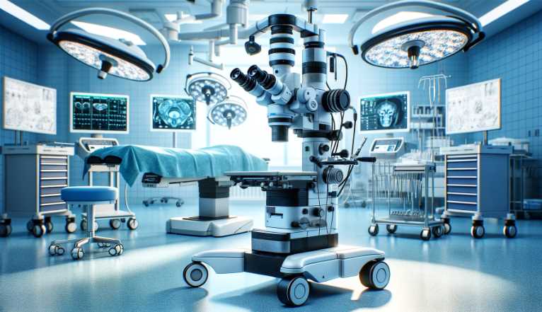
    This surgical microscope is an instrument that can be used to view objects inside the body. It uses a lens and a camera to magnify small parts of the examined object, making it possible to see details that would otherwise be difficult or impossible to see.

    Portable surgical microscopes are typically used for medical procedures like surgery, where it is essential to see the possible minor details of what’s happening inside the body. They are also used in dentistry, where they examine teeth and gums.

    Description and uses

    Portable surgical microscopes are miniature devices that view small objects and tissues inside the body. They are often used in medical procedures like surgery to help surgeons see challenging to see areas and to perform minor surgeries without having to go through an entire operation.

    The technology behind portable surgical microscopes has been improving rapidly over the past few years, becoming more affordable and user-friendly daily. Today, they’re even being used as mobile diagnostic tools for doctors in remote areas who can’t easily access a hospital.

    Here are six of its most popular uses:

    • To examine cells and tissues under a microscope. This can help diagnose diseases and rule out other possible causes for symptoms.
    • To perform surgery by viewing minor surgical procedures close up. This is especially helpful in cases where the disease or condition being treated is located near the body’s surface.
    • To examine foreign objects embedded in tissues or organs for diagnosis or treatment purposes.
    • Assess injuries and damage to internal organs during surgery or medical procedures related to childbirth or childbirth, such as Cesarean section deliveries and ultrasounds.
    • To diagnose genetic conditions and diseases using molecular techniques, such as DNA sequencing and protein analysis.
    • Examine specimens from extreme environments, such as outer space or deep sea depths.

    How to use

    A portable surgical microscope is a perfect tool if you’re looking to get a closer look at the world around you. Though it can be expensive and difficult to use, this microscope is worth the investment if you want to see things in detail. Here are six steps on how to use a portable surgical microscope:

    • Get an accurate measurement of your magnification setting. This will help ensure you get the best possible view of your specimens.
    • Place your specimen on the microscope’s stage, using as much surface area as possible. Try not to move it around too much, or you’ll lose resolution.
    • Adjust the focus until you have a clear image of your specimen. You may need to do this several times to get a good view.
    • Use the magnification controls on your microscope to zoom in and out as needed. Be sure to keep an eye on the exposure time – if it’s too short, parts of your specimen will be blurred; if it’s too long, images will become grainy.
    • Take notes or photos of what you’re seeing so you can later reference them when preparing your reports or presentations.

    Which type of microscope is best for medical use?

    Wall-hanging microscopes are perfect for general medical practice because they’re easy to use and portable. They also have a high magnification range, meaning you can see small details. They’re also versatile enough for other purposes, like studying plant cells or animal tissues.

    Advantages

    Portable surgical microscopes are an excellent tool for medical professionals who need to examine small objects in detail. Here are five reasons why you should consider using one:

    Easy to use- Most portable surgical microscopes are lightweight and easy to carry around. You can use them anywhere, even in difficult or confined spaces.

    Reliable- Portable surgical microscopes have been designed with durability and performance in mind, which means they’ll work well even under harsh conditions.

    Accurate– Because they’re equipped with high-resolution lenses, portable surgical microscopes can provide stunning images of small objects. This makes it easier for doctors to identify abnormalities and treat them effectively.

    Versatile- Portable surgical microscopes can be used for various tasks, including identifying disease symptoms and examining tissue samples.

    Cost-effective- Compared to other diagnostic tools, portable surgical microscopes are relatively affordable and practical for medical professionals.

    Disadvantages

    There are three main disadvantages to using a portable surgical microscope: its price, size, and the need for electricity.

    First, portable surgical microscopes can be expensive, ranging in price from around $1,000 to $10,000. This means they’re not suitable for everyone – especially those who only need them occasionally.

    Second, portable surgical microscopes are often relatively small and lightweight, making them difficult to use or transport. This is particularly true if they must be transported on a plane or in a car.

    Third, even if you get your hands on an excellent portable surgical microscope, you’ll still need to provide it with electricity – most likely via batteries. This means you’ll have to carry around extra equipment just in case you need to use it during an emergency.

    When should you use a portable surgical microscope?

    Suppose you are a nurse or other medical professional assisting doctors during surgery. In that case, you will also need to use a portable surgical microscope to provide them with accurate and up-to-date information about the status of the patient’s condition. Using a portable surgical microscope can prevent any potential complications from occurring during surgery.

    How does a portable surgical microscope differ from a traditional microscope?

    A portable surgical microscope differs from a traditional microscope in its design and functionality. While both serve the purpose of magnifying objects for detailed examination, a portable surgical microscope is specifically designed for medical procedures. It is lightweight, compact, and often features enhanced maneuverability, making it suitable for surgical applications where precision and mobility are crucial.

    FeaturePortable Surgical MicroscopeTraditional Microscope
    DesignCompact and lightweight for portabilityTypically larger and bulkier
    ApplicationPrimarily used in surgical settingsWidely used in laboratories and research
    ManeuverabilityEnhanced mobility for surgical useStationary, limited mobility
    Magnification LevelsAdjustable for various surgical needsFixed magnification levels

    What are the key advantages of using a portable surgical microscope in medical procedures?

    The use of a portable surgical microscope in medical procedures offers several advantages, contributing to improved surgical outcomes and enhanced patient care.

    AdvantageDescription
    Enhanced PrecisionEnables precise visualization during surgery
    PortabilityFacilitates mobility in different surgical settings
    VersatilityAdjustable magnification for diverse procedures
    Improved ErgonomicsDesigned for surgeon comfort and reduced fatigue

    How is the portability of surgical microscopes beneficial for healthcare professionals?

    The portability of surgical microscopes is highly beneficial for healthcare professionals, offering flexibility and ease of use in various medical scenarios.

    BenefitDescription
    Quick SetupEasy to assemble and disassemble for swift deployment
    AdaptabilitySuitable for both traditional and minimally invasive procedures
    Efficient Time ManagementReduces setup time, allowing more focus on patient care

    Can a portable surgical microscope be used in outpatient clinics?

    Yes, portable surgical microscopes are designed to be versatile and can be effectively utilized in outpatient clinics for minor surgical procedures. Their compact design and ease of setup make them suitable for a wide range of medical settings.

    What types of surgeries benefit from the use of portable surgical microscopes?

    Portable surgical microscopes find applications in various surgical specialties, enhancing precision and visualization in procedures such as:

    Surgical SpecialtyBenefit of Portable Microscope
    NeurosurgeryDetailed visualization of brain structures
    OphthalmologyEnhanced magnification for eye surgeries
    ENT (Ear, Nose, Throat)Improved precision in delicate procedures
    OrthopedicsClose examination of bones and joints

    How do surgeons maintain the hygiene of portable surgical microscopes?

    Maintaining a sterile environment is crucial in surgical settings. Surgeons ensure the hygiene of portable surgical microscopes through:

    1. Disposable Covers: Using disposable, sterile covers for the microscope to prevent contamination.
    2. Regular Cleaning: Employing approved cleaning agents to sanitize surfaces without damaging the equipment.
    3. Proper Storage: Storing the portable microscope in a designated, clean area when not in use.

    What features should one consider when choosing a portable surgical microscope for a medical facility?

    When selecting a portable surgical microscope, healthcare facilities should consider the following features:

    FeatureConsideration
    Magnification RangeEnsures compatibility with various surgical needs
    Lighting OptionsAdjustable illumination for optimal visibility
    Portability and WeightEasy to move and set up in different environments
    Documentation CapabilitiesIntegration with imaging and recording equipment

    Are there any safety precautions associated with the use of portable surgical microscopes?

    Yes, safety precautions are essential to ensure the well-being of both patients and healthcare professionals. Some key precautions include:

    1. Electrical Safety: Ensuring proper grounding and electrical safety measures during use.
    2. Proper Calibration: Regular calibration to maintain accurate magnification levels.
    3. User Training: Adequate training for medical staff on the correct usage and handling of the portable microscope.

    Can a portable surgical microscope be connected to imaging and recording devices?

    Yes, many portable surgical microscopes are equipped with features that allow seamless integration with imaging and recording devices. This capability aids in documentation, training, and reviewing surgical procedures.

    How do healthcare facilities ensure the proper maintenance of portable surgical microscopes?

    Maintaining the functionality of portable surgical microscopes involves:

    1. Regular Inspections: Scheduled checks for any signs of wear, damage, or malfunction.
    2. Calibration Checks: Ensuring that the magnification and focus settings are accurate.
    3. Prompt Repairs: Addressing any issues promptly with the assistance of qualified technicians.

    By adhering to these maintenance practices, healthcare facilities can prolong the lifespan of portable surgical microscopes and ensure optimal performance.

    How does the magnification and illumination of a portable surgical microscope compare to a traditional microscope?

    The magnification and illumination of a portable surgical microscope can vary depending on the specific model and manufacturer. However, in general, portable surgical microscopes are designed to provide similar magnification and illumination capabilities as traditional surgical microscopes.

    Portable surgical microscopes typically offer a range of magnification options, often starting at around 4x and going up to 40x or higher. The magnification can be adjusted during surgery to allow the surgeon to view the surgical site at the optimal level of detail.

    Similarly, portable surgical microscopes typically feature bright and adjustable illumination that provides clear and even lighting across the surgical site. This illumination can be provided by a variety of light sources, such as LED or fiber optic, and can often be adjusted to suit the surgeon’s preferences and the needs of the surgical procedure.

    While there may be some variations in the specific magnification and illumination capabilities of different portable surgical microscope models, these devices are designed to provide high-quality imaging and lighting that meet the needs of modern surgical procedures. As such, they can be an effective alternative to traditional surgical microscopes in a range of settings and applications.

    What are some of the potential drawbacks or limitations of using a portable surgical microscope?

    While portable surgical microscopes offer many benefits and advantages, there are also some potential drawbacks and limitations to consider. These may include:

    1. Limited magnification: Portable surgical microscopes may have a more limited range of magnification options compared to traditional surgical microscopes. This can make it more difficult to achieve extremely high levels of detail and resolution in some surgical procedures.
    2. Smaller field of view: The smaller size of portable surgical microscopes may result in a smaller field of view, which can limit the surgeon’s ability to see and work within the surgical site.
    3. Reduced stability: The compact and lightweight design of portable surgical microscopes can sometimes result in reduced stability, which can make it more difficult to achieve precise movements and adjustments during surgery.
    4. Lower image quality: Depending on the specific model and manufacturer, portable surgical microscopes may not offer the same level of image quality and clarity as traditional surgical microscopes. This can be due to factors such as the quality of the optics, the type of illumination used, and other design features.
    5. Limited accessory options: Portable surgical microscopes may have a more limited range of accessory options compared to traditional surgical microscopes. This can limit the surgeon’s ability to customize the microscope to their specific needs and preferences.
    6. Increased cost: Depending on the specific model and features, portable surgical microscopes can sometimes be more expensive than traditional surgical microscopes.

    Overall, while portable surgical microscopes offer many benefits and advantages, it’s important to carefully consider their potential limitations and drawbacks when deciding whether they are the right choice for a specific surgical procedure or application.

    What is the role of nurses and other support staff in using and maintaining a portable surgical microscope during surgical procedures?

    Nurses and other support staff play an important role in using and maintaining a portable surgical microscope during surgical procedures. Their responsibilities may include:

    1. Preparation: Nurses and other support staff may be responsible for setting up the portable surgical microscope before the surgery begins, including ensuring that it is properly calibrated and positioned for the surgeon’s needs.
    2. Assistance: During the surgery, nurses and support staff may assist the surgeon with adjusting the microscope’s magnification and illumination settings, as well as other controls as needed.
    3. Maintenance: Nurses and support staff may be responsible for maintaining and cleaning the portable surgical microscope, including ensuring that all components are properly sterilized before and after use.
    4. Troubleshooting: In the event of any technical issues or malfunctions with the portable surgical microscope, nurses and support staff may be responsible for identifying and addressing the problem or contacting technical support for assistance.
    5. Documentation: Nurses and support staff may be responsible for documenting the use of the portable surgical microscope during the surgery, including any adjustments made to the microscope’s settings or any issues that arise.

    Overall, nurses and support staff play a crucial role in ensuring that the portable surgical microscope is properly used and maintained during surgical procedures. By working closely with the surgeon and other members of the surgical team, they can help to ensure that the microscope functions effectively and contributes to the success of the surgery.

    Final Words

    It can be a little overwhelming for beginners to know about the different specifications and features of a good surgical microscope. Also, it depends on where you buy from, but some models also come with cameras so that you can capture your details too.

    In case you are looking for the best portable surgical microscope in India, we have curated a list of options that include popular brands like Olympus, Leitz, and Leica. These high-end brands produce amazing products that ensure your work is done accurately. So if you love lab experiments or want to improve their skills when it comes down to conducting surgeries, then these instruments may be worth checking out!