The most powerful optical microscope, such as the advanced super-resolution microscopes, can achieve resolutions down to a few nanometers, allowing researchers to visualize structures at the molecular level. These microscopes use techniques like structured illumination, stimulated emission depletion, or stochastic optical reconstruction microscopy to surpass the diffraction limit of conventional light microscopes. They enable unprecedented insights into cellular structures, protein interactions, and nanomaterials.
However, their high precision often requires specialized training and careful sample preparation. Despite their remarkable capabilities, they still face limitations in imaging deep within tissues due to light scattering, which is a key focus of ongoing research and development efforts.
After reading this article, you will better understand what to look for when purchasing one. So, without further ado, let’s get started!
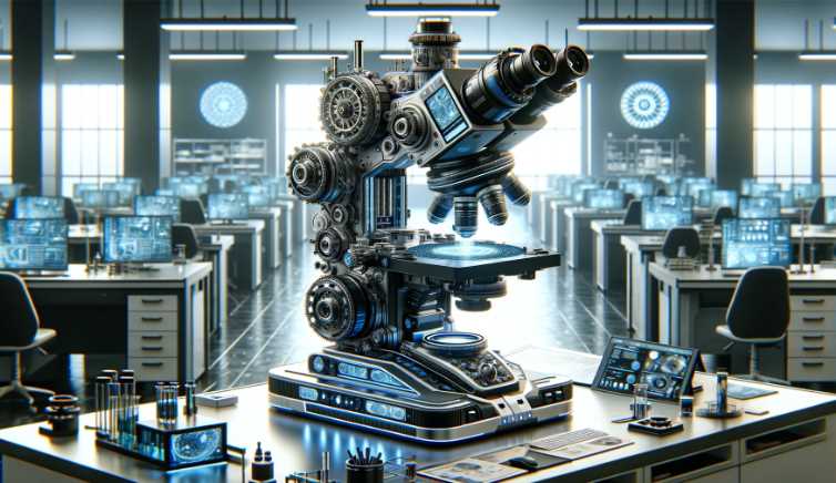
Which is the Most Powerful Optical Microscope?
In 2009, the development of the world’s most powerful optical microscope, the STED (stimulated emission depletion) microscope, revolutionized nanoscale imaging. Here are some key facts about this groundbreaking technology:
| Fact | Information |
|---|---|
| Year of Development | 1994 |
| Resolution | Can visualize structures as small as 20 nanometers |
| Principle | Utilizes a combination of fluorescence and stimulated emission to overcome the diffraction limit of light microscopy. |
| How it Works | A doughnut-shaped laser beam selectively de-excites fluorophores, resulting in highly localized fluorescence, allowing for super-resolution imaging. |
| Applications | Enables visualization of cellular structures, protein complexes, and molecular interactions at unprecedented detail, vital for biomedical research. |
| Nobel Prize | The inventors Stefan W. Hell, Eric Betzig, and William E. Moerner were awarded the Nobel Prize in Chemistry in 2014 for their contributions to the development of super-resolved fluorescence microscopy. |
The STED microscope’s ability to surpass the diffraction limit of light microscopy has paved the way for groundbreaking discoveries in various fields, from neuroscience to molecular biology. Its impact continues to be felt in advancing our understanding of the nanoscopic world within cells and tissues.
The ability to see 20 times smaller than a conventional optical microscope, down to 50 nanometers under normal light, is indeed impressive. However, it’s crucial to keep in mind that the field of microscopy is dynamic, and new breakthroughs may have occurred after my last update.
For the most up-to-date and accurate information, I recommend checking recent scientific literature, research publications, or the latest news in the field of microscopy to identify any advancements or newer technologies that may have surpassed the capabilities of the microsphere nanoscope.
| Microscope Type | Key Features | Applications |
|---|---|---|
| Confocal Microscope | – Uses a pinhole to eliminate out-of-focus light | – 3D imaging |
| – Provides optical sectioning | – Fluorescence microscopy | |
| – High spatial resolution | – Live cell imaging |
| Super-Resolution Microscope | Key Features | Applications |
|---|---|---|
| Structured Illumination Microscope (SIM) | – Resolves structures below the diffraction limit | – Live-cell imaging |
| – Faster imaging compared to traditional methods | – Subcellular structure analysis | |
| – 2D and 3D imaging capabilities | – Fluorescence microscopy |
| Stimulated Emission Depletion (STED) Microscope | Key Features | Applications |
|---|---|---|
| – Achieves sub-diffraction resolution | – Super-resolution imaging | – Nanoscale imaging of cellular structures |
| – Uses depletion laser to reduce the focal spot size | – 3D imaging capabilities | – Fluorescence microscopy |
| – Enables detailed investigation of nanostructures | – Live-cell imaging |
| Multiphoton Microscope | Key Features | Applications |
|---|---|---|
| – Uses two-photon or multi-photon excitation | – Reduced phototoxicity | – Deep tissue imaging |
| – Penetrates deeper into tissues | – 3D imaging capabilities | – In vivo imaging |
| – Minimizes out-of-focus light | – Imaging thick samples | – Neurobiology research |
Historical Overview
Embarking on a journey through the annals of optical microscopy unveils a rich tapestry of scientific progress, marked by pivotal milestones that have shaped our understanding of the microscopic world.
A. Milestones in Optical Microscopy
1. Invention of the Microscope
The inception of optical microscopy can be traced back to the early 17th century, with the groundbreaking invention of the microscope. Dutch spectacle maker Zacharias Janssen and his father Hans Janssen are often credited with creating the first compound microscope around 1595. This primitive microscope, consisting of convex lenses in a tube, laid the foundation for the exploration of the unseen.
2. Early Improvements and Discoveries
The 17th century witnessed the refinement of the microscope by notable figures such as Anton van Leeuwenhoek, who meticulously crafted single-lens microscopes and made groundbreaking observations of microscopic life. Leeuwenhoek’s detailed studies, including the first observation of bacteria, opened a new frontier in biology, unraveling a world that was previously hidden from human sight.
B. Transition to Powerful Optical Microscopes
1. Advancements in Optics
As scientific curiosity grew, so did the need for improved optical instruments. The 19th century marked a significant turning point with the development of achromatic lenses by Joseph Jackson Lister, effectively reducing chromatic aberrations. This advancement, coupled with the contributions of Carl Zeiss and Ernst Abbe, set the stage for enhanced optical clarity in microscopes.
2. Breakthrough Technologies
The 20th century ushered in a wave of breakthrough technologies that propelled optical microscopy to new heights. The development of phase-contrast microscopy by Frits Zernike in the 1930s allowed for the visualization of transparent specimens, while fluorescence microscopy, pioneered by Marvin Minsky in the 1950s, introduced a new dimension by illuminating specific structures with fluorescent dyes.
These innovations culminated in the modern era of powerful optical microscopes, where advancements in lens technology, illumination systems, and automated controls have collectively elevated the precision and capabilities of these instruments.
Types of Optical Microscopes
Exploring the diverse array of optical microscopes reveals a spectrum of instruments, each tailored to specific applications and offering unique advantages in scientific exploration.
A. Compound Microscopes
1. Definition and Basic Structure
Compound microscopes, the workhorses of many laboratories, are characterized by their dual-lens system. Comprising an objective lens near the specimen and an eyepiece for observation, these microscopes utilize magnification and illumination to reveal intricate details. The basic structure involves a sturdy frame supporting a stage for specimen placement and an adjustable focus mechanism for precise observations.
2. Applications and Limitations
The applications of compound microscopes span various scientific disciplines, from biology and medicine to materials science. They excel in revealing fine details of transparent specimens, making them invaluable for cellular and histological studies. However, their limitations include a relatively shallow depth of field and challenges in observing thicker, opaque samples.
B. Stereo Microscopes
1. Overview and Design
Stereo microscopes, also known as dissecting or binocular microscopes, offer a three-dimensional view of specimens. Their distinctive design incorporates two optical paths, providing depth perception. These microscopes typically have a zoom capability, allowing for variable magnification levels. The design features a stereoscopic eyepiece for comfortable viewing and a well-illuminated stage suitable for larger, opaque specimens.
2. Practical Applications
Stereo microscopes find applications in fields where a detailed, three-dimensional view is crucial. Biologists use them for dissections, entomologists study insects, and researchers in electronics benefit from their utility in soldering and intricate component assembly. Their practicality lies in the ability to inspect larger objects while maintaining a clear, magnified view.
C. Confocal Microscopes
1. Principle of Operation
Confocal microscopes revolutionize optical imaging through a unique principle of operation. They employ a pinhole aperture to eliminate out-of-focus light, allowing only the focal plane to contribute to the final image. This optical sectioning technique enhances clarity and contrast, enabling the visualization of fine structures within thick specimens. Laser illumination is commonly used in confocal microscopy.
2. Advantages in Imaging
The advantages of confocal microscopes are evident in their ability to capture sharp, high-contrast images. Researchers benefit from improved resolution, especially in three-dimensional reconstructions. Confocal microscopy is pivotal in fluorescence imaging, providing detailed insights into cellular structures and dynamic processes. However, the complexity and cost of confocal systems can pose limitations for some laboratories.
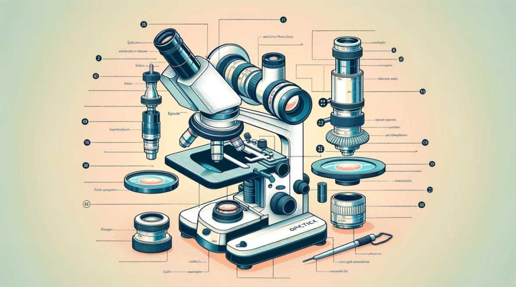
Features and Components of Powerful Optical Microscopes
The capabilities of powerful optical microscopes are intricately tied to their advanced features and components. From high-resolution optics to precision stage and focus mechanisms, each element plays a pivotal role in unlocking the mysteries of the microscopic world.
A. High-Resolution Optics
1. Importance of Resolution in Microscopy
Resolution stands as a cornerstone in microscopy, determining the clarity and detail with which microscopic structures can be observed. Powerful optical microscopes prioritize high-resolution optics to discern finer details, providing researchers with a level of precision crucial for accurate analysis. This emphasis on resolution ensures that even the subtlest cellular structures and molecular arrangements can be visualized, contributing to a deeper understanding of biological and material specimens.
2. Advances in Lens Technology
The journey toward powerful optical microscopes has been marked by continuous advancements in lens technology. The development of sophisticated lenses with reduced aberrations and enhanced light-gathering capabilities has significantly contributed to improved resolution. Cutting-edge lens designs, such as apochromatic and fluorite objectives, allow for unparalleled clarity in imaging, pushing the boundaries of what was once achievable in optical microscopy.
B. Enhanced Illumination Systems
1. Role of Illumination in Microscopy
Illumination serves as the guiding light in microscopy, playing a crucial role in revealing the details of specimens. Powerful optical microscopes feature enhanced illumination systems that go beyond mere brightness. These systems are designed to provide uniform and controlled lighting, minimizing artifacts and optimizing the contrast of observed structures. The importance of precise illumination cannot be overstated, particularly in fluorescence and phase-contrast microscopy.
2. Innovative Lighting Techniques
In the realm of powerful optical microscopes, innovative lighting techniques further elevate the quality of observations. Techniques like dark-field and differential interference contrast (DIC) illumination enhance contrast and reveal subtle variations in specimen composition. Advanced illumination methods, including LED and laser illumination, not only ensure optimal visibility but also open new possibilities in fluorescence imaging, enabling the study of specific cellular components with unprecedented clarity.
C. Precision Stage and Focus Mechanisms
1. Importance in Microscopic Analysis
The ability to precisely manipulate the position of a specimen is paramount in microscopic analysis. Powerful optical microscopes feature precision stage and focus mechanisms that facilitate accurate and controlled movement. This precision is particularly crucial when studying dynamic processes, such as live cell imaging, where maintaining the focal plane is essential for capturing meaningful data.
2. Automation and Digital Controls
Modern powerful optical microscopes embrace automation and digital controls to streamline the microscopic workflow. Automated stage movements, autofocus capabilities, and digitally controlled focus mechanisms enhance efficiency and reduce user-induced variability. These features not only save time but also contribute to the reproducibility of experiments, ensuring consistent and reliable results across multiple observations.
Applications of the Most Powerful Optical Microscopes
Powerful optical microscopes have revolutionized scientific research across various disciplines, offering unparalleled capabilities for detailed observation and analysis. The applications span from unraveling the complexities of cellular structures to delving into the intricacies of nanotechnology and material characterization.
A. Biomedical Research
1. Cellular and Molecular Imaging
Powerful optical microscopes have become indispensable tools in biomedical research, allowing for high-resolution cellular and molecular imaging. With advanced optics and imaging techniques, researchers can explore the intricate details of cells and subcellular structures. This capability is particularly crucial for understanding cellular processes, identifying anomalies, and advancing our knowledge of diseases at the microscopic level.
2. Live Cell Observation
The ability to observe living cells in real-time is a hallmark of the most powerful optical microscopes. Through techniques like fluorescence microscopy, these instruments enable researchers to track dynamic cellular events, monitor cell behavior, and gain insights into biological processes. Live cell observation has profound implications for fields such as pharmacology, where the effects of drugs on living cells can be directly visualized.
B. Material Science
1. Nanotechnology and Material Characterization
In the realm of material science, powerful optical microscopes play a pivotal role in nanotechnology and material characterization. These microscopes provide the resolution needed to visualize and analyze materials at the nanoscale. Researchers can investigate the properties of nanomaterials, study their behavior, and contribute to the development of innovative materials with tailored functionalities.
2. Surface Analysis
Powerful optical microscopes contribute significantly to surface analysis, allowing researchers to explore the topography and composition of materials. Techniques like confocal microscopy and advanced imaging modalities facilitate detailed examinations of surfaces, offering insights into the roughness, morphology, and even chemical composition of materials. This is particularly relevant in industries such as electronics and materials engineering, where surface properties directly impact performance.
Applications of the Most Powerful Optical Microscopes – Overview Table
| Discipline | Applications |
|---|---|
| Biomedical Research | – Cellular and Molecular Imaging |
| – Live Cell Observation | |
| Material Science | – Nanotechnology and Material Characterization |
| – Surface Analysis |
Comparison with Other Microscopy Techniques
Understanding the strengths and limitations of different microscopy techniques is crucial for researchers to choose the most suitable tool for their specific applications. Here, we compare powerful optical microscopy with electron microscopy and scanning probe microscopy.
A. Electron Microscopy
1. Contrasting Optical and Electron Microscopy
While powerful optical microscopes utilize visible light to observe specimens, electron microscopes employ electron beams. The key contrast lies in the wavelength of the imaging particles—photons for optical microscopes and electrons for electron microscopes. This fundamental difference results in varying resolutions and penetration depths.
2. Complementary Roles in Research
Optical and electron microscopy play complementary roles in research. Powerful optical microscopes excel in imaging living cells and soft materials with resolutions typically in the micrometer range. In contrast, electron microscopes provide nanoscale resolution and are ideal for imaging inorganic materials, nanomaterials, and ultrastructural details of biological specimens.
B. Scanning Probe Microscopy
1. Nanoscale Imaging and Manipulation
Scanning probe microscopy operates at the nanoscale, utilizing a sharp probe to scan surfaces. Unlike optical and electron microscopy, it offers both imaging and manipulation capabilities at the atomic and molecular levels. This technique provides detailed topographical information and the ability to manipulate individual atoms.
2. Unique Capabilities and Limitations
Scanning probe microscopy has unique capabilities but also limitations. It excels in nanoscale imaging with unparalleled resolution but is limited by slow imaging speeds. Additionally, it is sensitive to environmental conditions and may not be suitable for certain biological samples.
Comparison Table: Optical, Electron, and Scanning Probe Microscopy
| Aspect | Optical Microscopy | Electron Microscopy | Scanning Probe Microscopy |
|---|---|---|---|
| Imaging Particle | Photons (Visible Light) | Electrons | Probe Tip (Mechanical) |
| Resolution | Micrometer Range | Nanometer to Angstrom Range | Nanometer to Atomic Range |
| Specimen Type | Living Cells, Soft Materials | Inorganic, Nanomaterials, Ultrastructure of Biological Specimens | Surfaces, Nanomaterials |
| Speed | Fast | Moderate to Slow | Slow |
| Manipulation Capability | Limited | Limited (Manipulation of Electrons) | Yes (Atomic and Molecular Level) |
| Environmental Sensitivity | Moderate | High | High |
Tips for Effective Use of Powerful Optical Microscopes
Achieving optimal results with powerful optical microscopes requires attention to detail and adherence to best practices. Here are essential tips to enhance your microscopy experience:
A. Proper Sample Preparation
1. Importance of Sample Condition
The quality of sample preparation significantly impacts microscopic observations. Ensure specimens are well-preserved, properly fixed, and appropriately stained, considering the specific requirements of the imaging technique. Proper sample condition is crucial for obtaining accurate and meaningful results.
2. Common Techniques for Preparation
Utilize standard sample preparation techniques tailored to your specimen type and imaging modality. Techniques such as sectioning, staining, and mounting ensure the specimen is well-prepared for observation under the microscope. Following established protocols minimizes artifacts and enhances the clarity of microscopic images.
B. Maintenance and Calibration
1. Ensuring Optimal Performance
Regular maintenance is key to sustaining the performance of powerful optical microscopes. Keep optical components clean, inspect for any signs of wear, and address issues promptly. Routine cleaning and alignment procedures contribute to the longevity and reliability of the microscope.
2. Regular Checks and Adjustments
Perform regular checks and adjustments to guarantee accurate results. Calibration of objectives, eyepieces, and the stage is critical for maintaining precision. Periodic assessments of illumination sources and filters ensure consistent lighting conditions, minimizing potential variations in image quality.
C. Image Analysis and Documentation
1. Software Tools for Image Processing
Utilize advanced image processing software to enhance and analyze microscopic images. These tools allow for contrast adjustments, noise reduction, and three-dimensional reconstructions. Familiarize yourself with the features of the software to extract valuable information from captured images.
2. Record-Keeping and Documentation Practices
Establish systematic record-keeping practices for your microscopy sessions. Document important parameters such as magnification, illumination settings, and any adjustments made during imaging. Proper documentation facilitates reproducibility of experiments and aids in the comprehensive analysis of data over time.
Tips for Effective Use of Powerful Optical Microscopes – Summary Table
| Aspect | Tips |
|---|---|
| Proper Sample Preparation | – Emphasize Sample Condition |
| – Utilize Standard Preparation Techniques | |
| Maintenance and Calibration | – Ensure Regular Maintenance for Optimal Performance |
| – Perform Regular Calibration Checks and Adjustments | |
| Image Analysis and Documentation | – Use Advanced Image Processing Software |
| – Establish Comprehensive Record-Keeping Practices |
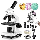
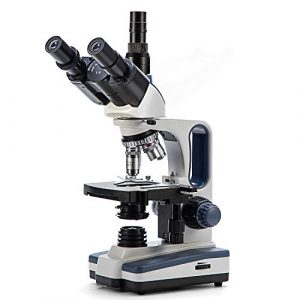
NATIONAL GEOGRAPHIC Dual LED Student Microscope
Are you looking for a microscope that can help you learn science or math in a fun and engaging way? Then the National Geographic dual LED student microscope is perfect for you! This educational tool is easy to use and comes with a carrying case that makes it easy to take with you wherever you go.
It also has a built-in light that makes it easy to see the details of your specimens. So, if you are looking for a microscope that can help you learn entertainingly and efficiently, the National Geographic microscope is the perfect option!

Main Features
1. Dual-LED illumination for superior image quality and contrast.
2. 10x and 20x magnifications for greater detail and precision.
3. Meets the latest safety standards for educational use.
4. Compact and portable design for easy mobility.
5. It Comes with a carrying case for easy storage and transport.
Superior image quality – Professional grade optics combine with dual LED illumination to produce clear and bright images, even in low-light conditions.
Portable – The lightweight and easy-to-carry design make it perfect for students and professionals who need to take their microscope with them wherever they go.
High-resolution imaging – National Geographic’s award-winning optics provide detailed images of live and fixed specimens that are ideal for scientific research.
Robust construction – The durable metal construction ensures that your microscope will last through years of use.
Affordable – At just reasonable price, the National Geographic dual-LED student microscope is an excellent value for your money
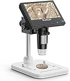
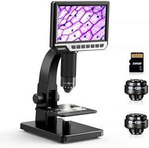
MOYSUWE MDM9 LCD Digital Microscope
Moysuwe MDM9 is a self-contained, portable microscope with a high-quality HD LCD monitor and a DC 12V power supply. It also features automatic brightness control, 10x and 40x eyepieces, 3.5mm audio output, and a smartphone camera connector.
Additionally, it has a built-in digital camera that can capture images at resolutions of up to 2 megapixels. So, if you are looking for a high-quality, portable microscope ideal for educational use, the Moysuwe MDM9 is the perfect option!
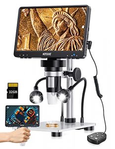
Main Features
– A digital microscope with high resolution and bright images.
– Use it to inspect your jewelry, watches, coins, and gemstones.
– Great for scientific use, like in biology labs.
1. Accurate imaging – The MDM9 has a resolution of up to 20,000x and a field of view of 100mm. Its high-quality optics make it an excellent choice for academic and research use.
2. Fast and easy photo scanning – This digital microscope makes it easy to capture high-resolution photos and videos with just a few taps.
3. Compact and portable – It is small and lightweight, making it easy to take with you wherever you go.
4. Easy to use – The microscope is user-friendly and easy to operate.
5. Flexible and versatile – The MDM9 LCD can examine biological and non-biological specimens.
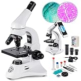
Swift SW350T Siedentopf Compound Lab Microscope
If you’re in the market for a microscope that can handle some big specimens, then the Swift SW350T lab microscope may be a perfect choice. With its high-quality optical system and advanced features, this microscope is ideal for scientists and research professionals who need a powerful tool to handle delicate specimens. This microscope review will look at some of its key features and help you decide if it’s suitable for your needs.
Main Features
– It is a high-performance microscope.
– Features high-speed expansion rates and focusing speeds.
– Provides accurate microscopy of biological samples.
1. With a magnification of 350x, the Swift SW350T Siedentopf Head Trinocular Compound Lab Microscope is the perfect instrument for studying small tissues and cells.
2. It offers fast, easy switching between objectives, with a range of 10x to 350x.
3. The compact and lightweight design make it easy to transport and use.
4. The built-in LED illumination provides clear viewing even in low-light conditions.
5. The SW350T Siedentopf Head Trinocular Compound Lab Microscope offers a lifetime warranty.
TOMLOV DM11 LCD 2000X Biological Microscope
If you’re looking for a high-quality microscope that’s easy to use, then you should consider the TOMLOV DM11 LCD digital microscope. It has a range of features that make it great for both educational and research purposes, and it’s also an excellent value for the money. This review will examine some of its key features and discuss how they can benefit you.
Main Features
– Digital microscope with 2000X magnification.
– Can see the minor details on a slide.
– Can be used for biological, medical, and veterinary applications.
– Easy to operate.
– Bright and clear images.
1. With a magnification of 2000X, the TOMLOV DM11 LCD digital microscope can observe details that are difficult or impossible to see with other microscopes.
2. It has a wide-angle lens that makes it easier to view more significant areas of the specimen.
3. The digital display makes it easy to view images and make accurate measurements.
4. The built-in speaker allows you to listen to the specimen without using headphones.
5. The microscope is compact and easy to carry around, making it an excellent choice for scientists and students who need to take specimens wherever they go.
AmScope SE305-P Binocular Stereo Microscope
If you’re in the market for a microscope that’ll help you see things in stunning clarity, the AmScope SE305-P should be at the top of your list. With its high-quality optics and responsive controls, this microscope is perfect for students, researchers, and anyone who wants to look closely at their specimens. Its compact design makes it easy to take with you wherever you go. Read our detailed review to find out more about this top-rated microscope!
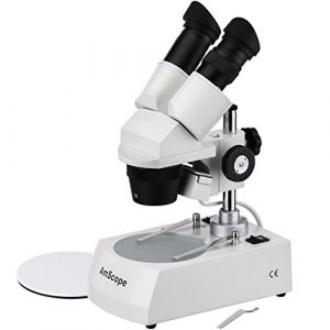
Main Features
– It’s an affordable, accurate microscope.
– Powerful and durable.
– Perfect for any microscopy application.
– High-quality stereo vision control system.
– Compact and lightweight design that is perfect for use on the go.
– Take to the field for a better view.
– Get more out of your project with a stereo microscope.
1. High resolution: With a resolving power of up to 1,000x, the AmScope SE305-P binocular stereo microscope can view incredibly detailed images.
2. Compact design: The AmScope SE305-P is a lightweight and compact microscope that is easy to take with you wherever you go.
3. Dual eyepieces: This model has a dual eyepiece design that allows you to view both the left and right images simultaneously. This allows for a completer and more accurate picture.
4. Built-in camera: It also has a built-in camera that allows you to take pictures and videos of your specimens.
5. 3-inch LCD screen: The 3-inch LCD screen makes it easy to see and navigate your images and videos.
What to Look For When Buying an Optical Microscope, Lenses, and Accessories?
After years of research and development, the most powerful optical microscope has finally been unveiled – the A&D! With its ability to magnify objects up to 200x, the microscope is perfect for researchers, doctors, biologists, and more. This buying guide will outline the features and benefits of the A&D microscope so that you can make an informed decision before buying it.
We will also provide a buyer’s guide for choosing the best place to buy it. So, without further ado, let’s get started! To determine if an optical microscope is the most powerful model for your needs, you should consider the following factors:
There are a few key things to keep in mind before purchasing a powerful optical microscope:
Magnification power is essential but not the only factor to consider. Other features, such as the quality of the optics and the type of lighting available, can also be crucial factors.
Make sure to consider the size and weight of the microscope, especially if you plan on transporting it frequently.
Level of magnification: This is an essential factor to consider when choosing an optical microscope. The most powerful microscopes can offer up to 1000x magnification, while less powerful models may offer only up to 400x magnification.
This is an essential factor to consider when choosing an optical microscope. The most powerful microscopes can offer up to 1000x magnification, while less powerful models may offer only up to 400x magnification.
The size of the sample that can be viewed: The most powerful microscopes can typically accommodate larger samples than less powerful models. If you need to view larger specimens, consider a more powerful microscope.
Optical microscopes can typically accommodate larger samples than less powerful models. If you need to view larger specimens, consider a more powerful microscope. The level of detail that can be observed: The most powerful microscopes offer the highest level of detail, allowing you to see minor features of your specimen. Consider a more powerful microscope if you need to keep the most intricate details.
The powerful optical microscopes offer the highest level of detail, allowing you to see the minor features of your specimen. Consider a more powerful microscope if you need to observe the most intricate details. The type of microscope: Not all optical microscopes are created equal.
Can you see atoms with an optical microscope?
No, optical microscopes cannot see atoms. Optical microscopes work by using a light beam to magnify an object. This allows the user to see details that would be impossible to see with the naked eye.
Which part of an optical microscope contains a magnifying lens?
The eyepiece of an optical microscope is an essential part of the instrument, as it contains the magnifying lens. The eyepiece is located at the front of the microscope and is where you view the specimen.
How should you carry a compound optical microscope?
Knowing how to carry it properly when carrying a compound optical microscope is essential. The best way to do this is to use a carrying case or backpack with a specially designed compartment for the microscope.
You should also ensure that the microscope is securely fastened to the case or backpack so that it does not move around. When transporting the microscope, please keep it clean and free from dust and debris.
Can an optical microscope see cancer cells?
Yes, an optical microscope can see cancer cells. However, it is essential to note that the produced images are not always accurate, and the magnification is not always sufficient to determine the characteristics of cancer cells.
The optical microscope is not a substitute for a surgical microscope used for a more accurate cancer diagnosis.
Can an optical microscope observe surface roughness?
Yes, an optical microscope can observe surface roughness. Surface roughness can be used to identify the materials that are being examined. By looking at the surface texture, you can determine the grain size and the degree of roughness. This information can help you understand the material’s properties and how it will react under various conditions.
For example, suppose you are studying a material that will be used in a medical device. In that case, it is essential to understand how the material will respond to stress and how it will degrade over time. By examining the surface texture, you can ensure that the material will meet your requirements.
Can you damage a microscope’s optical path?
Yes, you can damage a microscope’s optical path if you are not careful. Microscope optical paths are delicate and can be easily damaged if not handled with care.
This damage can often be corrected by replacing the microscope’s optical path, but at other times, the damage may be too extensive to be repaired. If you are unsure if your microscope’s optical path is damaged, you should consult a microscope technician.
Do I dampen the microscope optical lens wipe before using it?
No, you do not need to dampen the microscope optical lens wipe before using it. The wiping motion will cause the wipe to become damp, which will affect the quality of the captured image.
How does an optical microscope work?
An optical microscope is a type of microscope that uses lenses to magnify images. It operates by using a light source and a mirror that reflects the light onto an object being viewed. The object is then placed in a special chamber that allows the light to pass through it and onto a photodiode, which records the image. This image can then be displayed on a monitor or printed out.
How is the final magnification of an optical microscope calculated?
The final magnification of an optical microscope is calculated by taking the object’s magnification (x) and dividing it by the objective’s NA (number of Angstroms). For example, if the object’s magnification is 10x and the objective has a NA of 100, the final magnification would be 1x.
How much does an optical microscope cost?
An optical microscope can range in price from around $50 to $2000, depending on the desired features and specifications. The most popular options include the Nikon Eclipse TE200 and the Olympus BX61.
These microscopes offer excellent image quality and versatility, making them a good investment for businesses and researchers. Additionally, they are easy to use and can be operated by anyone with a basic understanding of chemistry and optics.
How to add a scale bar on an optical microscope?
Adding a scale bar to an optical microscope is a relatively simple task. It can help in enhancing the accuracy of measurements. The scale bar is a thin metal or plastic strip attached to the microscope eyepiece and calibrated to give accurate readings of distances. This is useful for measurements such as DNA or protein concentration or for measuring the size of cells or tissues under the microscope.
To add a scale bar to your microscope, first ensure that the eyepiece is properly aligned with the objective. Next, attach the scale bar using screws or magnets. Finally, adjust the eyepiece until the scale bar is centered on the measured object and take the reading.
How to use an optical microscope?
Using an optical microscope can be a fun and educational experience, and it can be used to view objects that are too small to see with the naked eye and can be an excellent tool for scientists and researchers. There are a few things that you need to know to use an optical microscope effectively:
Magnification level – The magnification level is the amount of magnification the microscope can achieve. The higher the magnification, the smaller the object that can be viewed.
Eyepiece is the microscope part that you look through to see the object. Choosing the right eyepiece for the object you are trying to view.
Objective lens – The objective lens is the lens at the front of the microscope that magnifies the image being seen by the eyepiece.
Light – Light is essentially energy that comes in particles called photons. Each point on an image contains the same amount of information, and only a single photon can reach each pixel. Still, multiple images are possible by combining the lights from different points (i.e., one image created from one source view would contain parts “in focus,” while another could show what was behind it).
Is an optical microscope stronger than a light microscope?
An optical microscope is typically more substantial than a light microscope, and this is because an optical microscope uses a higher magnifying power to see more minor details. Additionally, an optical microscope can view objects that are not visible to a light microscope.
What are optical microscopes used for?
Optical microscopes are used for various tasks, including studying biological tissues and cells, inspecting mineral samples, and analyzing environmental samples. They can be used for general or specific applications, such as cancer research.
Optical microscopes use a variety of techniques to image objects and cells. The most common is light interference microscopy, which uses a phase-contrast microscope to create an image of an object or cell by allowing light to pass through the object and be interference- canceled out by the light passing through the cell. This technique is used to view larger objects and see details that would be too small to see with a regular microscope.
Other techniques used in optical microscopy include super-resolution microscopy and fluorescence microscopy. Super-resolution microscopy uses high-resolution cameras to take pictures of small objects that are too small to be seen with a regular microscope.
What are the major types of optical microscopes?
There are three major types of optical microscopes: phase-contrast, fluorescence, and light-condensing.
Phase-contrast microscopes use phase contrast to see different elements of an image. This is done by splitting the light into two waves, one that travels through the sample and the other that is reflected. The waves are then combined, and the difference in energy is used to create an image. This type of microscopy is used to examine small objects to be seen individually.
Fluorescence microscopes use fluorescent dyes to make specific elements of an image visible. This is done by exciting the fluorescent dye with ultraviolet light and then light emission at different wavelengths. The emitted wavelength depends on the type of molecule that was excited. By using this technique, other structures within an image can be seen.
Light-condensing microscopes use light to see objects in an image. This is done with a mechanism that includes two mirrors that reflect the light towards an objective lens, which changes the angle of reflection and allows viewing images without interference from glass or other transparent obstructions between the object being considered and the camera.
What is an inverted optical microscope?
An inverted optical microscope is a microscope that uses a light source at the bottom of the telescope and a mirror at the top to collect light and direct it to a viewing lens. This microscope is advantageous because it is easier to use and gives a more comprehensive viewing range than a traditional microscope.
Additionally, inverted optical microscopes are often used to image cells and tissues under various conditions, including live cells and tissues.
This microscope is helpful for researchers looking for high-resolution images of cells and tissues. Additionally, inverted optical microscopes are often used in the medical field to diagnose conditions such as cancer. They are also used to research the structure and function of cells and tissues.
What is the difference between an optical microscope and an electron microscope?
The main difference between optical and electron microscopes is that optical microscopes use light to magnify an object, while electron microscopes use electrons.
An optical microscope is a type of microscope that uses a lens to magnify an object. It is usually used to view small objects such as cells and bacteria. An electron microscope is a type of microscope that uses an electron beam to see small objects. It is usually used to view larger objects such as viruses and minerals.
What is the maximum magnification of an optical microscope?
The maximum magnification of an optical microscope is 10x. This means that the observed object can be seen as small as ten times its original size. This is great for viewing small details and examining specimens that are difficult to view with the naked eye.
It is important to note that the magnification will decrease as the object is enlarged, so it is essential to use the correct lens for the task.
How to clean an optical microscope?
Cleaning an optical microscope can be daunting, but it can be a relatively easy process with a bit of preparation and diligence. Here are a few tips to help you clean an optical microscope:
Make sure to dust the microscope regularly with a soft, lint-free cloth.
Wipe down the objective lens with a clean, dry cloth.
Remove dust or dirt accumulated on the condenser and eyepieces with a clean, lint-free cloth.
Wipe down the front of the microscope with a clean, dry cloth.
If needed, use a microfiber cloth to remove any smudges or fingerprints.
Polish the lens with a soft cloth and a mild cleaning solution.
Place the microscope in a well-ventilated room to allow the cleaning solution to evaporate.
Avoid harsh chemicals, which can damage the lens and other delicate parts. If you encounter any problems while cleaning your microscope, do not hesitate to contact us for help; a qualified technician can help you clean it properly and restore its function.
Final Word
When it comes to optical microscopes, there are many to choose from. However, regarding features and user-friendliness, only one reigns supreme: the NATIONAL GEOGRAPHIC dual LED student microscope. With its incredible magnification and high resolution, this model is perfect for students interested in exploring the world of biology and medicine at a cellular level.
Additionally, its illumination capabilities ensure that objects are visible even in the darkest of environments, while its durable construction makes it a long-lasting investment. Do you have any suggestions on what other optical microscopes we should consider? Let us know in the comments below!
Facts
1. They can be used for various investigations, including examining biological samples, studying minerals and rocks, and viewing everything from plant cells to celestial objects.
2. Microscopes use lenses (or mirrors) to focus light onto an image sensor, producing an electronic or digital representation of what is being viewed.
3. The resolution of a microscope determines how good detail can be seen on the screen; the higher the number, the sharper and clearer images will be.
4. Most microscopes have built-in illumination that allows you to see things in low light conditions and help researchers capture high-resolution images without spending hours prepping specimens beforehand!
5. Some telescopic objectives also feature adjustable zooming power so scientists can get closer or farther away from their sample(s).
In this guide, you can read a comprehensive comparison between the top 5 optical microscopes on the market today. Furthermore, you will also learn about their different features, specs, and where to buy them. So, this guide is for you whether you are a scientist or an amateur microscopy enthusiast!
Resources and References
For those keen on delving deeper into the world of powerful optical microscopes, here are some recommended resources:
Books and Journals
- “Advanced Optical Microscopy: Principles and Applications” by P. Török
- “Handbook of Biological Confocal Microscopy” edited by James B. Pawley
Key Research Papers and Reviews
- Betzig, E., et al. (2006). “Imaging intracellular fluorescent proteins at nanometer resolution.” Science, 313(5793), 1642-1645.
- Hell, S. W., et al. (1994). “Breaking the diffraction resolution limit by stimulated emission: stimulated-emission-depletion fluorescence microscopy.” Optics Letters, 19(11), 780-782.

I am an enthusiastic student of optics, so I may be biased when I say that optics is one of the most critical fields. It doesn’t matter what type of optics you are talking about – optics for astronomy, medicine, engineering, or pleasure – all types are essential.
Last update on 2025-07-01 / Affiliate links / Images from Amazon Product Advertising API
Table of Contents

Pingback: A Guide To Optical Microscope: History, How Does it Work, Maintenance
Pingback: Which is the most Reliable Microscope for Coin Collectors and Numismatists
Pingback: Beyond the Naked Eye: Is a sperm microscope necessary for sperm analysis?
Pingback: Can I See My Sperm with Microscope