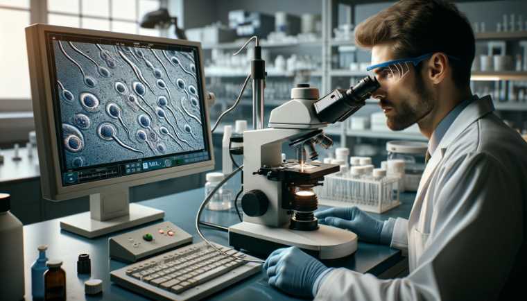
Yes, viruses are visible under a microscope. They are too small to be seen without magnification.
When viewed under a microscope, viruses appear as tiny particles with unique shapes and structures. Their size and features can vary depending on the type of virus. Scientists and researchers use powerful microscopes to study viruses and understand their characteristics, behavior, and impact on living organisms.
The ability to visualize viruses under a microscope is essential for studying and developing treatments and vaccines to combat viral infections. Through advanced imaging techniques, researchers continue to uncover valuable insights into the world of viruses and their potential effects on human health and the environment. Understanding the visual aspects of viruses plays a crucial role in combating various viral diseases and improving public health.
Virus Name
Average Size (nm)
Visualization Technique
Influenza Virus
80-120
Electron Microscope
SARS-CoV-2
50-200
Electron Microscope
Human Papillomavirus
50-60
Electron Microscope
HIV
80-120
Electron Microscope
Adenovirus
70-100
Electron Microscope
What Is A Virus?
A virus is a microscopic infectious agent that can only replicate within the cells of a living organism. It is composed of genetic material, either DNA or RNA, surrounded by a protein coat. Definition: Viruses are non-living entities that depend on a host for survival and reproduction. They are much smaller than bacteria and can only be seen under an electron microscope. Characteristics: Viruses lack the cellular structure of bacteria and cannot carry out metabolic processes on their own. They are known for their ability to cause various diseases in humans, animals, and plants, making them significant subjects of study in the field of microbiology.
Structure of A Virus
Viruses are not visible to the naked eye and can only be observed under a microscope. Their structure consists of a genetic material, either DNA or RNA, enclosed within a protein coat called a capsid. Some viruses also have an outer lipid envelope surrounding the capsid. The size and shape of viruses vary greatly, with some being spherical, helical, or complex in structure. Depending on the type of virus, their dimensions can range from 20 to 400 nanometers.
Microscopy Techniques For Visualizing Viruses
Microscopy techniques play a crucial role in visualizing viruses and understanding their characteristics. Light microscopy, also known as optical microscopy, is one such technique that uses visible light to observe samples. With the help of lenses and light sources, it allows scientists to magnify viruses and study their structure. However, due to the limited resolution of light microscopy, viruses may not always be visible at their actual size.
Electron microscopy, on the other hand, offers higher magnification and resolution. By using a beam of electrons instead of light, it enables researchers to examine viruses in greater detail. The high resolution provided by electron microscopy allows for visualization of even smaller viruses and their intricate structures.
Fluorescence microscopy is a specialized technique that involves the use of fluorescent dyes to label specific parts of the viruses. By emitting light at different wavelengths, these dyes help in visualizing and distinguishing different components of the viruses. Fluorescence microscopy is particularly useful in studying viral replication and understanding interactions between viruses and host cells.
Microscopy Technique
Advantages
Light Microscopy
Easy to use and allows observation of live viruses
Electron Microscopy
Provides high-resolution images and reveals intricate viral structures
Fluorescence Microscopy
Enables visualization of specific viral components and their interactions
Can Viruses Be Seen Under A Light Microscope?
Viruses are often too small to be seen under a light microscope due to their tiny size, typically ranging from 20 to 300 nanometers. However, there are some exceptions to this rule. Poxviruses and Papillomaviruses are examples of viruses that can be observed using a light microscope. These exceptional cases are possible because these viruses are relatively large compared to other viruses.
Despite these exceptions, light microscopy has limitations when it comes to visualizing viruses. The resolution power of a light microscope is limited by the wavelength of visible light, which is around 400 to 700 nanometers. Since viruses are much smaller than this wavelength, they are often not visible under a light microscope. For observing smaller viruses, such as influenza viruses or HIV, more advanced techniques such as electron microscopy or scanning electron microscopy are required.
It is important to note that even though viruses may not be visible under a light microscope, they play a significant role in many aspects of our lives. Understanding their structure and behavior is crucial to developing effective treatments and prevention strategies.
Visualizing Viruses Using Electron Microscopy
Electron microscopy enables the visualization of viruses due to its high magnification capabilities. This powerful tool can capture detailed images of viruses, allowing scientists to study their structure and characteristics. By using electron microscopy, researchers have been able to obtain striking images of viruses at an ultra-high resolution, providing valuable insights into their morphology and behavior. This technology has revolutionized the study of viruses by offering a closer look at their intricate features and allowing for a deeper understanding of their impact on living organisms.
Capabilities of electron microscopy
Examples of virus images obtained through electron microscopy
High magnification
Images of influenza virus, HIV, and herpesvirus
Ultra-high resolution
Detailed visualization of Zika virus and Ebola virus
Structural analysis
Capturing the morphology of SARS-CoV-2
Final Words:
Viruses are microscopic organisms that cannot be directly observed under a regular light microscope. However, with the use of electron microscopes, scientists have been able to capture detailed images of viruses, revealing their intricate structures. These technologies have greatly contributed to our understanding of viruses and their pathogenic characteristics.
By studying viruses at the microscopic level, researchers can develop effective strategies to combat and manage viral infections.
No, centrioles are not typically visible under a light microscope.
Centrioles are small, cylindrical structures found in animal cells, and they play a crucial role in the organization of microtubules during cell division. However, centrioles are very small and are below the resolution limit of a light microscope, which is approximately 200 nanometers. Therefore, they cannot be resolved and visualized using a standard light microscope.
Parameter
Value
Magnification Range
40x – 1000x
Resolution
Approximately 200 nanometers
Maximum Useful Magnifica
500x – 600x for most details
Illumination Source
Visible light
Sample Preparation
Usually requires staining
Cost
Relatively inexpensive
Common Applications
Biological and medical studies
Limitations
Limited resolution for small structures like centrioles
Understanding Centrioles
Centrioles, small but essential cellular structures, are key players in the intricate choreography of cell division and cytoskeletal organization. Comprising cylindrical tubes, centrioles are typically found in pairs near the cell’s nucleus. Understanding the structure and functions of centrioles provides insights into their significance in cellular processes.
What are Centrioles?
Centrioles are microtubule-based organelles, typically organized in pairs called centrosomes. Structurally, they consist of nine triplets of microtubules arranged in a cylindrical pattern, often likened to the blades of a turbine. This unique arrangement contributes to their role as microtubule organizers within the cell.
Centriole Functions
The primary role of centrioles lies in orchestrating cell division. During mitosis and meiosis, centrioles play a vital role in forming the spindle apparatus, a structure that segregates chromosomes into daughter cells. Additionally, centrioles are involved in the formation of cilia and flagella – cellular projections crucial for cell movement and sensory functions.
Centrioles and Cellular Organization
Beyond their role in cell division, centrioles contribute significantly to the organization of the cytoskeleton. The cytoskeleton is a dynamic network of protein filaments that gives the cell its shape, provides mechanical support, and facilitates intracellular transport. Centrioles help anchor microtubules, influencing the overall structural integrity of the cell.
Understanding the significance of centrioles in cellular processes sheds light on their indispensable functions. The intricacies of their structure and their involvement in vital cellular activities emphasize the importance of studying these organelles to unravel the mysteries of cell biology.
Challenges of Light Microscopy
Light microscopy, a cornerstone in the realm of biology, has been instrumental in studying various cellular structures. However, when it comes to observing structures as small as centrioles, inherent limitations arise. This section explores the challenges posed by light microscopy in the quest to visualize centrioles.
Limitations of Light Microscopy
One of the fundamental challenges lies in the resolution of light microscopes. The wavelength of visible light imposes a limit on the smallest structures that can be resolved. Centrioles, with their diminutive size and intricate structure, often fall below this limit, making them challenging to observe accurately.
To illustrate this limitation, consider the typical resolution of a light microscope, which is around 200 nanometers. Given that centrioles are on the order of 100 nanometers in diameter, pushing the boundaries of light microscopy, the fine details may remain elusive under conventional observation.
The Size of Centrioles
Centrioles, measuring approximately 200 to 250 nanometers in length, pose a size challenge when viewed through light microscopes. The optical limitations make it difficult to discern these structures with the clarity required for comprehensive analysis.
Let’s visualize this in a table:
Parameter
Centriole Diameter
Light Microscope Resolution
Approximate Size
200-250 nanometers
~200 nanometers
This comparison underscores the tight margins within which light microscopy operates concerning centriole observation.
Optical Challenges
The optical properties of centrioles further complicate their visibility under light microscopes. The refractive index mismatch between the centrioles and the surrounding cellular environment leads to distortions and reduced image clarity. This phenomenon hampers the accurate depiction of centriolar structures, making their observation a nuanced task.
Let’s summarize the optical challenges in a table:
Optical Challenge
Impact on Visibility
Refractive Index Mismatch
Distortions and reduced image clarity
Can Centrioles be Observed Using a Light Microscope?
Centrioles, unfortunately, are not readily visible under a standard light microscope. Their size and structure make them challenging to discern with the limited resolution of a light microscope, which typically ranges between 200 and 300 nanometers.
Microscope Type
Visibility of Centrioles
Light Microscope
Not Visible
Electron Microscope
Clearly Visible
Why are Centrioles Difficult to See with a Light Microscope?
Centrioles, being small organelles with dimensions below the resolving power of a light microscope, lack the contrast necessary for clear observation. Their size falls below the wavelength of visible light, making it challenging to distinguish them from the surrounding cellular structures.
Size of Centrioles
Light Microscope Resolution
< 200 nm
Limited Visibility
What Microscope Can be Used to Visualize Centrioles?
To observe centrioles effectively, an electron microscope is recommended. The higher resolution of electron microscopes, which can reach below 1 nanometer, allows for the detailed imaging of centrioles and other subcellular structures.
Microscope Type
Suitable for Centriole Observation
Light Microscope
Inadequate Resolution
Electron Microscope
Recommended
Can Special Staining Techniques Enhance Centriole Visibility?
Yes, employing specific staining techniques can enhance the visibility of centrioles under a light microscope. Dyes and stains that selectively bind to centriolar components can improve contrast, making these organelles more distinguishable.
Staining Technique
Effect on Centriole Visibility
Centriole Staining
Enhanced Contrast
Immunofluorescence
Improved Visibility
What is the Size of Centrioles, and How Does it Affect Visibility?
Centrioles typically have a diameter of about 250 nanometers and a length of approximately 500 nanometers. Their small size poses a challenge for light microscopy, where the resolution limit often exceeds the dimensions of these organelles.
Centriole Dimensions
Light Microscope Resolution
250 nm (diameter)
Limited Visibility
Are Centrioles Important Despite Their Limited Visibility?
Despite their challenge to observe directly, centrioles play a crucial role in cell division, organizing the microtubules of the mitotic spindle. Their significance lies in their contribution to the proper segregation of chromosomes during cell division, ensuring the formation of genetically identical daughter cells.
Centriole Importance
Visibility Challenge
Crucial for Cell Division
Limited Direct Observation
Can Advances in Microscopy Technology Improve Centriole Visibility?
Advancements in microscopy techniques, such as super-resolution microscopy, hold promise for improving centriole visibility. These techniques surpass the diffraction limit of traditional light microscopy, potentially allowing for clearer imaging of centrioles.
Microscopy Advancements
Potential for Improved Visibility
Super-Resolution Microscopy
Promising for Enhanced Clarity
Conclusion
To unravel the mysteries of centrioles, we have navigated through their intricate structures, vital functions in cell biology, and the challenges posed by light microscopy. The limitations inherent in the resolution of light microscopes, coupled with the small size and optical challenges presented by centrioles, underscore the complexity of studying these cellular components.
As we conclude, it is evident that pushing the boundaries of traditional light microscopy is essential for a comprehensive understanding of centrioles. Prospects hinge on advancements in microscopy techniques, with electron and super-resolution microscopy offering promising avenues. By surpassing light microscopy’s limitations, these methods provide more precise insights into the elusive world of centrioles.
As scientific knowledge expands and technology continues to evolve, the potential for studying centrioles through light microscopy may improve.
Fahim Foysal is a well-known expert in the field of binoculars, with a passion for exploring the great outdoors and observing nature up close. With years of experience in the field, Fahim has honed his skills as a binocular user and has become a go-to resource for those seeking advice on choosing the right binoculars for their needs.
Fahim’s love for the natural world began during his time at The Millennium Stars School and College and BIAM Laboratory School, where he spent much of his free time exploring the outdoors and observing the wildlife around him. This passion for nature led him to pursue a degree in Fine Arts from the University of Dhaka, where he gained a deep understanding of the importance of observation and attention to detail.
Throughout his career, Fahim has used his expertise in binoculars to help others discover the beauty of the natural world. His extensive knowledge of binocular technology and optics has made him a trusted advisor for amateur and professional wildlife observers alike. Whether you’re looking to spot rare birds or observe animals in their natural habitats, Fahim can help you choose the perfect binoculars for your needs. With his guidance, you’ll be able to explore the outdoors with a newfound appreciation for the beauty of the natural world.
No, electron microscopes cannot generally be used to observe living cells.
The preparation process for electron microscopy involves dehydration and vacuum conditions that are incompatible with maintaining living cells.
Electron microscopes use electron beams instead of light to achieve much higher resolution images than light microscopes. However, the sample preparation for electron microscopy involves fixing, dehydrating, and embedding the specimen in a resin. This process is unsuitable for preserving living cells’ delicate structures and functions. Dehydration and exposure to a vacuum can cause cellular structures to collapse and lose their natural state.
Other techniques, such as light or confocal microscopy, are typically used to visualize living cells. These methods allow real-time observation of dynamic cellular processes without extensive sample preparation.
Characteristic
Transmission Electron Microscope (TEM)
Scanning Electron Microscope (SEM)
Resolution
0.1 nanometers or better
1-10 nanometers
Magnification
Up to 50 million times
Up to 2 million times
Sample Preparation
Thin sectioning and staining
Coating with a thin layer of metal
3D Imaging Capability
Limited
Yes
Depth of Field
Narrow
Broad
Types of Specimens
Thin sections of biological samples
Solid surfaces
Image Type
2D cross-sectional images
3D surface images
Applications
Subcellular structures, viruses, etc.
Surface morphology, material science
Vacuum Requirement
High vacuum
High vacuum
Cost
Expensive
Expensive
Basics of Electron Microscopy
In microscopic exploration, electron microscopy stands as a paramount tool, revealing the hidden intricacies of the nanoscale world. This section will delve into the fundamental principles of electron microscopy, outlining the two primary types—Transmission Electron Microscopes (TEM) and Scanning Electron Microscopes (SEM).
Types of Electron Microscopes
Electron microscopes, owing to their exceptional resolution, are categorized into two main types: Transmission Electron Microscopes (TEM) and Scanning Electron Microscopes (SEM).
Transmission Electron Microscope (TEM)
TEM operates on the principle of transmission, where a beam of electrons passes through an ultrathin specimen. The interaction of electrons with the specimen provides detailed insights into its internal structures. This type of electron microscope has been instrumental in unraveling the ultrastructure of cells, showcasing cellular components with remarkable detail.
Scanning Electron Microscope (SEM)
On the other hand, SEM employs a different approach. Instead of transmitting electrons through the specimen, SEM scans its surface. This scanning process generates a 3D image, revealing the topography of the specimen. SEM is particularly useful for studying the surface features of cells and other microscopic structures.
Sample Preparation in Electron Microscopy
A critical aspect of electron microscopy is the preparation of biological samples, which significantly influences the quality of the obtained images.
Fixation and Staining Techniques
Researchers employ fixation and staining techniques to enhance contrast and reveal specific cellular structures. Chemical fixatives, such as formaldehyde, stabilize cellular structures, preventing degradation during the imaging process. Additionally, heavy metal stains, like osmium tetroxide, interact with specific cellular components, further enhancing contrast.
Types of Electron Microscopes
Microscope Type
Operating Principle
Main Application
TEM
Transmission of Electrons through Specimen
Internal Structure of Cells
SEM
Scanning Surface with Electrons
Surface Topography of Cells
Fixation and Staining Techniques
Technique
Purpose
Examples
Chemical Fixation
Stabilize Cellular Structures
Formaldehyde
Heavy Metal Stains
Enhance Contrast
Osmium Tetroxide
Advances in Live Cell Imaging with Electron Microscopy
Cryo-Electron Microscopy (Cryo-EM)
Innovative techniques have propelled advancements in live cell imaging, and one notable breakthrough is cryo-electron microscopy (Cryo-EM). This method addresses the challenges faced by traditional electron microscopy when observing living cells.
Preservation of Cell Structures at Low Temperatures
Cryo-EM involves rapidly freezing biological samples, preserving them at ultra-low temperatures. This process, known as vitrification, retains cellular structures in a near-native state. By avoiding the distortions caused by conventional sample preparation methods, Cryo-EM provides a unique opportunity to capture dynamic cellular processes without compromising structural integrity.
Cryo-Electron Microscopy (Cryo-EM)
Advancement
Description
Preservation of Cell Structures
Rapid freezing preserves cellular structures at low temps.
Near-Native State Imaging
Retains cellular integrity for studying dynamic processes.
Cryo-EM Applications in Studying Living Cells
Cryo-EM has found diverse applications in the realm of live cell imaging. From unraveling the intricacies of cell division to examining viral infection mechanisms, this technique offers a new dimension in understanding the dynamic nature of living cells.
Applications of Cryo-Electron Microscopy
Cellular Process
Cryo-EM Application
Cell Division
Study of dynamic events during cell division.
Viral Infection
Unraveling mechanisms of viral entry and replication.
Correlative Light and Electron Microscopy (CLEM)
Another significant stride in live cell imaging is the integration of correlative light and electron microscopy (CLEM). This approach seamlessly merges the strengths of light microscopy with the high resolution of electron microscopy.
Integration of Light and Electron Microscopy
CLEM begins with live-cell imaging using light microscopy, capturing dynamic events in real time. Once specific structures of interest are identified, the specimen transitions to an electron microscope for high-resolution imaging. This integration allows researchers to correlate real-time observations with detailed structural information.
Correlative Light and Electron Microscopy (CLEM)
Integration Process
Benefits
Light Microscopy Observation
Real-time capture of dynamic cellular events.
Transition to Electron Microscopy
High-resolution imaging for detailed structural information.
Real-time Imaging of Living Cells
The real strength of CLEM lies in its ability to provide a comprehensive understanding of living cells. By bridging the gap between dynamic processes observed through light microscopy and detailed structural analysis via electron microscopy, CLEM has become an invaluable tool in the study of cellular dynamics.
Real-time Imaging in Correlative Light and Electron Microscopy
Observation Technique
Strengths
Light Microscopy
Real-time observation of dynamic cellular processes.
Electron Microscopy
High-resolution imaging for detailed structural analysis.
Limitations and Future Prospects
While the recent advancements in live cell electron microscopy have been groundbreaking, certain limitations still hinder its widespread application.
Technical Constraints
One primary challenge is the existence of technical constraints. The resolution achieved in live cell electron microscopy is often lower compared to static samples. This limitation poses challenges in capturing the intricate details of dynamic cellular processes.
Resolution Challenges
The quest for higher resolution remains an ongoing pursuit. Achieving atomic-level resolution in live cell imaging proves to be particularly challenging due to the dynamic nature of biological samples and the potential damage caused by electron beams.
Table 1: Current Limitations of Live Cell Electron Microscopy
Challenges
Description
Technical Constraints
Limitations in resolution for dynamic cellular processes.
Resolution Challenges
Difficulty in achieving atomic-level resolution in vivo.
Emerging Technologies and Future Directions
Despite the current limitations, the field of live cell electron microscopy is dynamic, with ongoing research and emerging technologies pointing towards a promising future.
Advancements in Microscopy Techniques
Researchers are actively exploring advancements in microscopy techniques, aiming to overcome current limitations. Adaptive optics and novel sample preparation methods are being investigated to enhance resolution and minimize potential damage to living cells.
Potential Breakthroughs in Live Cell Imaging
The future holds exciting prospects for live cell imaging. Continued innovation may lead to breakthroughs that enable researchers to explore cellular dynamics with unprecedented clarity. The integration of artificial intelligence (AI) for real-time analysis and improved environmental control chambers are among the potential avenues that could shape the future of live cell electron microscopy.
Future Directions in Live Cell Electron Microscopy
Technological Advances
Potential Impact
Adaptive Optics
Enhanced resolution for dynamic cellular processes.
AI Integration
Real-time analysis for improved understanding of dynamics.
Advanced Environmental Control
Minimizing environmental impact on living cell integrity.
Conclusion
Electron microscopes have revolutionized our understanding of living cells by allowing us to observe their intricate structures and processes. These powerful instruments have pushed the boundaries of scientific exploration, enabling researchers to delve deeper into the world of microscopic organisms.
Through their high-resolution imaging capabilities, electron microscopes have provided invaluable insights into the behavior and characteristics of living cells. With continued advancements in technology, we can expect even greater discoveries and advancements in our knowledge of these fascinating building blocks of life.
Fahim Foysal is a well-known expert in the field of binoculars, with a passion for exploring the great outdoors and observing nature up close. With years of experience in the field, Fahim has honed his skills as a binocular user and has become a go-to resource for those seeking advice on choosing the right binoculars for their needs.
Fahim’s love for the natural world began during his time at The Millennium Stars School and College and BIAM Laboratory School, where he spent much of his free time exploring the outdoors and observing the wildlife around him. This passion for nature led him to pursue a degree in Fine Arts from the University of Dhaka, where he gained a deep understanding of the importance of observation and attention to detail.
Throughout his career, Fahim has used his expertise in binoculars to help others discover the beauty of the natural world. His extensive knowledge of binocular technology and optics has made him a trusted advisor for amateur and professional wildlife observers alike. Whether you’re looking to spot rare birds or observe animals in their natural habitats, Fahim can help you choose the perfect binoculars for your needs. With his guidance, you’ll be able to explore the outdoors with a newfound appreciation for the beauty of the natural world.
Staphylococcus bacteria appear as spherical clusters or grape-like structures under a microscope.
Staphylococcus, commonly known as staph, is a genus of bacteria characterized by its round shape and tendency to form clusters. When viewed under a microscope, these bacteria appear as spherical clusters resembling grapes or bunches. The name “staphylococcus” is derived from the Greek words “staphyle,” meaning bunch of grapes, and “kokkos,” meaning berry.
Staphylococcus bacteria are Gram-positive, meaning they retain the violet stain in the Gram-staining process. This is due to the thick layer of peptidoglycan in their cell walls. The grape-like clusters result from the bacteria dividing into multiple planes.
Characteristic
Description
Shape
Spherical (cocci)
Arrangement
Clustered (staphylo arrangement)
Gram Staining
Gram-positive
Size
Approximately 0.5 to 1.5 micrometers in diameter
Color (Gram Staining)
Purple
Understanding Staphylococcus
Structure of Staph Bacteria
Staphylococcus bacteria exhibit a distinct spherical cluster structure, resembling a bunch of grapes under the microscope. The cells are arranged in irregular, non-motile clusters, a defining feature that aids in their identification. Each individual Staph cell possesses a cell wall, membrane, and cytoplasm.
Types of Staph Bacteria
Staphylococcus aureus is the most well-known species among the Staph genus. Recognizing its golden appearance on agar plates is a common method of identification. However, various other Staph species exist, each with unique characteristics and health implications. Differentiation between these species becomes crucial in understanding and treating Staph infections.
Staph bacteria, when observed under a microscope, exhibit distinct morphological features crucial for identification. Their spherical shapes, often arranged in characteristic clusters, set them apart from other bacterial species.
Gram Staining and Its Significance in Staph Identification
Staphylococcus Type
Gram Staining Result
Cell Wall Characteristics
Staphylococcus aureus
Gram-Positive
Thick peptidoglycan layer
Staphylococcus epidermidis
Gram-Positive
Thinner peptidoglycan layer, biofilm-forming
Staphylococcus saprophyticus
Gram-Positive
Thicker peptidoglycan layer, uropathogenic
Gram staining is a fundamental technique aiding in the categorization of staphylococci based on their cell wall composition. Staphylococcus aureus, a Gram-positive bacterium, possesses a thick peptidoglycan layer, contributing to its pathogenicity. Other staphylococci, like Staphylococcus epidermidis and Staphylococcus saprophyticus, exhibit variations in peptidoglycan thickness, influencing their roles in infections.
Detailed Examination of Staph Cells and Clusters
Microscopic analysis allows for a detailed inspection of various components within staph cells. The cytoplasmic content, presence of appendages, and the arrangement of cells in clusters provide additional insights.
Microscopic Features of Different Staph Strains
Staphylococcus Type
Cytoplasmic Details
Cluster Arrangement
Staphylococcus aureus
Abundant cytoplasm, potential for toxins
Grape-like clusters (staphylococcal clusters)
Staphylococcus epidermidis
Sparse cytoplasm, biofilm-producing
Irregular clusters (biofilm matrix)
Staphylococcus saprophyticus
Plentiful cytoplasm, uropathogenic
Small clusters with uropathogenic features
The examination of cytoplasmic content allows differentiation between strains. Staphylococcus aureus, with abundant cytoplasm, may produce toxins influencing its virulence. Staphylococcus epidermidis, known for biofilm formation, shows sparse cytoplasm emphasizing biofilm matrix production. Staphylococcus saprophyticus, associated with urinary tract infections, exhibits distinctive uropathogenic features.
Highlighting Unique Features Based on Staph Type
Different staph strains may present unique microscopic features that impact their clinical significance. For instance, methicillin-resistant Staphylococcus aureus (MRSA) may exhibit altered cell structures or arrangements not found in other staphylococci. These variations become crucial markers for both identification and the development of targeted treatment strategies, especially in the context of antibiotic resistance.
Microscopic Variances in MRSA
MRSA Characteristics
Microscopic Features
Altered cell wall structure
Irregular cell wall thickness
Unique cluster arrangements
Clusters with irregular shapes
Potential presence of toxins
Increased cytoplasmic complexity
Microscopic analysis of MRSA reveals deviations in cell wall structures, cluster arrangements, and cytoplasmic content. Recognizing these unique features aids in accurately identifying MRSA strains and informs clinicians about potential challenges in treatment due to antibiotic resistance.
Visualizing staph under a microscope provides invaluable insights into the diverse world of these bacteria. Microscopic analysis and advanced staining techniques allow for precise identification and characterization, enabling healthcare professionals to tailor effective treatment strategies for staph infections. Understanding the microscopic nuances of different staph strains, including MRSA, is pivotal in the ongoing battle against antibiotic-resistant bacteria.
The Gram stain is a critical test performed in microbiology to classify bacteria into two groups based on the characteristics of their cell walls, giving clues to their identification. Staphylococcus species are Gram-positive, which means they hold onto the primary dye (crystal violet) and appear purple under the microscope. Here’s a look at the Gram stain procedure:
Step
Description
1. Crystal Violet
Application of the crystal violet dye which is taken up by all bacteria.
2. Iodine Treatment
Iodine is applied to form a complex with the crystal violet, which becomes trapped in the thick peptidoglycan layer of Gram-positive bacteria.
3. Alcohol Decolorization
Alcohol is used to wash the slide; it decolorizes Gram-negative bacteria while Gram-positive bacteria retain the crystal violet-iodine complex.
4. Counterstain (Safranin)
A counterstain, typically safranin, is applied, which dyes the now colorless Gram-negative bacteria a different color for differentiation.
Visual Characteristics Under High Magnification
Staphylococcus appear as purple, round cells in clusters when stained with Gram stain.
They may also display a golden color on agar plates, leading to the name Staphylococcus aureus for some strains.
Their cluster formation can sometimes be differentiated from streptococci, which are typically found in chains.
Common Species Of Staphylococcus
While Staphylococcus aureus is the most renowned pathogenic species due to its association with numerous infections, there are other species that are also important in clinical diagnostics:
Species
Commonly Associated With
Staphylococcus epidermidis
Skin flora, contaminant in blood cultures, device-related infections
Staphylococcus saprophyticus
Urinary tract infections, particularly in young women
Challenges and Limitations in Microscopic Staph Analysis
Microscopic analysis of Staphylococcus encounters several challenges and limitations that may impact identification accuracy and subsequent treatment strategies.
Challenges in Microscopic Staph Analysis
Challenges
Impact on Microscopic Analysis
Variation in Sample Preparation
Inconsistent staining and visualization of cellular structures
Overlapping Morphological Features
Difficulty in distinguishing between different staphylococcal strains
Limited Resolution in Light Microscopy
Inability to discern finer details, affecting accurate identification
Artifacts in Electron Microscopy
Presence of distortions or artifacts, compromising image reliability
Consistent sample preparation is crucial, as variations can lead to unreliable staining and visualization. Overlapping morphological features pose challenges in distinguishing closely related staphylococcal strains. Light microscopy’s limited resolution can hinder detailed examination, while artifacts in electron microscopy may compromise the reliability of captured images. Addressing these challenges is essential for enhancing the precision of microscopic staph analysis and improving diagnostic and therapeutic outcomes.
How does Staphylococcus appear under a microscope?
Staphylococcus bacteria, commonly known as Staph, is observable under a microscope in clusters resembling grapes. These clusters have a distinctive round shape, appearing as cocci, or spherical cells. The typical arrangement of Staphylococcus cells is in irregular, nonuniform clusters.
Characteristics
Description
Shape
Spherical (cocci)
Arrangement
Irregular clusters resembling grapes
What staining techniques are used to visualize Staph under a microscope?
Gram staining is commonly employed to visualize Staphylococcus bacteria under a microscope. Staphylococci can be classified as either Gram-positive or Gram-negative based on their response to this staining technique. Staphylococci typically appear purple when stained, indicating a positive Gram reaction.
Staining Technique
Result for Staphylococcus
Gram Staining
Purple (Gram-positive reaction)
What is the size of Staph cells when observed under a microscope?
Staphylococcus cells are generally small, with an average diameter ranging from 0.5 to 1.0 micrometers when viewed under a microscope. The relatively compact size of these cocci contributes to their ability to form clusters and survive in various environments.
Size Range (Diameter)
Description
0.5 to 1.0 micrometers
Small spherical cells
Can the appearance of Staph under a microscope vary among different species?
Yes, the appearance of Staphylococcus can vary among different species. While the general characteristics such as spherical shape and cluster arrangement remain consistent, there may be subtle differences in size and specific arrangements depending on the Staph species.
Variations in Appearance
Description
Size and Arrangement
Slight variations depending on Staph species
How does Methicillin-Resistant Staphylococcus aureus (MRSA) appear under a microscope?
MRSA, a strain of Staphylococcus aureus resistant to many antibiotics, retains the typical appearance of Staph under a microscope. However, due to its resistance to methicillin, it is specifically identified through additional laboratory tests. Under a microscope, MRSA appears as Gram-positive cocci in clusters, similar to other Staph species.
MRSA under Microscope
Description
Gram Staining
Purple (Gram-positive reaction)
Cocci in Clusters
Similar to other Staph species
How does Staphylococcus epidermidis differ in appearance from other Staph species under a microscope?
Staphylococcus epidermidis, a common skin bacterium, shares the general characteristics of Staph under a microscope. However, subtle differences may exist in its arrangement and size. Staphylococcus epidermidis tends to form more regular clusters and may have a slightly smaller size compared to some other Staph species.
Staphylococcus epidermidis
Description
Cluster Arrangement
More regular clusters
Size
Slightly smaller than some other Staph species
Final Words
The microscopic world of bacteria is complex and fascinating. When viewed under a microscope, Staphylococcus species can be readily identified by their grape-like clusters and purple hue following a Gram stain. Understanding what these microscopic organisms look like and how to identify them plays a crucial role in microbiology and medical diagnostics, ultimately aiding in the effective treatment and control of infections.
Fahim Foysal is a well-known expert in the field of binoculars, with a passion for exploring the great outdoors and observing nature up close. With years of experience in the field, Fahim has honed his skills as a binocular user and has become a go-to resource for those seeking advice on choosing the right binoculars for their needs.
Fahim’s love for the natural world began during his time at The Millennium Stars School and College and BIAM Laboratory School, where he spent much of his free time exploring the outdoors and observing the wildlife around him. This passion for nature led him to pursue a degree in Fine Arts from the University of Dhaka, where he gained a deep understanding of the importance of observation and attention to detail.
Throughout his career, Fahim has used his expertise in binoculars to help others discover the beauty of the natural world. His extensive knowledge of binocular technology and optics has made him a trusted advisor for amateur and professional wildlife observers alike. Whether you’re looking to spot rare birds or observe animals in their natural habitats, Fahim can help you choose the perfect binoculars for your needs. With his guidance, you’ll be able to explore the outdoors with a newfound appreciation for the beauty of the natural world.