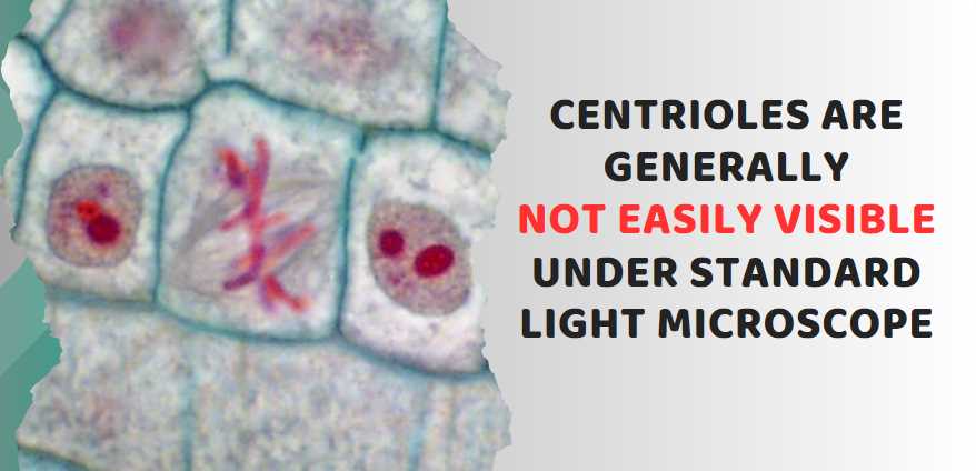
No, centrioles are not typically visible under a light microscope.
Centrioles are small, cylindrical structures found in animal cells, and they play a crucial role in the organization of microtubules during cell division. However, centrioles are very small and are below the resolution limit of a light microscope, which is approximately 200 nanometers. Therefore, they cannot be resolved and visualized using a standard light microscope.
| Image | Product | Detail | Price |
|---|---|---|---|
 | Carson MicroBrite Plus 60x-120x LED Lighted Pocket Microscope |
| See on Amazon |
 | Elikliv LCD Digital Coin Microscope |
| See on Amazon |
 | AmScope M150 Series Portable Compound Microscope |
| See on Amazon |
 | PalliPartners Compound Microscope for Adults & Kids |
| See on Amazon |
 | Skybasic 50X-1000X Magnification WiFi Portable Handheld Microscopes |
| See on Amazon |
| Parameter | Value |
|---|---|
| Magnification Range | 40x – 1000x |
| Resolution | Approximately 200 nanometers |
| Maximum Useful Magnifica | 500x – 600x for most details |
| Illumination Source | Visible light |
| Sample Preparation | Usually requires staining |
| Cost | Relatively inexpensive |
| Common Applications | Biological and medical studies |
| Limitations | Limited resolution for small structures like centrioles |
Understanding Centrioles
Centrioles, small but essential cellular structures, are key players in the intricate choreography of cell division and cytoskeletal organization. Comprising cylindrical tubes, centrioles are typically found in pairs near the cell’s nucleus. Understanding the structure and functions of centrioles provides insights into their significance in cellular processes.
What are Centrioles?
Centrioles are microtubule-based organelles, typically organized in pairs called centrosomes. Structurally, they consist of nine triplets of microtubules arranged in a cylindrical pattern, often likened to the blades of a turbine. This unique arrangement contributes to their role as microtubule organizers within the cell.
Centriole Functions
The primary role of centrioles lies in orchestrating cell division. During mitosis and meiosis, centrioles play a vital role in forming the spindle apparatus, a structure that segregates chromosomes into daughter cells. Additionally, centrioles are involved in the formation of cilia and flagella – cellular projections crucial for cell movement and sensory functions.
Centrioles and Cellular Organization
Beyond their role in cell division, centrioles contribute significantly to the organization of the cytoskeleton. The cytoskeleton is a dynamic network of protein filaments that gives the cell its shape, provides mechanical support, and facilitates intracellular transport. Centrioles help anchor microtubules, influencing the overall structural integrity of the cell.
Understanding the significance of centrioles in cellular processes sheds light on their indispensable functions. The intricacies of their structure and their involvement in vital cellular activities emphasize the importance of studying these organelles to unravel the mysteries of cell biology.
Challenges of Light Microscopy
Light microscopy, a cornerstone in the realm of biology, has been instrumental in studying various cellular structures. However, when it comes to observing structures as small as centrioles, inherent limitations arise. This section explores the challenges posed by light microscopy in the quest to visualize centrioles.
Limitations of Light Microscopy
One of the fundamental challenges lies in the resolution of light microscopes. The wavelength of visible light imposes a limit on the smallest structures that can be resolved. Centrioles, with their diminutive size and intricate structure, often fall below this limit, making them challenging to observe accurately.
To illustrate this limitation, consider the typical resolution of a light microscope, which is around 200 nanometers. Given that centrioles are on the order of 100 nanometers in diameter, pushing the boundaries of light microscopy, the fine details may remain elusive under conventional observation.
The Size of Centrioles
Centrioles, measuring approximately 200 to 250 nanometers in length, pose a size challenge when viewed through light microscopes. The optical limitations make it difficult to discern these structures with the clarity required for comprehensive analysis.
Let’s visualize this in a table:
| Parameter | Centriole Diameter | Light Microscope Resolution |
|---|---|---|
| Approximate Size | 200-250 nanometers | ~200 nanometers |
This comparison underscores the tight margins within which light microscopy operates concerning centriole observation.
Optical Challenges
The optical properties of centrioles further complicate their visibility under light microscopes. The refractive index mismatch between the centrioles and the surrounding cellular environment leads to distortions and reduced image clarity. This phenomenon hampers the accurate depiction of centriolar structures, making their observation a nuanced task.
Let’s summarize the optical challenges in a table:
| Optical Challenge | Impact on Visibility |
|---|---|
| Refractive Index Mismatch | Distortions and reduced image clarity |
Can Centrioles be Observed Using a Light Microscope?
Centrioles, unfortunately, are not readily visible under a standard light microscope. Their size and structure make them challenging to discern with the limited resolution of a light microscope, which typically ranges between 200 and 300 nanometers.
| Microscope Type | Visibility of Centrioles |
|---|---|
| Light Microscope | Not Visible |
| Electron Microscope | Clearly Visible |
Why are Centrioles Difficult to See with a Light Microscope?
Centrioles, being small organelles with dimensions below the resolving power of a light microscope, lack the contrast necessary for clear observation. Their size falls below the wavelength of visible light, making it challenging to distinguish them from the surrounding cellular structures.
| Size of Centrioles | Light Microscope Resolution |
|---|---|
| < 200 nm | Limited Visibility |
What Microscope Can be Used to Visualize Centrioles?
To observe centrioles effectively, an electron microscope is recommended. The higher resolution of electron microscopes, which can reach below 1 nanometer, allows for the detailed imaging of centrioles and other subcellular structures.
| Microscope Type | Suitable for Centriole Observation |
|---|---|
| Light Microscope | Inadequate Resolution |
| Electron Microscope | Recommended |
Can Special Staining Techniques Enhance Centriole Visibility?
Yes, employing specific staining techniques can enhance the visibility of centrioles under a light microscope. Dyes and stains that selectively bind to centriolar components can improve contrast, making these organelles more distinguishable.
| Staining Technique | Effect on Centriole Visibility |
|---|---|
| Centriole Staining | Enhanced Contrast |
| Immunofluorescence | Improved Visibility |
What is the Size of Centrioles, and How Does it Affect Visibility?
Centrioles typically have a diameter of about 250 nanometers and a length of approximately 500 nanometers. Their small size poses a challenge for light microscopy, where the resolution limit often exceeds the dimensions of these organelles.
| Centriole Dimensions | Light Microscope Resolution |
|---|---|
| 250 nm (diameter) | Limited Visibility |
Are Centrioles Important Despite Their Limited Visibility?
Despite their challenge to observe directly, centrioles play a crucial role in cell division, organizing the microtubules of the mitotic spindle. Their significance lies in their contribution to the proper segregation of chromosomes during cell division, ensuring the formation of genetically identical daughter cells.
| Centriole Importance | Visibility Challenge |
|---|---|
| Crucial for Cell Division | Limited Direct Observation |
Can Advances in Microscopy Technology Improve Centriole Visibility?
Advancements in microscopy techniques, such as super-resolution microscopy, hold promise for improving centriole visibility. These techniques surpass the diffraction limit of traditional light microscopy, potentially allowing for clearer imaging of centrioles.
| Microscopy Advancements | Potential for Improved Visibility |
|---|---|
| Super-Resolution Microscopy | Promising for Enhanced Clarity |
Conclusion
To unravel the mysteries of centrioles, we have navigated through their intricate structures, vital functions in cell biology, and the challenges posed by light microscopy. The limitations inherent in the resolution of light microscopes, coupled with the small size and optical challenges presented by centrioles, underscore the complexity of studying these cellular components.
As we conclude, it is evident that pushing the boundaries of traditional light microscopy is essential for a comprehensive understanding of centrioles. Prospects hinge on advancements in microscopy techniques, with electron and super-resolution microscopy offering promising avenues. By surpassing light microscopy’s limitations, these methods provide more precise insights into the elusive world of centrioles.
As scientific knowledge expands and technology continues to evolve, the potential for studying centrioles through light microscopy may improve.

Fahim Foysal is a well-known expert in the field of binoculars, with a passion for exploring the great outdoors and observing nature up close. With years of experience in the field, Fahim has honed his skills as a binocular user and has become a go-to resource for those seeking advice on choosing the right binoculars for their needs.
Fahim’s love for the natural world began during his time at The Millennium Stars School and College and BIAM Laboratory School, where he spent much of his free time exploring the outdoors and observing the wildlife around him. This passion for nature led him to pursue a degree in Fine Arts from the University of Dhaka, where he gained a deep understanding of the importance of observation and attention to detail.
Throughout his career, Fahim has used his expertise in binoculars to help others discover the beauty of the natural world. His extensive knowledge of binocular technology and optics has made him a trusted advisor for amateur and professional wildlife observers alike. Whether you’re looking to spot rare birds or observe animals in their natural habitats, Fahim can help you choose the perfect binoculars for your needs. With his guidance, you’ll be able to explore the outdoors with a newfound appreciation for the beauty of the natural world.
Table of Contents

Pingback: Can You See Golgi Apparatus under a Light Microscope