Epithelial tissue under a microscope appears as tightly packed cells with distinct borders, forming sheets or layers. The cells often exhibit a regular arrangement and can be either simple (single layer) or stratified (multiple layers).
Epithelial tissue is one of the four basic tissue types in the human body, responsible for covering and lining surfaces. When observed under a microscope, the appearance of epithelial tissue depends on its classification as either simple or stratified.
- Simple Epithelium:
- Consists of a single layer of cells.
- Cells are tightly packed with minimal variations in shape.
- Provides a thin barrier for absorption and diffusion.
- Found in areas where filtration, absorption, or secretion occurs, such as the lining of the digestive tract.
- Stratified Epithelium:
- Comprises multiple layers of cells stacked on top of each other.
- Cells in the basal layer are more cuboidal or columnar, while those in the outer layers may be squamous (flattened).
- Offers protection against wear and tear.
- Located in regions exposed to mechanical stress, like the skin (epidermis).
Table: Comparison of Simple and Stratified Epithelium
| Characteristic | Simple Epithelium | Stratified Epithelium |
|---|---|---|
| Layer Arrangement | Single layer of cells | Multiple layers of cells |
| Cell Shape | Mostly squamous (flat) or cuboidal/columnar | Basal cells are cuboidal/columnar, outer cells may be squamous |
| Function | Facilitates diffusion, absorption, secretion | Provides protection against mechanical stress |
| Location | Lining of blood vessels, air sacs in lungs, etc. | Skin (epidermis), lining of the mouth, esophagus, and vagina, etc. |
Structure of Epithelial Tissue
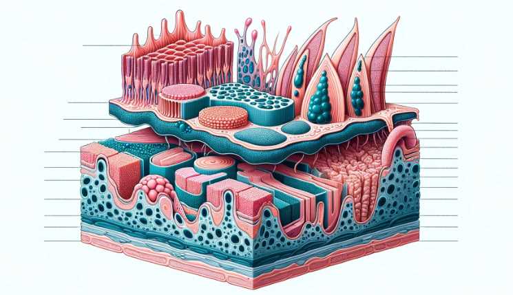
General Characteristics of Epithelial Tissue
Epithelial tissue, like a well-organized army, exhibits distinct characteristics under the microscope.
Cell Arrangement
Within this tissue, cells align in a strategic manner, forming a robust barrier. Squamous, cuboidal, and columnar cells, each with its unique structure, contribute to the tissue’s overall architecture.
| Cell Type | Description |
|---|---|
| Squamous | Thin and flat, suitable for specialized functions. |
| Cuboidal | Cube-shaped, often found in specific body locations. |
| Columnar | Tall and rectangular, dominating various organs. |
Cell Polarity
Epithelial cells showcase polarity, emphasizing the distinct apical and basal surfaces. This polarity is vital for their functional roles within the tissue.
Basement Membrane
Hidden from the naked eye, the basement membrane acts as the tissue’s anchor, providing structural support.
Cell Types within Epithelial Tissue
The microscopic exploration of epithelial tissue unveils the diverse roles played by different cell types.
Squamous Epithelial Cells
Squamous cells, resembling flattened pancakes, carry out specialized functions critical for the tissue’s overall function.
| Characteristic | Role within the Tissue |
|---|---|
| Thin and Flat | Allows for efficient diffusion and filtration. |
| Specialized Functions | Varies based on the organ or tissue type. |
Cuboidal Epithelial Cells
Cuboidal cells, with their cube-like structure, find their niche in specific body locations.
| Characteristic | Role within the Tissue |
|---|---|
| Cube-shaped | Provides structural support and secretion. |
| Locations | Kidney tubules, glands, and ducts. |
Columnar Epithelial Cells
Columnar cells, standing tall and rectangular, play crucial roles in various organs.
| Characteristic | Role within the Tissue |
|---|---|
| Tall and Rectangular | Facilitates absorption and secretion. |
| Locations | Lining of the digestive tract, respiratory tract. |
Microscopic Techniques for Observing Epithelial Tissue
Microscopic Techniques for Observing Epithelial Tissue
The microscopic exploration of epithelial tissue involves various techniques, each offering a unique perspective.
Staining Procedures
In the world of microscopic observation, staining procedures bring clarity to the seemingly transparent tissue.
| Staining Method | Purpose |
|---|---|
| Hematoxylin and Eosin (H&E) | Highlights cellular structures and differentiation. |
| Specialized Stains | Target specific components, aiding in detailed analysis. |
Resolution and Magnification
Light microscopy, with its intricate dance of resolution and magnification, transforms the invisible into the visible.
| Microscopic Feature | Importance |
|---|---|
| Enhanced Resolution | Reveals finer details of cellular structures. |
| Increased Magnification | Amplifies the overall view of the tissue. |
Electron Microscopy
Taking the exploration a step further, electron microscopy unveils details unreachable by traditional light microscopes.
Transmission Electron Microscopy (TEM)
TEM provides a peek into the internal structures of cells, akin to an X-ray for cellular anatomy.
| Aspect | Insight Provided |
|---|---|
| Internal Structures | Detailed visualization at the subcellular level. |
| Ultra-High Resolution | Enables observation of molecular structures. |
Scanning Electron Microscopy (SEM)
SEM captures the three-dimensional beauty of cell surfaces, turning microscopic images into intricate landscapes.
| Aspect | Insight Provided |
|---|---|
| Surface Topography | Detailed visualization of cellular surfaces. |
| 3D Imaging | Provides a holistic view of cellular structures. |
Preparation of Epithelial Tissue for Microscopic Examination
Preparation of Epithelial Tissue
Before the microscope unveils its secrets, meticulous preparation is essential.
Chemical Fixatives
Tissue fixation, achieved through chemical fixatives, is the initial step in preserving the delicate structures.
| Fixative Type | Purpose |
|---|---|
| Chemical Fixatives | Maintain cellular structures for microscopic examination. |
| Importance | Preserves cellular details for accurate observation. |
Sectioning Techniques
Creating thin sections for microscopic examination involves precise sectioning techniques.
| Technique | Purpose |
|---|---|
| Paraffin Embedding | Produces thin sections for light microscopy. |
| Cryosectioning | Enables the creation of frozen tissue sections. |
Artifacts and Distortions
In the delicate dance of slide preparation, artifacts and distortions may arise, demanding attention.
| Challenge | Mitigation Strategies |
|---|---|
| Folding or Tearing | Careful handling and meticulous sectioning. |
| Minimizing Distortions | Attention to detail during the preparation process. |
Limitations of Techniques
While microscopy unveils much, it also has its limitations, acknowledging which is crucial for accurate portrayal.
| Limitation | Impact on Microscopic Observation |
|---|---|
| Resolution Challenges | May hinder the clarity of microscopic images. |
| Overcoming Limitations | Utilizing complementary techniques for a comprehensive view. |
Observing Epithelial Tissue Under the Microscope
Observing Epithelial Tissue
With the tissue prepared, the microscope becomes our portal into the unseen world, unraveling the microscopic drama.
Slide Preparation
As the tissue takes center stage, meticulous slide preparation becomes an art form.
| Step | Description |
|---|---|
| Mounting Procedure | Carefully positioning the tissue on the slide. |
| Coverslipping | Sealing the microscopic world for observation. |
Common Stains Used in Epithelial Tissue Observation
Stains like Hematoxylin and Eosin (H&E) transform the microscopic view into a vivid tapestry.
| Staining Method | Visual Effect |
|---|---|
| H&E Staining | Highlights nuclei in blue and cytoplasm in pink. |
| Specialized Stains | Adds nuance, revealing specific cell types. |
Interpretation of Microscopic Features
As the microscope unveils the details, interpreting microscopic features becomes a captivating endeavor.
| Aspect | Interpretation |
|---|---|
| Identifying Cell Types | Based on morphological characteristics and arrangement. |
| Recognizing Pathological Changes | Identifying tumor cells and inflammatory responses. |
What is Epithelial Tissue?
Epithelial tissue is a type of tissue that lines the surfaces of the body, both inside and out. It serves as a protective barrier, helping to prevent dehydration and the entry of pathogens. This tissue also plays a crucial role in absorption, secretion, and sensation. The questions below explore various aspects of what epithelial tissue reveals about the body.
How is Epithelial Tissue Classified?
| Type of Epithelial Tissue | Characteristics |
| Simple Squamous | Single layer of flat cells; facilitates diffusion |
| Stratified Squamous | Multiple layers; provides protection against abrasion |
| Simple Cuboidal | Single layer of cube-shaped cells; involved in secretion and absorption |
| Simple Columnar | Single layer of elongated cells; functions in absorption and secretion |
| Pseudostratified Columnar | Appears stratified but all cells touch the basement membrane; often has cilia |
Epithelial tissue is classified based on the shape of cells and the number of layers present. This classification reveals the tissue’s specific functions within the body.
Where is Epithelial Tissue Found in the Body?
Epithelial tissue is found throughout the body, covering internal and external surfaces. Some key locations include:
| Location | Epithelial Tissue Type |
| Skin | Stratified Squamous Epithelium |
| Lungs Alveoli | Simple Squamous Epithelium |
| Kidney Tubules | Simple Cuboidal Epithelium |
| Small Intestine | Simple Columnar Epithelium |
| Trachea Lining | Pseudostratified Columnar Epithelium |
This distribution reveals the diverse roles of epithelial tissue in different organs and systems.
How Does Epithelial Tissue Contribute to Homeostasis?
Epithelial tissue plays a crucial role in maintaining internal balance (homeostasis) in the body. Through selective permeability and active transport, epithelial cells regulate the passage of substances, helping to control factors such as pH and ion concentrations. This contribution is vital for overall physiological stability.
What Happens to Epithelial Tissue During Wound Healing?
| Stage | Description |
| Inflammatory | Blood clotting, inflammation, and immune response |
| Proliferative | Epithelial cell migration and tissue reconstruction |
| Remodeling | Maturation and strengthening of new tissue |
During wound healing, epithelial tissue undergoes a series of stages, ensuring the restoration of damaged areas. This process involves the collaboration of various cell types and molecular signals.
Can Epithelial Tissue Undergo Cancerous Changes?
Yes, epithelial tissue can undergo cancerous changes, leading to the formation of tumors. The abnormal growth of cells may result from genetic mutations or exposure to carcinogens. Regular check-ups and early detection are crucial for effective cancer management.
How Does Aging Affect Epithelial Tissue?
Aging can impact epithelial tissue in several ways. The regenerative capacity of epithelial cells may decrease, leading to slower wound healing. Additionally, changes in skin elasticity and the mucous membranes are common. Maintaining a healthy lifestyle can mitigate some age-related effects on epithelial tissue.
Are There Diseases Specifically Affecting Epithelial Tissue?
| Disease | Description |
| Psoriasis | Chronic skin condition causing red, scaly patches |
| Cystic Fibrosis | Genetic disorder affecting respiratory and digestive systems |
| Peptic Ulcer Disease | Ulcers in the stomach lining or upper part of the small intestine |
| Barrett’s Esophagus | Precancerous condition of the esophagus due to chronic acid reflux |
Several diseases specifically affect epithelial tissue, highlighting the tissue’s vulnerability to various health conditions.
Can Epithelial Tissue Regenerate?
Epithelial tissue has a remarkable regenerative capacity. The constant turnover of cells allows for the repair of damaged areas. However, the regenerative ability varies among different types of epithelial tissue, with some being more proficient at regeneration than others.
How is Epithelial Tissue Studied in Research?
Research on epithelial tissue involves various techniques, including:
| Method | Description |
| Histology | Microscopic examination of tissue |
| Cell Culture | Growing epithelial cells in a controlled environment |
| Molecular Analysis | Studying genetic and molecular factors |
| Imaging Techniques | Visualizing epithelial tissue in vivo |
These research methods provide valuable insights into the structure and function of epithelial tissue, contributing to advancements in medical science.
Conclusion
The journey into the microscopic world of epithelial tissue is a testament to the marvels of science. From the meticulous preparation of tissue to the vivid colors painted by stains, each step in the process reveals hidden beauty. As the microscope becomes a storyteller, narrating the tale of cellular architecture, the significance of understanding epithelial tissue microscopy becomes evident.
This microscopic exploration not only aids in diagnostics but also contributes to educational and research advancements, paving the way for a deeper understanding of our cellular landscape. The microscope, a tool for both discovery and learning, becomes a bridge connecting the visible and invisible realms.
Ross MH, Pawlina W. (2010). “Histology: A Text and Atlas.” 6th edition
Kumar V, Abbas AK, Aster JC. (2014). “Robbins and Cotran Pathologic Basis of Disease.” 9th edition

Fahim Foysal is a well-known expert in the field of binoculars, with a passion for exploring the great outdoors and observing nature up close. With years of experience in the field, Fahim has honed his skills as a binocular user and has become a go-to resource for those seeking advice on choosing the right binoculars for their needs.
Fahim’s love for the natural world began during his time at The Millennium Stars School and College and BIAM Laboratory School, where he spent much of his free time exploring the outdoors and observing the wildlife around him. This passion for nature led him to pursue a degree in Fine Arts from the University of Dhaka, where he gained a deep understanding of the importance of observation and attention to detail.
Throughout his career, Fahim has used his expertise in binoculars to help others discover the beauty of the natural world. His extensive knowledge of binocular technology and optics has made him a trusted advisor for amateur and professional wildlife observers alike. Whether you’re looking to spot rare birds or observe animals in their natural habitats, Fahim can help you choose the perfect binoculars for your needs. With his guidance, you’ll be able to explore the outdoors with a newfound appreciation for the beauty of the natural world.
Table of Contents
