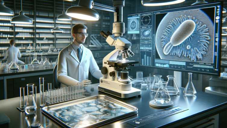
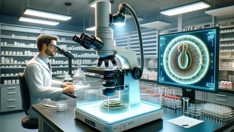
Giardia is a microscopic parasite that causes a diarrheal illness known as giardiasis in humans. The organism exists in two forms: a motile, pear-shaped trophozoite and a non-motile, oval-shaped cyst. When examining Giardia under a microscope, you would typically observe the trophozoite and cyst stages. Here’s a brief description of each:
| Image | Product | Detail | Price |
|---|---|---|---|
 | Carson MicroBrite Plus 60x-120x LED Lighted Pocket Microscope |
| See on Amazon |
 | Elikliv LCD Digital Coin Microscope |
| See on Amazon |
 | AmScope M150 Series Portable Compound Microscope |
| See on Amazon |
 | PalliPartners Compound Microscope for Adults & Kids |
| See on Amazon |
 | Skybasic 50X-1000X Magnification WiFi Portable Handheld Microscopes |
| See on Amazon |

- Trophozoite Stage:
- Shape: The trophozoite is pear-shaped or teardrop-shaped.
- Size: It is relatively large for a single-celled organism, measuring about 10 to 20 micrometers in length.
- Features: The trophozoite has a characteristic appearance with a pair of nuclei that are visible under the microscope. The organism is flagellated, meaning it has hair-like structures called flagella that it uses for movement.
- Cyst Stage:
- Shape: The cyst is oval or round.
- Size: It is smaller than the trophozoite, typically around 8 to 12 micrometers in diameter.
- Features: The cyst is the dormant, resistant form of Giardia. It has a protective outer shell that allows it to survive outside the host in harsh conditions. Inside the cyst, you can find the infective structures that, when ingested, can cause infection.

When examining a sample under the microscope, special staining techniques are often used to enhance the visibility of Giardia. One commonly used staining method is the trichrome stain, which helps highlight the characteristic features of the trophozoite and cyst stages.
Key Characteristics of Giardia under Microscope:
- Shape: Giardia trophozoites, the active and feeding form of the parasite, typically have a pear or teardrop shape. They are approximately 10 to 20 micrometers in length and 5 to 15 micrometers in width.
- Nuclei: Giardia trophozoites have two distinct nuclei, which are often visible under high magnification. The nuclei are positioned close to the center of the organism.
- Flagella: Giardia possesses flagella, whip-like appendages that extend from the body. These flagella are used for movement and contribute to the parasite’s distinctive appearance. There are four pairs of flagella: two anterior, two lateral, one caudal, and one ventral.
- Attachment Disk: The ventral side of Giardia trophozoites contains an adhesive structure called the ventral disk, which helps the parasite attach to the intestinal lining.
- Cysts: In addition to the trophozoite form, Giardia can also exist in a cyst form, which is a dormant and more resistant stage. Cysts are typically round and have a thick, protective wall. The cyst form is responsible for the transmission of Giardia between hosts.
When diagnosing giardiasis, stool samples are often examined under a microscope to detect the presence of Giardia trophozoites or cysts. The appearance of Giardia under the microscope can vary slightly, but the characteristics mentioned above are typical for this parasitic organism. Keep in mind that the exact details may vary based on the staining methods used and the specific conditions of the microscope examination.
Giardia: A Microscopic Perspective
Giardia, a microscopic protozoan parasite, belongs to the genus Giardia and falls under the family Giardiidae. This classification places it among diplomonads, highlighting its unique biological features. Understanding the general characteristics of Giardia is crucial for effective microscopic observation and comprehensive knowledge of its behavior.
Table: General Characteristics of Giardia
| Characteristic | Details |
|---|---|
| Classification and Taxonomy | Genus: Giardia; Family: Giardiidae |
| Habitat and Prevalence | Intestinal tracts of humans and animals; Global prevalence, especially in areas with poor sanitation and water treatment |
Classification and Taxonomy
Giardia’s taxonomic classification places it within the genus Giardia, highlighting its distinct biological characteristics. The family Giardiidae further categorizes it among diplomonads, showcasing its evolutionary relationships within the microbial world.
Habitat and Prevalence
Giardia predominantly inhabits the intestinal tracts of humans and various animals. Its prevalence is notable on a global scale, with a higher incidence observed in regions characterized by inadequate sanitation and water treatment. This distribution emphasizes the relevance of understanding Giardia’s general characteristics for global health considerations.
Lifecycle of Giardia
Giardia’s lifecycle is a fascinating process involving two main stages: trophozoites and cysts. This intricate life cycle plays a pivotal role in the transmission and infection dynamics of Giardia.
Table: Lifecycle of Giardia
| Stage | Description |
|---|---|
| Trophozoite Stage | Active, feeding stage; pear-shaped with flagella and adhesive discs; resides in the small intestine of the host |
| Cyst Stage | Inactive, survival stage; oval-shaped with a protective cyst wall; formed as a response to harsh environmental conditions |
Trophozoite and Cyst Stages
- Trophozoite Stage: This is the active, feeding stage of Giardia. Trophozoites are pear-shaped, measuring approximately 10-20 micrometers in length. They possess flagella and adhesive discs, aiding in attachment to the host’s intestinal wall.
- Cyst Stage: The cyst stage is an inactive, survival form of Giardia. Cysts are oval-shaped and exhibit a robust cyst wall, providing protection against harsh environmental conditions. Cysts are formed as a response to factors like dehydration, facilitating transmission between hosts.
Transmission and Infection
The transmission of Giardia primarily occurs through the ingestion of cysts, which are resistant to environmental challenges. Once ingested, cysts release trophozoites in the host’s small intestine, leading to infection. Understanding these stages is vital for developing effective strategies for prevention, diagnosis, and treatment of Giardia infections.
Microscopic Techniques for Giardia Observation
Microscopic observation of Giardia requires a careful and systematic approach to ensure accurate identification and analysis. This section explores the essential techniques involved in observing Giardia under a microscope, encompassing sample collection, preparation, staining methods, and the overall importance of employing proper microscopy techniques.
A. Sample Collection and Preparation
Table: Sample Collection and Preparation
| Technique | Details |
|---|---|
| Sample Collection | Fecal samples are commonly collected for Giardia observation, ensuring representation of the intestinal environment. |
| Sample Preservation | Immediate fixation or refrigeration helps prevent deterioration of the sample, preserving the integrity of Giardia cysts and trophozoites. |
| Concentration Techniques | Centrifugation or sedimentation may be employed to concentrate parasites, enhancing their visibility during microscopy. |
Proper sample collection and preparation are foundational steps in the microscopic observation of Giardia. Fecal samples, often the primary source, should be collected meticulously to ensure representative specimens. Immediate fixation or refrigeration of samples is crucial to prevent degradation and maintain the viability of Giardia cysts and trophozoites. Additionally, concentration techniques such as centrifugation help enhance the concentration of parasites, facilitating more accurate observations under the microscope.
B. Staining Methods for Enhanced Visibility
Table: Staining Methods for Giardia Observation
| Staining Method | Description |
|---|---|
| Direct Wet Mount | Involves placing a fresh sample directly on a microscope slide with a cover slip; provides a quick observation of motile trophozoites. |
| Modified Iron-Hematoxylin Staining | Utilizes a staining solution containing iron and hematoxylin to enhance contrast and visibility of Giardia cysts and trophozoites. |
| Immunofluorescence Staining | Utilizes specific antibodies labeled with fluorescent dyes to target Giardia antigens, allowing for highly specific and sensitive detection under fluorescence microscopy. |
- Direct Wet Mount: This technique offers a rapid observation method by placing a fresh sample directly on a microscope slide with a cover slip. It allows for the visualization of motile trophozoites, providing quick insights into Giardia activity.
- Modified Iron-Hematoxylin Staining: In this method, a staining solution containing iron and hematoxylin is used to enhance the contrast and visibility of Giardia cysts and trophozoites. This staining technique improves the clarity of cellular structures for more detailed microscopic examination.
- Immunofluorescence Staining: Immunofluorescence staining employs specific antibodies labeled with fluorescent dyes. This highly targeted approach allows for the specific and sensitive detection of Giardia antigens under fluorescence microscopy. Immunofluorescence staining is particularly valuable for enhancing specificity in identifying Giardia.
C. Importance of Proper Microscopy Techniques
The success of Giardia observation hinges on employing proper microscopy techniques.
Table: Importance of Proper Microscopy Techniques
| Aspect | Details |
|---|---|
| Accuracy in Identification | Proper techniques enhance accuracy in identifying Giardia cysts and trophozoites, reducing the risk of misdiagnosis. |
| Timely Diagnosis | Efficient microscopy techniques contribute to timely diagnosis, enabling prompt initiation of appropriate treatment for giardiasis. |
| Research Advancements | Continuous refinement of microscopy techniques supports ongoing research, leading to advancements in our understanding of Giardia and related diseases. |
Proper microscopy techniques are paramount for accurate identification and timely diagnosis of Giardia. The use of accurate methods ensures precision in differentiating Giardia from other microorganisms, reducing the likelihood of misdiagnosis. Additionally, these techniques contribute to ongoing research advancements, fostering a deeper understanding of Giardia and its impact on human health.
Microscopic Techniques for Giardia Observation
Microscopic observation of Giardia is a meticulous process that involves specific techniques for sample collection, preparation, and staining to enhance visibility. These techniques are crucial for accurate identification, aiding in the diagnosis and understanding of Giardia-related diseases.
A. Sample Collection and Preparation
Sample Collection and Preparation Table
| Technique | Details |
|---|---|
| Fecal Sample Collection | Collect fecal samples meticulously to ensure a representative specimen. |
| Sample Preservation | Immediately fix or refrigerate samples to prevent degradation and maintain viability. |
| Concentration Techniques | Utilize centrifugation or sedimentation to enhance the concentration of parasites. |
Proper sample collection is fundamental for successful Giardia observation. Fecal samples, commonly used for this purpose, should be collected carefully to ensure they represent the intestinal environment accurately. Immediate fixation or refrigeration of samples is essential to prevent degradation, preserving the integrity of Giardia cysts and trophozoites. Concentration techniques such as centrifugation enhance the visibility of parasites under the microscope.
B. Staining Methods for Enhanced Visibility
Staining Methods Table
| Staining Method | Description |
|---|---|
| Direct Wet Mount | Place a fresh sample directly on a microscope slide with a cover slip for a quick observation of motile trophozoites. |
| Modified Iron-Hematoxylin Staining | Use a staining solution containing iron and hematoxylin to enhance contrast and visibility of Giardia cysts and trophozoites. |
| Immunofluorescence Staining | Utilize specific antibodies labeled with fluorescent dyes to target Giardia antigens, allowing for highly specific and sensitive detection under fluorescence microscopy. |
- Direct Wet Mount: This technique involves placing a fresh sample directly on a microscope slide with a cover slip. It offers a rapid observation method, allowing for the visualization of motile trophozoites and providing quick insights into Giardia activity.
- Modified Iron-Hematoxylin Staining: This method employs a staining solution containing iron and hematoxylin to enhance the contrast and visibility of Giardia cysts and trophozoites. The staining improves the clarity of cellular structures for more detailed microscopic examination.
- Immunofluorescence Staining: This technique uses specific antibodies labeled with fluorescent dyes. It allows for the specific and sensitive detection of Giardia antigens under fluorescence microscopy, enhancing specificity in identifying Giardia.
C. Importance of Proper Microscopy Techniques
Importance of Proper Microscopy Techniques Table
| Aspect | Details |
|---|---|
| Accuracy in Identification | Proper techniques enhance accuracy in identifying Giardia cysts and trophozoites, reducing the risk of misdiagnosis. |
| Timely Diagnosis | Efficient microscopy techniques contribute to timely diagnosis, enabling prompt initiation of appropriate treatment for giardiasis. |
| Research Advancements | Continuous refinement of microscopy techniques supports ongoing research, leading to advancements in our understanding of Giardia and related diseases. |
Proper microscopy techniques play a pivotal role in the accurate identification of Giardia. These techniques contribute to reducing the risk of misdiagnosis by enhancing accuracy in differentiating Giardia from other microorganisms. Timely diagnosis is facilitated through efficient microscopy techniques, enabling the prompt initiation of appropriate treatment for giardiasis.
What Does Giardia Look Like?
A. Detailed Description of Giardia Morphology
Understanding the detailed morphology of Giardia is essential for accurate identification under a microscope. Giardia exists in two primary forms: trophozoites and cysts.
1. Trophozoite Appearance
a. Size and Shape
Trophozoites, the active and feeding stage of Giardia, typically measure between 10-20 micrometers in length. Their pear-shaped bodies are easily distinguishable, and this size range allows for efficient movement within the host’s small intestine.
b. Flagella and Adhesive Discs
Giardia trophozoites exhibit characteristic flagella—hair-like structures that protrude from the body. These flagella play a crucial role in the motility of the parasite. Additionally, adhesive discs located at the anterior end of the trophozoite aid in attachment to the host’s intestinal wall, facilitating colonization.
2. Cyst Characteristics
a. Wall Structure
Giardia cysts represent the dormant, survival stage of the parasite. They possess a resilient cyst wall that provides protection against environmental challenges. This cyst wall is essential for the transmission of Giardia between hosts.
b. Size and Shape
Cysts are typically smaller than trophozoites and exhibit an oval shape. Their smaller size contributes to the ease of transmission and dissemination in various environments.
B. High-Resolution Microscopy Images
To provide a visual representation of Giardia morphology, high-resolution microscopy images are invaluable. These images offer a closer look at the intricate details of trophozoites and cysts, allowing for a more comprehensive understanding of their structural features.
High-Resolution Microscopy Images Table
| Stage | Image Description |
|---|---|
| Trophozoite | Pear-shaped trophozoite with visible flagella and discs. |
| Cyst | Oval-shaped cyst with a distinct and protective wall. |
C. Comparison with Other Microscopic Organisms
Giardia exhibits unique features that distinguish it from other microscopic organisms commonly encountered in various environments. A comparative analysis highlights these distinctions.
Comparison Table
| Characteristic | Giardia | Other Microorganisms |
|---|---|---|
| Motility | Flagella-driven motility | Varied modes of locomotion |
| Attachment | Adhesive discs for host attachment | Attachment mechanisms vary widely |
| Life Cycle | Alternation between trophozoite and cyst stages | Diverse life cycles among different organisms |
| Size | 10-20 micrometers (trophozoites) | Size ranges widely across microorganisms |
Giardia’s flagella-driven motility, adhesive discs for host attachment, and unique life cycle set it apart from other microscopic organisms. Size variations, attachment mechanisms, and diverse life cycles among different organisms highlight the diversity within the microscopic world.
Identifying Giardia-Associated Diseases
A. Giardiasis and Its Symptoms
Giardiasis, the disease caused by the protozoan parasite Giardia, manifests with a range of symptoms affecting the gastrointestinal system. Recognizing these symptoms is crucial for prompt diagnosis and effective treatment.
Giardiasis Symptoms Table
| Symptom | Description |
|---|---|
| Diarrhea | Frequent, loose, and often foul-smelling bowel movements |
| Abdominal Cramps | Intermittent or continuous discomfort in the abdomen |
| Nausea | Feeling of queasiness or an urge to vomit |
| Dehydration | Reduced fluid levels in the body due to persistent diarrhea |
| Weight Loss | Unintentional weight loss resulting from malabsorption |
B. Link Between Giardia Morphology and Disease Severity
The morphology of Giardia plays a significant role in determining the severity of associated diseases. Variations in the appearance of trophozoites under microscopic observation may correlate with the intensity of infection and clinical symptoms.
Understanding Giardia morphology allows healthcare professionals to assess the potential impact on the patient’s health. For instance, an increased number of trophozoites or specific morphological characteristics may indicate a more severe infection, guiding clinicians in tailoring appropriate treatment strategies.
C. Importance of Early Detection Through Microscopy
Early detection of Giardia through microscopy is paramount for several reasons. Microscopic observation allows for the identification of Giardia cysts and trophozoites in clinical samples, confirming the presence of the parasite in the patient’s gastrointestinal tract.
Importance of Early Detection Table
| Aspect | Details |
|---|---|
| Prompt Treatment | Early detection enables timely initiation of specific anti-Giardia medications. |
| Prevention of Transmission | Identifying Giardia early helps implement preventive measures to limit further spread. |
| Reduction of Disease Severity | Early intervention may mitigate the severity of giardiasis, preventing complications. |
| Public Health Surveillance | Swift identification supports public health efforts in monitoring and controlling outbreaks. |
Swift identification of Giardia through microscopy facilitates the prompt initiation of specific anti-Giardia medications, reducing the duration and severity of symptoms. Additionally, early detection aids in implementing preventive measures to limit further transmission, protecting both individual patients and the broader community. By reducing disease severity, early intervention can prevent complications associated with giardiasis, contributing to improved patient outcomes.
3 Tips for Efficient Giardia Observation
A. Proper Microscope Usage
Efficient Giardia observation begins with mastering microscope usage. Regular calibration and maintenance ensure optimal performance. Adjusting lighting and focus settings enhances clarity, aiding in the identification of Giardia cysts and trophozoites.
B. Sample Handling and Preparation Tips
Meticulous sample handling is crucial. Ensure accurate representation by collecting fecal samples carefully. Immediate fixation or refrigeration prevents sample degradation, preserving Giardia integrity. Utilize concentration techniques like centrifugation for enhanced visibility during microscopy.
C. Common Challenges and Troubleshooting
Be prepared to tackle common challenges encountered during Giardia observation. Issues such as debris interference or insufficient staining require troubleshooting. Regularly check equipment and adjust techniques to overcome challenges, ensuring accurate and reliable results in Giardia identification.
Facts and Statistics
A. Key Facts about Giardia
- Ubiquitous Parasite: Giardia is a ubiquitous protozoan parasite that infects the small intestine of humans and animals, causing giardiasis.
- Waterborne Transmission: The primary mode of transmission is through contaminated water sources, emphasizing the importance of water hygiene.
- Resilient Cysts: Giardia exists in two stages, with cysts being the dormant, environmentally resistant form, allowing for survival outside a host.
B. Statistics on Global Prevalence and Incidence
- Worldwide Distribution: Giardia has a global presence, affecting both developed and developing countries, with varying degrees of prevalence.
- High Incidence in Developing Regions: Developing regions often experience higher incidences due to inadequate sanitation and limited access to clean water.
- Common Waterborne Outbreaks: Waterborne outbreaks contribute significantly to the prevalence of giardiasis, emphasizing the importance of clean water initiatives.
C. Impact on Vulnerable Populations
- Children and Immunocompromised Individuals: Giardia infections can have severe consequences for vulnerable populations, particularly children and those with compromised immune systems.
- Malnutrition and Growth Impairment: Chronic giardiasis in children may lead to malnutrition and growth impairment, affecting long-term health.
- Increased Susceptibility in Immunocompromised Individuals: Immunocompromised individuals, such as those with HIV/AIDS, face a higher risk of prolonged and severe Giardia infections, posing additional health challenges.
Understanding these facts and statistics is crucial for public health initiatives, emphasizing the need for global efforts in sanitation, clean water provision, and targeted interventions to protect vulnerable populations.
Final Words
In exploring Giardia under a microscope, we unveiled its distinct trophozoite and cyst stages, each with unique morphological features. High-resolution microscopy images provided a visual journey into the microscopic world of Giardia, showcasing its pear-shaped trophozoites and oval-shaped cysts.
Microscopic studies are pivotal in deciphering Giardia-related diseases. Accurate identification through microscopy guides effective treatment, early detection prevents transmission, and ongoing research enhances our understanding. The microscope remains a powerful tool in unraveling the mysteries of Giardia, contributing significantly to public health efforts worldwide.
Resources and References
A. Recommended Textbooks and Journals
- Textbooks:
- “Parasitic Diseases” by Despommier et al.
- “Medical Microbiology” by Murray et al.
- Journals:
- “Journal of Parasitology”
- “Clinical Microbiology Reviews”
B. Online Resources and Databases
- Websites:
- Databases:
C. Citations for Scientific Studies and Research Papers
- Scientific Studies:
- Thompson RCA, Hopkins RM, Homan WL. “Nomenclature and genetic groupings of Giardia infecting mammals.” Parasitology Today, 2000.
- Research Papers:
- Adam RD. “Biology of Giardia lamblia.” Clinical Microbiology Reviews, 2001.
These resources provide a comprehensive foundation for studying Giardia, from authoritative textbooks and journals to online databases and key scientific studies.

Fahim Foysal is a well-known expert in the field of binoculars, with a passion for exploring the great outdoors and observing nature up close. With years of experience in the field, Fahim has honed his skills as a binocular user and has become a go-to resource for those seeking advice on choosing the right binoculars for their needs.
Fahim’s love for the natural world began during his time at The Millennium Stars School and College and BIAM Laboratory School, where he spent much of his free time exploring the outdoors and observing the wildlife around him. This passion for nature led him to pursue a degree in Fine Arts from the University of Dhaka, where he gained a deep understanding of the importance of observation and attention to detail.
Throughout his career, Fahim has used his expertise in binoculars to help others discover the beauty of the natural world. His extensive knowledge of binocular technology and optics has made him a trusted advisor for amateur and professional wildlife observers alike. Whether you’re looking to spot rare birds or observe animals in their natural habitats, Fahim can help you choose the perfect binoculars for your needs. With his guidance, you’ll be able to explore the outdoors with a newfound appreciation for the beauty of the natural world.
Table of Contents
