Nail fungus, though invisible to the naked eye, unravels its secrets when subjected to the scrutiny of a microscope. This detailed examination brings to light three fundamental structures that define the microscopic landscape of nail fungus.
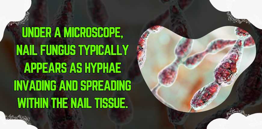
Fungal Hyphae Under the lens, fungal hyphae emerge as the silent architects of nail fungus. These microscopic threads, resembling delicate strands, weave their way into the intricate terrain of the nail bed. The examination of fungal hyphae provides a crucial diagnostic tool, allowing experts to pinpoint the specific type of fungus responsible for the infection. Dermatophytes, molds, and yeasts each leave a unique imprint, visible only through the lens of a microscope. Witnessing the invasive nature of these hyphae underscores the challenge of eradicating nail fungus completely.
Spores and Conidia Moving deeper into the microscopic realm, we encounter the reproductive units of the fungus – spores and conidia. These minute entities serve as the seeds of dissemination, enabling the fungus to spread its influence. Spores, encapsulated in protective structures, appear as tiny, round entities under the microscope. Concurrently, conidia, a form of asexual spore, present an alternative avenue for fungal reproduction. Observing these reproductive structures not only unveils the fungus’s lifecycle but also provides insights into strategies for curbing its relentless propagation.
Mycelium Formation Further exploration reveals the mesmerizing formation of mycelium. Mycelium represents the interconnected mass of hyphae, creating an intricate network within the nail. This web-like structure serves as the lifeblood of the fungal colony, enabling it to extract nutrients from the nail and thrive. Understanding mycelium formation is pivotal in assessing the severity of the infection and devising targeted treatment strategies. Like a microscopic tapestry, the mycelium showcases the adaptability and resilience of the fungus.
Differentiation from Healthy Nail Structures
To truly comprehend the impact of nail fungus, a comparative analysis between healthy and infected nail structures is essential. This differentiation underlines the microscopic disparities that aid in accurate diagnosis and effective treatment.
Under the microscope, healthy nails boast a well-organized and uniform structure. The absence of abnormal hyphae, spores, or mycelium distinguishes the microscopic landscape of healthy nails from their infected counterparts. The invading fungal elements, marked by disorganized hyphae, clusters of spores, and the intricate mycelium network, paint a vivid picture of the fungal colonization.
Images and Comparisons for Clarity
To bridge the gap between scientific knowledge and lay understanding, including images and side-by-side comparisons becomes paramount.
Images captured through the microscope serve as visual guides, providing a firsthand view of the stark differences between healthy and infected nail structures. These visuals, akin to microscopic narratives, demystify the complexities of nail fungus for the general audience. Side-by-side comparisons enhance clarity, elucidating the nuances of fungal hyphae, spores, and mycelium in contrast to their healthy counterparts.
How does Nail Fungus Develop?
The development of nail fungus involves a series of stages, each contributing to the progression of the infection. The following stages provide a comprehensive overview:
Stage
Description
Initial Contamination
Fungal spores come into contact with the nail, often through a warm and moist environment.
Adhesion and Invasion
The spores adhere to the nail surface and invade the nail plate through tiny cracks or separations.
Hyphal Growth
Fungal hyphae extend within the nail structure, causing damage and triggering the characteristic symptoms.
Spore Production
As the infection progresses, the fungus produces spores, contributing to the spread of the infection to adjacent nails.
Immune Response Activation
The immune system reacts to the infection, leading to inflammation and further complications in the surrounding tissues.
Understanding the stages of nail fungus development is essential for implementing effective prevention and treatment strategies.
How is Nail Fungus Diagnosed?
Diagnosing nail fungus involves a combination of clinical examination, laboratory tests, and, in some cases, microscopic analysis. The diagnostic process can be outlined as follows:
Diagnostic Step
Description
Clinical Assessment
A healthcare professional examines the affected nails, assessing color, texture, and any associated symptoms.
Microscopic Examination
Nail clippings or scrapings are collected and examined under a microscope to identify characteristic fungal elements.
Cultural Tests
Nail samples may be cultured in a laboratory to identify the specific type of fungus, aiding in targeted treatment approaches.
PCR (Polymerase Chain Reaction)
Molecular techniques like PCR may be employed for precise identification of fungal species, especially in challenging cases.
Wood’s Lamp Examination
In some instances, a Wood’s lamp may be used to detect fungal infections by observing fluorescence in the affected area.
Combining these diagnostic methods ensures accurate identification of nail fungus, guiding healthcare professionals in crafting effective treatment plans.
What are the Treatment Options for Nail Fungus?
Several treatment options are available for nail fungus, ranging from topical medications to oral antifungal drugs. The choice of treatment depends on the severity of the infection. Here’s an overview:
Treatment Type
Description
Topical Antifungals
Over-the-counter or prescription creams, ointments, or nail lacquers containing antifungal agents applied directly to the affected nails.
Oral Antifungal Medications
Prescription medications taken orally, reaching the bloodstream to target the fungus systemically. Common examples include terbinafine and itraconazole.
Laser Therapy
Laser devices target the fungal infection, promoting the destruction of the fungus without harming the surrounding tissues.
Surgical Removal
In severe cases, surgical intervention may be considered to remove the infected nail, allowing for the application of antifungal treatments.
Home Remedies
Some individuals explore natural remedies like tea tree oil or vinegar soaks, although their effectiveness varies, and professional advice is recommended.
Tailoring the treatment approach to the specific characteristics of the nail fungus is crucial for achieving optimal results.
How Long Does it Take to Cure Nail Fungus?
The duration of nail fungus treatment varies based on several factors, including the severity of the infection, the chosen treatment method, and individual response. A general timeline can be outlined as follows:
Treatment Phase
Duration
Onset of Improvement
Visible improvement may begin within a few weeks to a couple of months, with a reduction in symptoms.
Complete Cure
Achieving a complete cure may take several months, often ranging from six months to a year or longer.
Preventive Measures
Continued use of preventive measures, such as antifungal creams, even after apparent cure, is recommended to minimize the risk of recurrence.
Consistent adherence to the prescribed treatment plan and preventive measures is essential for successful and lasting results.
How Can Nail Fungus be Prevented?
Preventing nail fungus involves adopting good foot hygiene practices and minimizing exposure to risk factors. Consider the following preventive measures:
Preventive Measure
Description
Keep Feet Clean and Dry
Regularly wash and thoroughly dry feet, paying attention to spaces between toes where moisture can accumulate.
Use Antifungal Powders or Sprays
Applying antifungal powders or sprays can help prevent fungal growth, especially in shoes and socks.
Choose Breathable Footwear
Opt for breathable footwear made of materials like leather to allow proper ventilation and reduce moisture retention.
Avoid Sharing Personal Items
Refrain from sharing items such as towels or nail clippers to prevent the spread of fungal infections.
Protect Feet in Public Areas
Wear shower shoes or sandals in public places like gyms and swimming pools to reduce the risk of fungal exposure.
Incorporating these preventive measures into daily routines can significantly reduce the likelihood of developing nail fungus.
When Should I Consult a Healthcare Professional?
Consulting a healthcare professional is crucial when dealing with nail fungus, especially in the following situations:
Situation
Description
Persistent Symptoms
If symptoms persist despite home remedies or over-the-counter treatments, seeking professional advice is advisable.
Worsening Condition
If the condition worsens, with increased pain, spreading of the infection, or additional nail involvement, prompt consultation is necessary.
Underlying Health Conditions
Individuals with diabetes or compromised immune systems should promptly consult a healthcare professional due to the heightened risk of complications.
Uncertain Diagnosis
If there is uncertainty about the diagnosis or if other nail conditions are suspected, seeking professional evaluation is recommended.
Early intervention and professional guidance enhance the chances of successful treatment and prevent complications associated with nail fungus.
Conclusion
In my personal experience, dealing with nail fungus was a challenging journey. The microscopic examination of my nail samples provided a deeper understanding of the infection, leading to a more targeted treatment plan. Early detection and intervention are paramount, underscoring the importance of regular check-ups and maintaining good nail hygiene.
Fahim Foysal is a well-known expert in the field of binoculars, with a passion for exploring the great outdoors and observing nature up close. With years of experience in the field, Fahim has honed his skills as a binocular user and has become a go-to resource for those seeking advice on choosing the right binoculars for their needs.
Fahim’s love for the natural world began during his time at The Millennium Stars School and College and BIAM Laboratory School, where he spent much of his free time exploring the outdoors and observing the wildlife around him. This passion for nature led him to pursue a degree in Fine Arts from the University of Dhaka, where he gained a deep understanding of the importance of observation and attention to detail.
Throughout his career, Fahim has used his expertise in binoculars to help others discover the beauty of the natural world. His extensive knowledge of binocular technology and optics has made him a trusted advisor for amateur and professional wildlife observers alike. Whether you’re looking to spot rare birds or observe animals in their natural habitats, Fahim can help you choose the perfect binoculars for your needs. With his guidance, you’ll be able to explore the outdoors with a newfound appreciation for the beauty of the natural world.