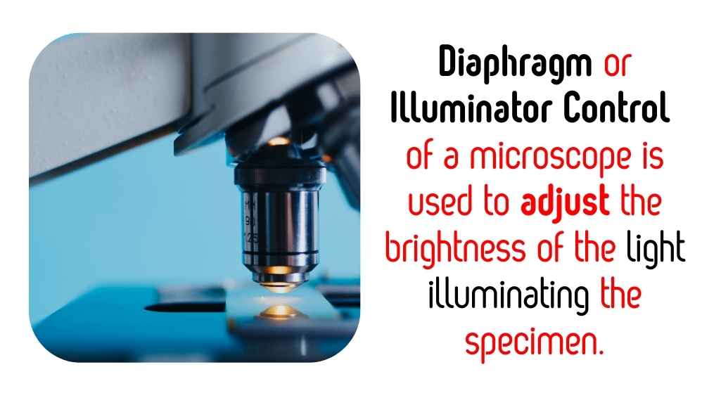
The diaphragm or illuminator control of a microscope is used to adjust the brightness of the light illuminating the specimen.
Microscopes require proper lighting to clearly view specimens. The diaphragm, often located beneath the stage, regulates the amount of light passing through the slide by adjusting its aperture size. Meanwhile, the illuminator control, typically an electronic dial or switch, directly adjusts the intensity of the light source. Together, these components help create the optimal lighting conditions needed for clear and detailed observations.
| Image | Product | Detail | Price |
|---|---|---|---|
 | Carson MicroBrite Plus 60x-120x LED Lighted Pocket Microscope |
| See on Amazon |
 | Elikliv LCD Digital Coin Microscope |
| See on Amazon |
 | AmScope M150 Series Portable Compound Microscope |
| See on Amazon |
 | PalliPartners Compound Microscope for Adults & Kids |
| See on Amazon |
 | Skybasic 50X-1000X Magnification WiFi Portable Handheld Microscopes |
| See on Amazon |
Proper brightness adjustment is critical; too much light can wash out the image, while too little can obscure fine details.
| Parameter | Effect on Image Quality | Optimal Adjustment Method |
|---|---|---|
| Illuminator Intensity (Lux) | Too high: Washed out details | Use the illuminator control to reduce brightness. |
| Diaphragm Aperture (mm) | Too large: Excessive light | Gradually narrow the aperture for clarity. |
| Brightness Setting (%) | Ideal range: 40–70% for most samples | Adjust to balance contrast and visibility. |
| Image Contrast (%) | Poor with incorrect brightness | Optimize using both diaphragm and light intensity. |
| Specimen Type | Opaque: Requires higher brightness | Transparent: Lower brightness for better contrast. |

Brightness control is vital in microscopy as it ensures that the sample is neither underexposed nor overexposed. Proper illumination enhances image clarity and detail, making the observation process more efficient. Without adequate brightness adjustment, the sample may appear too dark or washed out, hindering the identification of important features.
Parts of a Microscope That Help Adjust Brightness
Microscopes are invaluable tools in scientific research, medicine, and education. Proper illumination is one of the most critical factors for effective microscopic observation. Brightness adjustments allow the user to illuminate specimens adequately, ensuring the best clarity and detail. Several components in a microscope work together to manage and adjust brightness. Below, we will explore these components in detail and their specific roles in brightness control.
1. Light Source
The light source is the primary provider of illumination in most modern microscopes. Typically, it consists of an LED or halogen bulb located beneath the microscope stage. These light sources are chosen for their brightness, energy efficiency, and durability.
How the Light Source Works
The light source emits light that passes upward through the condenser and onto the specimen. Modern microscopes often include an adjustable light source, allowing users to control the intensity of the light for optimal viewing. This adjustability is particularly useful when switching between different magnifications or specimen types, as each may require varying levels of brightness.
Common Adjustments
- Intensity Control: The light source is equipped with a rheostat or slider that modifies the brightness.
- Angle Adjustments: In some advanced microscopes, the angle of the light source can be altered to provide oblique illumination, enhancing certain specimen details.
Advantages of Modern Light Sources
- LED Bulbs: These bulbs produce consistent, cool light, reducing the risk of heat damage to delicate specimens.
- Halogen Bulbs: Known for their brightness and wide spectrum, they provide more natural illumination.
2. Rheostat (Light Intensity Control Knob)
The rheostat is an integral component of brightness adjustment. It is typically found near the base of the microscope and functions as a control dial or slider. The primary role of the rheostat is to regulate the intensity of the light source.
How the Rheostat Adjusts Brightness
By turning the rheostat, users can increase or decrease the voltage supplied to the light source, which directly affects its brightness. For example:
- Turning the knob clockwise increases brightness.
- Turning it counterclockwise decreases brightness.
This control is essential for achieving the right illumination for different magnifications. Lower magnifications often require less light, while higher magnifications benefit from greater brightness.
Why the Rheostat is Critical
- Precision: Allows fine-tuning of brightness to avoid underexposure or overexposure.
- Versatility: Adapts the microscope to different specimen types and viewing conditions.
3. Condenser
The condenser is positioned beneath the stage and above the light source. Its primary function is to focus the light beam onto the specimen. This focusing process ensures that the light is concentrated on the sample, enhancing brightness and clarity.
Adjusting the Condenser
The condenser is adjustable in height, which affects how the light is distributed across the specimen:
- Lowering the Condenser: Spreads light more broadly, reducing brightness.
- Raising the Condenser: Focuses the light more tightly, increasing brightness.
Condenser Components
The condenser often includes an internal lens system that directs light toward the specimen with precision. Additionally, it works in conjunction with the diaphragm to refine brightness and contrast.
Applications of the Condenser
- Brightfield Microscopy: A properly adjusted condenser is essential for even illumination.
- Special Techniques: Advanced condensers can support methods like phase-contrast or darkfield microscopy.
4. Diaphragm (Iris or Disc)
The diaphragm is a critical component for managing the amount of light that passes through the specimen. Located as part of the condenser assembly, it works by adjusting the aperture size.
Types of Diaphragms
- Iris Diaphragm: Consists of overlapping metal blades that form a circular aperture. It provides smooth and precise control over the aperture size.
- Disc Diaphragm: A rotating disc with multiple holes of different sizes that can be selected to adjust the aperture.
How the Diaphragm Affects Brightness
- Smaller Aperture: Reduces brightness but increases contrast, useful for detailed observations.
- Larger Aperture: Increases brightness but reduces contrast, ideal for viewing larger or less detailed specimens.
Tips for Using the Diaphragm
- Start with a smaller aperture to observe finer details.
- Gradually open the diaphragm to balance brightness and contrast as needed.
5. Mirror (in Older Microscopes)
Before the advent of built-in light sources, microscopes relied on mirrors to reflect external light toward the specimen. Though less common today, mirrors are still found in some basic or non-electric microscopes, particularly in educational settings or regions without reliable electricity.
How the Mirror Works
The mirror, usually a flat or concave surface, captures light from an external source (like a lamp or sunlight) and reflects it into the condenser. The angle of the mirror determines the direction and concentration of light.
Adjusting the Mirror
- Flat Side: Produces even illumination, suitable for most specimens.
- Concave Side: Concentrates light for brighter illumination, useful for high-magnification observations.
Advantages of Mirrors
- Simplicity: Requires no power source, making it ideal for portable or field microscopes.
- Durability: Less prone to malfunction compared to electrical components.
Parts of a Microscope and Their Role in Brightness Adjustment
| Component | Function in Brightness Adjustment | Additional Notes |
|---|---|---|
| Light Source | Provides primary illumination. | Typically an LED or halogen bulb. |
| Rheostat | Controls light intensity. | Found near the base of the microscope. |
| Condenser | Focuses light onto the specimen. | Adjusts concentration and focus of light. |
| Diaphragm | Regulates the amount of light passing through the sample. | Impacts both brightness and contrast. |
| Mirror | Reflects external light into the condenser. | Found in older or non-electric microscopes. |
How These Components Work Together
Achieving optimal brightness in a microscope involves coordination between several components. Each part has a unique role, but their combined adjustments ensure the specimen is adequately illuminated for detailed observation. Understanding how these components work together simplifies the process of brightness control. Below is an explanation of their interplay and a recommended sequence for adjustments.
Coordination of Components
- Rheostat and Light Source
- The rheostat manages the intensity of the light source by controlling the electrical supply to the bulb.
- Adjusting the rheostat ensures the base illumination is appropriate for the sample and magnification level.
- Condenser and Diaphragm
- The condenser focuses the light beam onto the specimen, determining the evenness and concentration of illumination.
- The diaphragm fine-tunes the light by regulating the aperture size, balancing brightness and contrast.
- Overall Adjustment
- These components interact dynamically; increasing light intensity with the rheostat may require adjustments to the condenser or diaphragm to avoid overexposure.
- Conversely, changes to the diaphragm’s aperture size may necessitate altering the condenser’s position to maintain uniform illumination.
Recommended Sequence for Adjustments
Proper brightness adjustment is achieved by following a systematic sequence. This ensures that all components work in harmony:
- Turn on the Light Source
- Activate the microscope’s light source and set it to a moderate intensity using the rheostat.
- Avoid starting with maximum brightness to prevent glare or specimen damage.
- Adjust the Rheostat
- Gradually increase or decrease the light intensity based on the specimen’s requirements and magnification level.
- Position the Condenser
- Raise or lower the condenser to concentrate the light beam on the specimen. This step enhances clarity and minimizes uneven illumination.
- Fine-Tune the Diaphragm
- Adjust the diaphragm’s aperture size to balance the light intensity with contrast.
- Start with a smaller aperture for better contrast and expand as needed for increased brightness.
- Recheck and Refine
- Revisit the rheostat, condenser, and diaphragm settings to ensure uniform and optimal illumination across the field of view.
5 Tips for Proper Brightness Adjustment
Achieving the right brightness in microscopy is essential for clear and accurate observations. Below are practical tips to help users effectively adjust brightness while avoiding common pitfalls:
Start with Low Intensity
- Why: Starting with the light source at its lowest intensity prevents overexposure and allows for gradual adjustments.
- How: Turn on the light source and slowly increase the intensity using the rheostat until the specimen becomes visible without glare.
Use the Diaphragm Effectively
- Why: The diaphragm is key to balancing brightness and contrast. Proper adjustments enhance image quality without compromising detail.
- How:
- For high contrast, reduce the diaphragm aperture.
- For brighter illumination, open the diaphragm slightly, ensuring that light does not wash out fine details.
Consider the Magnification Level
- Why: Higher magnifications require more light as the field of view becomes smaller and the specimen’s details are magnified.
- How:
- Increase the light intensity with the rheostat at higher magnifications.
- Adjust the condenser to ensure the focused light beam matches the smaller field size.
Avoid Glare
- Why: Excessive brightness can cause discomfort and reduce the visibility of specimen details.
- How:
- Adjust the rheostat, condenser, and diaphragm in tandem to maintain balanced lighting.
- Recheck for uniform illumination across the field of view.
Common Issues with Brightness Adjustment and Solutions
While adjusting brightness in microscopy, users may encounter common problems that hinder clear observation. Below are typical issues, their causes, and practical solutions to address them:
Problem: Image Appears Too Dark
- Cause:
- Light intensity is set too low.
- The diaphragm aperture is too small, restricting the amount of light passing through.
- Solution:
- Gradually increase the rheostat setting to boost light intensity.
- Open the diaphragm slightly to allow more light through, ensuring the specimen remains adequately illuminated.
Problem: Image Is Too Bright
- Cause:
- The light source intensity is excessively high.
- The diaphragm is overly open, letting in too much light.
- Solution:
- Lower the rheostat setting to reduce light intensity.
- Close the diaphragm slightly to balance brightness and avoid overexposure.
Problem: Uneven Illumination
- Cause:
- The condenser is misaligned or not correctly positioned under the stage.
- The light source is improperly positioned, leading to uneven light distribution.
- Solution:
- Adjust the condenser position to center the light beam on the specimen.
- Verify that the light source is aligned with the condenser for uniform illumination.
Problem: Glare on the Image
- Cause:
- Excessively intense light causes glare, obscuring specimen details.
- The diaphragm setting is not optimized for contrast.
- Solution:
- Reduce the light intensity using the rheostat to eliminate excessive brightness.
- Adjust the diaphragm aperture to improve contrast and minimize glare.
How Does the Illuminator Work in a Microscope?
The illuminator works by providing a steady light that illuminates the sample on the stage. In most microscopes, the light is directed through a lens called the condenser to focus the light onto the specimen. Some microscopes allow you to adjust the intensity of the light to enhance visibility, making it easier to see different features of the sample.
Can the Brightness Be Adjusted on All Microscopes?
Most modern microscopes come with a way to adjust the brightness, but the method may vary. Some have a rheostat dial that controls the intensity of the light. Others may have a knob to adjust the amount of light that enters the condenser or an external light source that can be dimmed or brightened as needed. Not all older microscopes offer the same level of brightness control.
What Is the Difference Between Adjusting the Illuminator and the Condenser?
The illuminator controls the overall light intensity, while the condenser focuses and directs the light onto the specimen. Adjusting the illuminator affects how much light is emitted, while adjusting the condenser optimizes the way light is focused on the sample, affecting the clarity and contrast of the image.
What Should You Do if the Brightness Isn’t Working?
If the brightness on your microscope isn’t working, first check the light source to ensure it’s functioning properly. If the light bulb is burnt out or the power is off, the brightness may not adjust. Next, check if the rheostat or light control dial is set correctly. If these steps don’t resolve the issue, inspect the condenser to make sure it’s properly aligned, as an improper setup could affect how the light is focused.
Can Poor Brightness Affect Viewing Quality?
Yes, poor brightness can significantly affect your ability to see the specimen clearly. If the light is too dim, it can be difficult to make out fine details or differentiate features of the sample. On the other hand, too much brightness can cause glare, making it harder to focus. Proper brightness adjustment ensures that you can observe the specimen in optimal conditions.
Is There a Way to Increase the Brightness for High Magnification?
At high magnifications, more light is needed to clearly view the sample. In this case, the aperture diaphragm and condenser should be adjusted to allow more light through. Increasing the light intensity through the illuminator can also help. However, it’s important to ensure that the light is evenly distributed to avoid overexposure, which could cause the image to wash out.
Why Is It Important to Adjust the Brightness Correctly?
Adjusting the brightness correctly is important because it allows for clearer, more accurate observations of the specimen. Proper lighting helps bring out the details of the sample, reducing eye strain and improving the quality of the work. Too little or too much light can distort the image, making it difficult to analyze or observe specific features.
What If the Microscope Doesn’t Have an Adjustable Brightness Feature?
If your microscope doesn’t have an adjustable brightness feature, you can try adjusting the light source itself. Some older models or basic microscopes might have a fixed light intensity, but adding an external light source or using a brighter bulb could help enhance visibility. Additionally, adjusting the condenser or using different objectives might help improve image quality without needing to change the light.
Final Thoughts
Brightness adjustment is a fundamental aspect of microscopy, directly impacting the quality and accuracy of observations. The ability to control light intensity ensures that the specimen is neither too bright nor too dim, allowing clear visibility of even the smallest details. Each component of the microscope, such as the illuminator, iris diaphragm, condenser, and light intensity control knob, plays a unique role in achieving optimal brightness. These elements work together to regulate and focus light, creating the perfect balance for effective viewing.
Properly adjusting brightness not only enhances the clarity and contrast of the image but also reduces eye strain during prolonged use. This is especially important in fields like research, education, and medicine, where precision is critical. Regular practice and attention to technique help users fine-tune their skills in adjusting brightness, ensuring consistent results. Additionally, maintaining these components through cleaning and alignment contributes to the longevity of the microscope, preserving its functionality over time.
By understanding and mastering brightness adjustment, users can maximize the potential of their microscope. This knowledge not only improves observation quality but also fosters confidence and proficiency in microscopy, making it an invaluable skill for both beginners and experienced users.

I am an enthusiastic student of optics, so I may be biased when I say that optics is one of the most critical fields. It doesn’t matter what type of optics you are talking about – optics for astronomy, medicine, engineering, or pleasure – all types are essential.
Table of Contents
