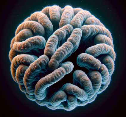
Under a microscope, telophase appears as a stage of mitosis where separated chromatids or chromosomes reach the opposite poles of the cell. Two distinct nuclei start forming, and the cell begins to undergo cytokinesis, forming two separate daughter cells.
Telophase is the final stage of mitosis or meiosis, during which the separated chromatids or chromosomes reach the opposite poles of the dividing cell. The key microscopic features of telophase include:
| Image | Product | Detail | Price |
|---|---|---|---|
 | Carson MicroBrite Plus 60x-120x LED Lighted Pocket Microscope |
| See on Amazon |
 | Elikliv LCD Digital Coin Microscope |
| See on Amazon |
 | AmScope M150 Series Portable Compound Microscope |
| See on Amazon |
 | PalliPartners Compound Microscope for Adults & Kids |
| See on Amazon |
 | Skybasic 50X-1000X Magnification WiFi Portable Handheld Microscopes |
| See on Amazon |
- Chromosomes at Opposite Poles: Chromosomes, which were duplicated during earlier stages of cell division, are now clearly visible as distinct structures. They align at opposite poles of the cell.
- Nuclear Envelope Formation: As the chromosomes reach their respective poles, a new nuclear envelope or membrane begins to form around each set of chromosomes. This process marks the reformation of the nuclei in the daughter cells.
- Cell Division Completion: Telophase is followed by cytokinesis, where the cell undergoes physical division to form two separate daughter cells, each with its nucleus enclosed within a nuclear envelope.
To provide a clearer understanding, here is a table summarizing the microscopic features of telophase:
| Microscopic Feature | Description |
|---|---|
| Chromosome Arrangement | Chromosomes align at opposite poles of the cell. |
| Nuclear Envelope Formation | New nuclear envelopes begin to form around each set of chromosomes. |
| Nuclei | Two distinct nuclei become visible as the cell prepares to divide. |

Cell Division Process: Mitosis and Meiosis
Cell division is a fundamental process that ensures the growth, development, and reproduction of organisms. There are two primary types of cell division: mitosis, responsible for the formation of somatic cells, and meiosis, which is specific to the generation of gametes.
Key Stages Leading Up to Telophase:
Both mitosis and meiosis share common stages leading up to Telophase, where the nucleus divides, and two daughter cells are formed.
Key Stages of Mitosis:
Mitosis is the process by which a single eukaryotic cell divides into two identical daughter cells. The key stages leading up to Telophase in mitosis include:
- Prophase: Chromosomes condense, the nuclear envelope disintegrates, and spindle fibers begin to form.
- Metaphase: Chromosomes align along the cell’s equator, known as the metaphase plate, ensuring an equal distribution of genetic material.
- Anaphase: Sister chromatids separate and move toward opposite poles of the cell, pulled by spindle fibers.
- Telophase: Chromosomes de-condense, a nuclear envelope reforms around each set of chromosomes, and the cell undergoes cytokinesis, resulting in two daughter cells.
Key Stages of Meiosis:
Meiosis is the process of cell division that produces haploid gametes (sperm and egg cells). The stages leading up to Telophase in meiosis are:
- Prophase I: Homologous chromosomes pair up in a process called synapsis, and genetic recombination occurs through crossing-over.
- Metaphase I: Homologous chromosome pairs align at the metaphase plate.
- Anaphase I: Homologous chromosomes separate and move to opposite poles, reducing the chromosome number by half.
- Telophase I: Chromosomes may partially de-condense, and the cell undergoes cytokinesis, resulting in two haploid cells.
- Prophase II, Metaphase II, Anaphase II, and Telophase II: These stages mirror those of mitosis, resulting in the formation of four haploid daughter cells, each with half the chromosome number of the original cell.
Outcome of Telophase:
The culmination of Telophase in both mitosis and meiosis is the formation of two daughter cells, each genetically identical in mitosis and genetically diverse in meiosis. This process is fundamental for organismal development, growth, and the generation of reproductive cells.
Sample Preparation for Microscopic Observation
Telophase microscopic observation requires meticulous sample preparation to ensure clear and detailed imaging. The process involves several key steps that preserve cellular structures and enhance visibility under the microscope.
6 Steps in Sample Preparation:
- Cell Harvesting: Begin by carefully harvesting the cells of interest. This may involve cell culture techniques, tissue sectioning, or other methods depending on the sample type.
- Fixation: The crucial step of fixation involves preserving the cellular structure by arresting cellular activities and preventing decay. Common fixatives include formaldehyde or glutaraldehyde.
- Permeabilization: To allow staining agents to penetrate the cells, permeabilization is employed. This step is particularly important for fluorescent microscopy, enabling dyes to access cellular components.
- Staining: Staining enhances the contrast and visibility of cellular structures. Various dyes target specific components, such as DAPI for DNA, and Phalloidin for actin filaments.
- Mounting: After staining, the sample is carefully mounted on a slide using a mounting medium. This not only secures the sample but also helps maintain its three-dimensional structure.
- Cover Slipping: A cover slip is placed over the sample, minimizing dehydration and protecting it during microscopic observation.
Importance of Proper Fixation:
Proper fixation is a cornerstone in sample preparation as it preserves cellular morphology and prevents degradation. It serves the following crucial purposes:
- Structural Integrity: Fixation immobilizes cellular structures, preventing distortions or alterations during subsequent processing.
- Halting Cellular Processes: Fixation halts cellular activities, ensuring that the sample represents a snapshot of the cells at the moment of fixation. This is vital for accurate observation.
- Preventing Autolysis and Putrefaction: Without fixation, cellular components would undergo autolysis and putrefaction, compromising the integrity of the sample.
Importance of Staining Techniques:
Staining techniques enhance the contrast of cellular structures, making them more visible under a microscope. The significance of staining includes:
- Visualization of Specific Structures: Different stains target specific cellular components, allowing researchers to focus on particular structures, such as chromosomes during Telophase.
- Identification of Cell Types: Staining helps distinguish between various cell types and subcellular structures, aiding in the accurate identification of cells undergoing Telophase.
- Quantitative Analysis: Staining facilitates quantitative analysis, enabling researchers to measure aspects like DNA content, cell proliferation, and protein expression.
In summary, the preparation of samples for microscopic observation involves a series of meticulous steps, with fixation and staining playing pivotal roles. Proper fixation preserves cellular integrity, while staining techniques enhance visibility, providing a clearer understanding of Telophase events under the microscope. These techniques collectively contribute to the success of microscopic studies and the accurate analysis of cellular processes.
Common Challenges in Observing Telophase
Observing Telophase under a microscope presents certain challenges that researchers must navigate to obtain accurate and reliable data.
Sample Distortion:
Issues Related to Sample Distortion:
- Dehydration: During sample preparation, dehydration can occur, leading to shrinkage and distortion of cellular structures.
- Fixation Artifacts: Improper fixation may result in artifacts, causing morphological changes and affecting the accurate representation of Telophase.
Minimizing Distortion for Accurate Observation:
- Optimal Fixation: Ensuring the use of suitable fixatives and adhering to recommended fixation times minimizes distortion.
- Balanced Dehydration: Carefully controlling the dehydration process helps prevent excessive shrinkage and distortion.
Limited Resolution:
Challenge of Limited Resolution:
- Microscope Limitations: Traditional light microscopy may have limitations in resolving fine details during Telophase.
Techniques to Enhance Resolution:
- Super-Resolution Microscopy: Utilizing advanced techniques like super-resolution microscopy enhances resolution, allowing for finer details to be observed.
- Immunofluorescence Staining: Employing specific fluorescent markers in immunofluorescence staining can improve visibility and resolution.
Suboptimal Contrast:
Challenges in Achieving Suboptimal Contrast:
- Inadequate Staining: Poorly applied or insufficient staining can result in low contrast, making it challenging to distinguish cellular structures.
Techniques to Enhance Contrast:
- Optimized Staining Protocols: Following established staining protocols and using appropriate dyes improves contrast.
- Fluorescence Microscopy: Utilizing fluorescence microscopy with fluorescent dyes enhances contrast and specificity in observing Telophase.
Photobleaching:
Impact of Photobleaching:
- Loss of Fluorescence: Prolonged exposure to light can lead to the loss of fluorescence signal, hindering continuous observation.
Strategies to Minimize Photobleaching:
- Timed Observation: Limiting the exposure time during observation sessions helps minimize photobleaching.
- Antifade Reagents: Using antifade reagents in fluorescence microscopy can mitigate the effects of photobleaching, preserving fluorescence signals.
Addressing these common challenges in observing Telophase enhances the accuracy and reliability of microscopic studies, providing researchers with a clearer understanding of the intricate events during this crucial stage of cell division.
Live Cell Imaging
In recent years, live cell imaging has emerged as a groundbreaking technique, revolutionizing the study of Telophase and providing unprecedented insights into dynamic cellular events.
Advancements in Live Cell Imaging Techniques:
- Real-Time Observations: Live cell imaging allows researchers to observe Telophase events in real-time, capturing the dynamic nature of cellular processes.
- Longitudinal Studies: Continuous monitoring of cells over extended periods enables longitudinal studies, offering a comprehensive view of Telophase progression.
Impact on Our Understanding:
- Dynamic Visualization: Live cell imaging facilitates the dynamic visualization of Telophase events, shedding light on the intricacies of chromosome de-condensation, nuclear envelope reformation, and cytokinesis.
- Cellular Dynamics: The ability to observe cellular dynamics in a live context enhances our understanding of the timing and coordination of events during Telophase.
Challenges and Future Prospects:
Technical Challenges:
- Phototoxicity: Prolonged exposure to light during live cell imaging can induce phototoxicity, affecting cell viability and confounding observations.
- Data Processing: Handling large volumes of live cell imaging data poses challenges in terms of storage, analysis, and interpretation.
Future Prospects:
- Improved Imaging Modalities: Ongoing advancements aim to develop imaging modalities with reduced phototoxicity and enhanced resolution for more accurate live cell observations.
- Integration with Other Techniques: Integrating live cell imaging with complementary techniques, such as super-resolution microscopy, holds promise for comprehensive Telophase analysis.
Live cell imaging has undeniably transformed our approach to Telophase research, providing a dynamic window into the previously inaccessible realm of real-time cellular events. While technical challenges persist, ongoing innovations and future prospects in live cell imaging promise to further deepen our understanding of Telophase and contribute to the broader field of cell biology.
How does Telophase appear under a microscope?
Telophase, the final stage of mitosis, exhibits distinct characteristics when observed under a microscope. A typical telophase cell appears as two separate nuclei forming within a single cell membrane. Chromosomes, which were condensed during earlier stages, start to de-condense and elongate. The microtubules that facilitated chromosome movement begin to disassemble, allowing the formation of two distinct daughter cells. Below is a table summarizing key features observed during telophase:
| Telophase Characteristics | Description |
|---|---|
| Nuclei Formation | Two distinct nuclei forming within a shared cell membrane |
| Chromosome State | De-condensation and elongation of chromosomes |
| Microtubule Disassembly | Disintegration of microtubules aiding chromosome movement |
What is the role of microtubules in Telophase?
Microtubules play a crucial role in telophase, facilitating the movement of chromosomes and the formation of daughter cells. During this phase, microtubules that were responsible for separating chromosomes begin to disassemble. This disassembly allows the chromosomes to move freely, leading to the formation of two distinct nuclei within the cell. Here’s a table summarizing the role of microtubules in telophase:
| Microtubule Function | Description |
|---|---|
| Chromosome Separation | Microtubules aid in pulling apart condensed chromosomes |
| Daughter Cell Formation | Disassembly of microtubules allows the formation of cells |
Can telophase be easily distinguished from other mitotic stages under a microscope?
Yes, telophase can be distinguished from other mitotic stages under a microscope due to its unique characteristics. The presence of two distinct nuclei within a single cell membrane, along with the de-condensation and elongation of chromosomes, sets telophase apart from the preceding stages. Microtubule disassembly is also a key indicator of telophase. The following table highlights the distinguishing features of telophase:
| Distinguishing Features | Description |
|---|---|
| Two Nuclei Formation | Presence of two distinct nuclei within a shared membrane |
| Chromosome Changes | De-condensation and elongation of chromosomes |
| Microtubule Disassembly | Disintegration of microtubules aiding chromosome movement |
What is the significance of observing telophase under a microscope?
Observing telophase under a microscope provides valuable insights into the process of cell division and the formation of new cells. It allows researchers and scientists to study the intricate details of how chromosomes are distributed among daughter cells and how cellular structures, such as nuclei and microtubules, undergo dynamic changes. This understanding is crucial for various fields, including cell biology and medical research. No table is needed for this question.
Are there any specific staining techniques recommended for observing telophase?
Various staining techniques can enhance the visibility of cellular structures during telophase. Dyes such as DAPI (4′,6-diamidino-2-phenylindole) are commonly used to highlight DNA, making chromosomes more visible. Immunofluorescence staining can also be employed to target specific proteins involved in telophase processes. Here’s a table summarizing recommended staining techniques:
| Staining Technique | Description |
|---|---|
| DAPI (4′,6-diamidino-2-phenylindole) | Highlights DNA, making chromosomes more visible |
| Immunofluorescence Staining | Targets specific proteins involved in telophase processes |
References:
- Alberts B, Johnson A, Lewis J, et al. Molecular Biology of the Cell. 4th edition. New York: Garland Science; 2002.
- Lodish H, Berk A, Zipursky SL, et al. Molecular Cell Biology. 4th edition. New York: W. H. Freeman; 2000

Fahim Foysal is a well-known expert in the field of binoculars, with a passion for exploring the great outdoors and observing nature up close. With years of experience in the field, Fahim has honed his skills as a binocular user and has become a go-to resource for those seeking advice on choosing the right binoculars for their needs.
Fahim’s love for the natural world began during his time at The Millennium Stars School and College and BIAM Laboratory School, where he spent much of his free time exploring the outdoors and observing the wildlife around him. This passion for nature led him to pursue a degree in Fine Arts from the University of Dhaka, where he gained a deep understanding of the importance of observation and attention to detail.
Throughout his career, Fahim has used his expertise in binoculars to help others discover the beauty of the natural world. His extensive knowledge of binocular technology and optics has made him a trusted advisor for amateur and professional wildlife observers alike. Whether you’re looking to spot rare birds or observe animals in their natural habitats, Fahim can help you choose the perfect binoculars for your needs. With his guidance, you’ll be able to explore the outdoors with a newfound appreciation for the beauty of the natural world.
Table of Contents
