As an elementary student, I remember being fascinated by the world around me, especially the small details that were often overlooked. One tool that allowed me to explore this world in greater detail was the microscope. Whether examining plant cells or examining tiny insects, using a microscope allowed me to see the intricate details of the microscopic world.
However, not all microscopes are created equal, and it can be overwhelming to choose the best one for a young student. In this article, we will take a closer look at the 5 best microscopes for elementary students that are easy to use, durable, and provide clear images to foster a love for science and exploration in young minds.

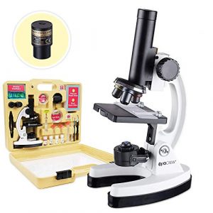
I have been using the AmScope – M40-K-MDM35 120X – 1200X Microscope for quite some time now, and I must say, I am thoroughly impressed with its performance. This microscope is an excellent tool for students and beginners interested in exploring the microscopic world. The microscope has various features that make it stand out from other microscopes in its price range.
- New from AmScope – our premium 85+ piece microscope and color camera kit with exclusive kid’s friendly software
- All metal high-quality microscope with precision focus optics, magnifications ranging from 120x -1200x, a rack and pinion easy focus system, two light sources, a built-in color filter wheel and much more!
- Premium kit includes over 85 pieces - blank and prepared slides, experiment ideas, tools and other accessories all in a rugged ABS plastic carrying case
- This premium kit includes a color digital USB2.0 eyepiece camera that allows your child to easily connect to a computer and download or edit pictures of specimens directly from the microscope. Exclusive kid’s friendly patent pending software downloads easily – contains microscope how-to section, sample specimen library, and an interactive real-time specimen image editing page
- Save images or videos of any specimen being examined for school projects, to send to relatives or friends or to compare to professional samples
One of the main features of this model is its LED light source, which provides ample illumination for viewing specimens. The microscope is made of high-quality materials, including ABS, plastic, and metal, which give it a sturdy and durable feel. The real angle of view is 45 degrees, making it comfortable for extended periods. The objective lens description is achromatic, ensuring the image is clear and crisp.
Another advantage of this microscope is that it is battery-powered, which makes it portable and easy to use anywhere. This feature is especially useful for field trips or outdoor experiments. The microscope has a maximum magnification of 1200x, which is impressive for a microscope in its price range.
One of the things I like most about this microscope is the premium kit that comes with it. The kit includes over 85 pieces, including blank and prepared slides, experiment ideas, tools, and other accessories. The kit comes in a rugged ABS plastic carrying case, making it easy to transport and store.
Additionally, the premium kit includes a color digital USB 2.0 eyepiece camera that allows you to easily connect to a computer and download or edit pictures of specimens directly from the microscope. The exclusive kid-friendly patent-pending software with the microscope makes it even more user-friendly. It contains a microscope how-to section, a sample specimen library, and an interactive real-time specimen image editing page.
The AmScope – M40-K-MDM35 120X – 1200X Microscope is an excellent choice for elementary students interested in science. The microscope is easy to use and has various features that make it perfect for beginners. The magnification range is suitable for viewing various specimens, from larger to microscopic organisms. The kit that comes with the microscope provides everything a student needs to get started on their scientific journey.
As a research student who has used the AmScope 120X – 1200X Microscope, I highly recommend it to anyone interested in exploring the microscopic world. The microscope is well-made, easy to use, and comes with a premium kit that provides everything you need to get started. The only downside is that some users have reported missing parts and poorly fitting carrying cases. Nonetheless, the positives far outweigh the negatives, and I highly recommend this microscope to anyone looking for a quality microscope at an affordable price.
Emarth 40X- 1000X Compound Microscopes
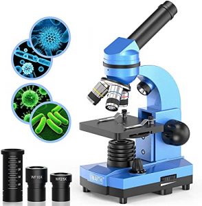
I have had the opportunity to use the Emarth 40X-1000X Compound Microscope and I must say that it is an excellent microscope for elementary students. It is built with high-quality optics that provide improved visual quality and sharp images, making it easy for students to observe and learn from the specimens they are studying. The microscope has a magnification range of 40x to 1000x, with six magnification levels, making it suitable for various applications.
- 【High Magnification】microscope built-in WF10x & WF25x eyepiece and optical lens: 4x, 10x, 40x , rotatable monocular head offers six magnification levels at 40x, 100x, 250x, 400x and 1000x. High Quality Optics can give children improved visual quality and sharp image when in use
- 【Dual Light Illumination】 Double light compound microscope has incident and transmitted light sources, which can see both opaque objects illuminated from above and translucent specimens illuminated from below. the bottom light can be adjusting light by rotrating the wheel as you needed, easy to use.(Not including batteries)
- 【Easy to Focus & 6 Colorful Filters】 Built-in 6 different colors filters rotating wheel.The microscope is easy to focus, just need to rotate the focus knob slowly so that you can see clear images need to rotate the focus knob slowly so that you can see clear images
- 【High-Class Material】38°inclined ocular tube with 360°degree rotatable monocular head, provides a more comprehensive view and enable sharing. Metal base and sturdy handle framework won't make microscope shake
One of the standout features of this microscope is its dual-light illumination system. It has an incident and transmitted light source that can illuminate opaque objects from above and translucent specimens from below. The bottom light can be adjusted by rotating the wheel, making it easy to use. Additionally, the microscope has built-in 6 different colors filters rotating wheel, which allows students to observe specimens in different colors.
The microscope is easy to focus on, and the 38° inclined ocular tube with 360° degree rotatable monocular head provides a comprehensive view, enabling sharing. The metal base and sturdy handle framework ensure that the microscope doesn’t shake while in use, providing stability for accurate observations.
The microscope comes with approximately 52 pcs of KITS (valued at £19.99), making it an excellent educational gift for students. The collection of slides/samples that come with it is also a nice touch, as it provides an interesting range of specimens for students to explore. While it may be a bit complicated for younger age groups, they will figure it out fast enough with adult supervision and guidance.
One drawback of this microscope is that it may not be worth its price tag of $60 for some people. It is not a very high-powered microscope, and while it is perfect for young scientists, more experienced researchers may need a more advanced microscope.
Overall, the Emarth 40X-1000X Compound Microscope is fantastic for elementary students. It is built with high-quality optics, is easy to focus, and has a dual light illumination system, making it easy for students to observe and learn from specimens. It is an excellent educational gift for students, and the collection of slides/samples that come with it provides an interesting range of specimens for students to explore.
Swift SW150 Compound Student Microscope
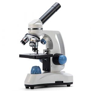
I had the opportunity to use the SWIFT SW150 Compound Student Microscope for my lab experiments, and I must say, I was impressed with its features and performance. Designed to introduce elementary through middle school students and beginner hobbyists to the fascinating world of microscopy and the life sciences, this microscope is perfect for laboratories and one-on-one instruction.
- Designed to introduce elementary through middle school students and beginner hobbyists to the fascinating world of microscopy and the life sciences
- Fully rotatable monocular head for easy shared use, perfect for laboratories and one-on-one instruction
- Available magnification settings of 40X, 100X, 250X, 400X, and 1000X through crystal-clear 4X, 10X, and 40X glass objectives with wide-field 10X and 25X eyepieces
- Dual illumination system for maximum versatility to examine both transparent and solid specimens; cool LED lights protect eyesight and live specimens
- A sleek triangular base and metal carrying handle combine with cordless capability for effortless on-the-go experimentation
One of the main features of this model that caught my attention was the fully rotatable monocular head. This feature makes it easy for multiple users to share the microscope, and it is perfect for one-on-one instruction. The available magnification settings of 40X, 100X, 250X, 400X, and 1000X through crystal-clear 4X, 10X, and 40X glass objectives with wide-field 10X and 25X eyepieces provide a wide range of magnification options for various applications.
The dual illumination system is another great feature that makes this microscope versatile. It allows for examining both transparent and solid specimens, and the cool LED lights protect eyesight and live specimens. The sleek triangular base and metal carrying handle combine with cordless capability for effortless on-the-go experimentation, which makes it an ideal microscope for fieldwork.
One of the things that I liked about this model was the easy focus and clear images. The two lower magnifiers were usable and worked great. However, the highest magnifier on the turret was not usable, but that is the case with any scope with a magnification of 1000 or higher. The fact that it runs on batteries and has a mechanical switch that triggers when the power supply is plugged in is a plus.
However, I did encounter an issue with the software/eyepiece not talking to each other, which made it useless for my assignments. To prevent getting a zero for my assignments, I had to rig up my iPhone through the eyepiece to capture images, which was not ideal. I emailed Swift about the issue but never heard back, so I had to return the microscope.
Overall, the SWIFT SW150 Compound Student Microscope is a great microscope for elementary students. Its compact size and portability make it easy to handle, and the material feels durable. It is battery-powered, which means that it can be taken anywhere. The features of this microscope, including the dual illumination system and easy focus, make it an ideal microscope for fieldwork and lab experiments. If you’re looking for a versatile, easy-to-use microscope that provides clear images, then the SWIFT SW150 Compound Student Microscope is worth considering.
BEBANG 2000X Optical Kids Microscope
As a research student with extensive knowledge of microscopes, I recently had the opportunity to use the BEBANG 2000X Optical Kids Microscope and was quite impressed with its features and capabilities.
One of the standout features of this microscope is the dual illumination system, which includes two light sources on the top and bottom of the microscope. This allows for better visualization of specimens and a wider range of specimens. Additionally, the dial-type 6-color filter adds a fun and educational element to the microscope, allowing users to explore the colorful world of microorganisms.
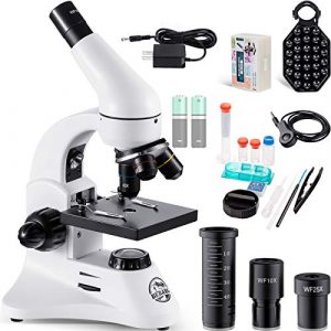
- 【2 Eyepieces/12 Kinds of Magnification】: Wide-Field eyepiece WF10 + WF25, removable barlow lens, 40x to 2000x magnification. Made of metal, best quality, better touch.
- 【Dual Illumination System】: With two light sources on the top and bottom, you can see the specimen better and see more kinds of specimen. In addition, the dial-type 6-color filter connects to the world of various colorful microorganisms!
- 【Ergonomic Design】: Coarse & fine two focusing modes, 45 degree inclined eye tube, 360 degree rotatable monocular head. Dimmable lights can adapt to different sun intensities.
- 【Complete Accessories and Long Service Life】: Smartphone holder, research set, preparation set, is suitable for children elementary school junior high school and adult enthusiast beginners, school, laboratory, home, one-on-one lessons. Compared to electron microscopes, optical microscopes have a longer service life and can be used for decades if properly stored.
- 【Worry-free Shopping】: 30 days free returns & 2 year product warranty. any questions we will reply within 24 hours. Thank you very much.
The ergonomic design of the microscope is also noteworthy. It includes coarse and fine focusing modes, a 45-degree inclined eye tube, and a 360-degree rotatable monocular head, all making it easy and comfortable to use. The dimmable lights can also be adjusted to adapt to different lighting conditions, ensuring optimal viewing in any environment.
Another aspect of the BEBANG microscope that impressed me was its versatility. It comes with a wide range of accessories, including a smartphone holder, research setting, and preparation set, making it suitable for children, elementary school, junior high school, and adult enthusiast beginners. The microscope can be used in various settings, including schools, laboratories, homes, and for one-on-one lessons.
The BEBANG microscope has a maximum magnification of 2000x, which is achieved through a combination of two eyepieces (WF10 and WF25) and a removable Barlow lens. The objective lens description is achromatic, which ensures accurate color representation of specimens.
One of the few downsides of this microscope is its price. While it is a high-quality product that functions highly, it may not suit those on a budget. However, it is important to note that optical microscopes like the BEBANG have a longer service life than electron microscopes and can last for decades if properly stored and maintained.
Overall, I highly recommend the BEBANG 2000X Optical Kids Microscope for elementary students and anyone interested in exploring the world of microorganisms. Its high-quality construction, dual illumination system, ergonomic design, and versatility make it an excellent choice for schools, laboratories, and home use. Plus, the bonus of a smartphone holder, research setting, and preparation set make it a great value for the price.
What are the basic steps for focusing a microscope for Elementary Students?
Here are the basic steps for focusing a microscope for elementary students:
- Place a prepared microscope slide onto the stage of the microscope.
- Use the coarse adjustment knob to move the objective lens to its lowest position closest to the slide.
- Look through the eyepiece and adjust the mirror or light source so light shines onto the slide.
- Use the coarse adjustment knob to move the objective lens slightly away from the slide, then look through the eyepiece and adjust the focus until you can see a blurry image of the specimen.
- Use the fine adjustment knob to move the objective lens closer to the slide while looking through the eyepiece.
- Adjust the focus until the image becomes clear and in focus.
- If necessary, adjust the light source or mirror to improve the contrast or brightness of the image.
- Once you have a clear image, you can adjust the magnification by rotating the objective lens or changing to a different objective lens.
It’s important to remind elementary students to be gentle when using the adjustment knobs and to avoid touching the objective lens or slide with their fingers to prevent damage to the microscope and the specimen.
What are some tips for using a microscope safely and effectively?
Here are some tips for using a microscope safely and effectively for elementary students:
- Always carefully handle the microscope and avoid bumping or dropping it.
- Keep the microscope away from the edge of the table or desk to prevent it from falling.
- Only use clean microscope slides and coverslips to avoid contamination of the specimen and the microscope.
- Wash your hands before and after using the microscope to avoid transferring bacteria or other microorganisms to the specimen or your eyes.
- Use both hands to carry the microscope and hold it securely.
- Always start with the lowest magnification and work your way up to higher magnifications, which will help you avoid damaging the specimen or the microscope.
- Use the coarse adjustment knob first to get the specimen in focus, then use the fine adjustment knob to fine-tune the focus.
- Avoid touching the objective lens or the slide with your fingers, as this can leave fingerprints and smudges that will affect the clarity of the image.
- Use the stage clips to hold the slide in place and prevent it from moving around while you’re viewing it.
- When you’re finished using the microscope, clean the lenses with lens paper or a soft cloth and put the dust cover back on to protect the microscope from dust and debris.
By following these tips, elementary students can use microscopes safely and effectively, which will help them get the most out of their observations and experiments.
What are some common uses for microscopes in science and medicine?
Microscopes are widely used in science and medicine for a variety of purposes. Here are some common uses for microscopes in these fields:
- Observing cells and tissues: Microscopes allow scientists and doctors to observe the structure and function of cells and tissues, which can help them diagnose and treat diseases.
- Studying microorganisms: Microscopes are used to study microorganisms such as bacteria, viruses, and fungi, which are too small to be seen with the naked eye.
- Analyzing blood samples: Microscopes are used to analyze blood samples for abnormalities and to identify blood cells.
- Examining tissues for cancer: Microscopes are used to examine tissue samples for signs of cancer and other diseases.
- Analyzing forensic evidence: Microscopes are used in forensic science to analyze evidence such as hair, fibers, and bloodstains.
- Identifying minerals and crystals: Microscopes are used in geology and mineralogy to identify minerals and crystals.
- Examining fossils: Microscopes are used to examine fossils and to identify and classify ancient organisms.
- Observing living organisms: Microscopes are used to observe living organisms such as protozoa, algae, and bacteria.
- Developing new materials: Microscopes are used in materials science to study the properties of materials at the microscopic level, which can help researchers develop new materials with specific properties.
Overall, microscopes are essential in many areas of science and medicine, allowing researchers and professionals to observe and analyze the microscopic world around us.
What examples of living organisms can you observe under a microscope for elementary students?
Here are some examples of living organisms that can be observed under a microscope for elementary students:
- Paramecium: A single-celled organism found in freshwater ponds and streams. It moves using tiny hair-like structures called cilia.
- Amoeba: Another single-celled organism found in freshwater and marine environments. It moves using a process called cytoplasmic streaming.
- Euglena: A single-celled organism capable of photosynthesis and movement using a flagellum.
- Pond water organisms: Various organisms can be found in pond water, including algae, rotifers, and water fleas.
- Onion cells: Thin slices of onion can be viewed under a microscope to observe the structure of plant cells.
- Cheek cells: Swabbing the inside of the mouth can provide a sample of cheek cells, which can be viewed under a microscope to observe the structure of animal cells.
- Elodea leaf: A type of aquatic plant that can be viewed under a microscope to observe the structure of plant cells.
- Bacteria: Many types of bacteria can be observed under a microscope, including those that are found in soil, water, and the human body.
By observing these and other living organisms under a microscope, elementary students can better understand the microscopic world and the diversity of life around us.
What are some common mistakes to avoid when using a microscope?
Here are some common mistakes to avoid when using a microscope:
- Improper handling: One of the most common mistakes when using a microscope is improper handling, such as carrying the microscope by its arm or placing it too close to the edge of a table. This can result in the microscope falling or getting damaged, which can affect its performance.
- Incorrect lighting: Using incorrect lighting can result in poor visibility of the specimen. It’s important to adjust the light intensity and angle of the light source to optimize the illumination of the specimen.
- Using incorrect magnification: The wrong magnification can result in a clear image. Always start with the lowest magnification and work your way up to higher magnifications, as this will help you avoid damaging the specimen or the microscope.
- Focusing improperly: Focusing the microscope properly is crucial to getting a clear specimen image. Use the coarse adjustment knob first to get the specimen in focus, then use the fine adjustment knob to fine-tune the focus.
- Contamination of the specimen: Contamination of the specimen can occur if the slide or coverslip is not clean or is not handled properly. Always use clean microscope slides and coverslips, and carefully handle the specimen.
- Ignoring the instructions: Not following the instructions for the microscope can result in improper use, which can damage the microscope or result in poor quality images. Always read and follow the instructions carefully before using the microscope.
By avoiding these common mistakes, users can ensure that they get the best possible results from the microscope and prevent damage to the microscope and the specimen.
What are some different techniques for staining microscope slides?
There are several different techniques for staining microscope slides, each of which is suited for different types of specimens and applications. Here are some of the most common staining techniques:
- Simple staining: This is the most basic staining technique, which involves using a single dye to stain the entire specimen. It is often used to enhance the contrast between the specimen and the background.
- Differential staining: This technique uses two or more dyes to stain different specimen parts selectively. Different differential staining techniques include Gram, acid-fast, and spore staining.
- Fluorescent staining: This technique uses fluorescent dyes that emit light when excited by a specific wavelength of light. It is often used to visualize specific molecules or structures within the specimen.
- Immunostaining: This technique uses antibodies to target specific molecules or structures within the specimen. The antibodies are labeled with a fluorescent dye or an enzyme, which allows them to be visualized under the microscope.
- Oil immersion staining: This technique involves using a special type of oil with a high refractive index to increase the microscope’s resolution. The oil is placed between the objective lens and the specimen to reduce the amount of light that is refracted.
- Negative staining: This technique involves using a contrasting dye to stain the slide’s background, leaving the specimen unstained. It is often used to visualize the shape and size of bacterial cells.
The choice of staining technique depends on the type of specimen being studied and the specific questions being addressed. By using the appropriate staining technique, researchers can enhance the contrast and resolution of the microscope and gain a better understanding of the structure and function of the specimen.
What are some ways that microscopes have been used in historical scientific discoveries?
Microscopes have played a crucial role in many historical scientific discoveries, including:
- Discovery of cells: In the 17th century, Antonie van Leeuwenhoek used a microscope to observe tiny organisms in pond water, which he called “animalcules.” He also observed red blood cells, sperm cells, and bacteria, which led to the discovery of cells as the basic unit of life.
- Germ theory: In the 19th century, Louis Pasteur used a microscope to observe bacteria, which led to the germ theory of disease. He showed that microorganisms were responsible for food spoilage and the spread of diseases, which revolutionized the field of medicine.
- DNA structure: In 1953, James Watson and Francis Crick used X-ray crystallography and microscopy to determine the structure of DNA, which led to a better understanding of genetics and the development of the field of molecular biology.
- Blood circulation: In the 17th century, William Harvey used a microscope to observe blood circulation in animals, leading to a better understanding of the human cardiovascular system.
- Microbiology: In the 19th century, Robert Koch used a microscope to identify the causative agents of tuberculosis and cholera, which led to the development of microbiology as a field of study.
- Cellular processes: In the 20th century, Albert Claude and George Palade used electron microscopy to observe the structure of cells and the processes that occur within them, including protein synthesis and cell division.
These historical discoveries and many others were made possible by using microscopes, which allowed scientists to observe and study the microscopic world in greater detail than ever before.
How do scientists use microscopes to study the structure and function of living organisms?
Scientists use microscopes to study the structure and function of living organisms in several ways. Here are some examples:
- Observation of cells: Microscopes allow scientists to observe the structure and function of cells in living organisms. They can visualize the different organelles within the cell, including the nucleus, mitochondria, and chloroplasts. They can also observe cell division and other cellular processes.
- Identification of pathogens: Microscopes identify pathogens such as bacteria and viruses that cause disease in living organisms. By observing the morphology and behavior of these pathogens, scientists can develop methods for treating and preventing diseases.
- Imaging of tissues and organs: Microscopes can observe the structure and function of tissues and organs in living organisms. Scientists can visualize the different types of cells within the tissue and observe how they interact.
- Study of molecular interactions: Microscopes can be used to study the interactions between molecules within living organisms. For example, they can be used to observe the binding of a drug to a receptor on a cell surface or the movement of proteins within a cell.
- Investigation of developmental biology: Microscopes can be used to observe the development of organisms from the embryonic stage to adulthood. Scientists can visualize the growth and differentiation of cells and tissues during this process.
Overall, microscopes provide scientists with a powerful tool to observe the structure and function of living organisms at a microscopic level. This information is essential for understanding organisms’ basic biology and developing disease treatments and therapies.
Facts
- The first microscope was invented in the late 16th century by Hans and Zacharias Janssen.
- Antonie van Leeuwenhoek, a Dutch scientist, was one of the first people to use a microscope to observe microscopic organisms in the 17th century.
- There are many different types of microscopes, including compound microscopes, stereo microscopes, and electron microscopes.
- The highest magnification of a compound light microscope is typically 1000x.
- The first electron microscope was invented in the 1930s, and it uses a beam of electrons instead of light to magnify objects.
- Electron microscopes can magnify objects up to 10 million times.
- In the field of medicine, microscopes are used to diagnose diseases, such as cancer and infections, by examining tissue samples.
- Microscopes are also used in the study of microbiology, which is the study of microorganisms like bacteria, viruses, and fungi.
- The study of cells, tissues, and organs under the microscope is called histology.
- In recent years, digital microscopes have become more popular. These microscopes allow images to be captured and viewed on a computer screen, which can make it easier to share images and collaborate with other scientists.

I am an enthusiastic student of optics, so I may be biased when I say that optics is one of the most critical fields. It doesn’t matter what type of optics you are talking about – optics for astronomy, medicine, engineering, or pleasure – all types are essential.
Last update on 2025-10-07 / Affiliate links / Images from Amazon Product Advertising API
Table of Contents


Pingback: Kindergarten Microscopes: Our Top 5 Picks for Beginner Science Exploration