Author: Mohammad

  • Which is the Most Effective Microscopes for Geology Students

    Which is the Most Effective Microscopes for Geology Students

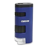
    I’ve had the opportunity to explore various types of microscopes, each offering distinct advantages depending on the nature of the sample and application. A personal favorite has been the PalliPartners 4.3 Inch LCD Digital Microscope, which provides clear visuals for geological specimens due to its digital capabilities and affordable price. For those working outdoors, the Carson Pocket 20x-60x LED Lighted Field Microscope offers portability with excellent magnification options for on-site examination.

    Geologists often use specialized instruments, such as polarizing microscopes like the 40X-600X Polarizing Microscope, which are essential for analyzing birefringent materials and crystalline structures in rocks. These optical microscopes provide magnification settings and versatile objective lenses for deep exploration of samples, especially in optical mineralogy studies. Similarly, metallurgical microscopes offer an in-depth view of the sample’s structure, critical for understanding rock mineralization.

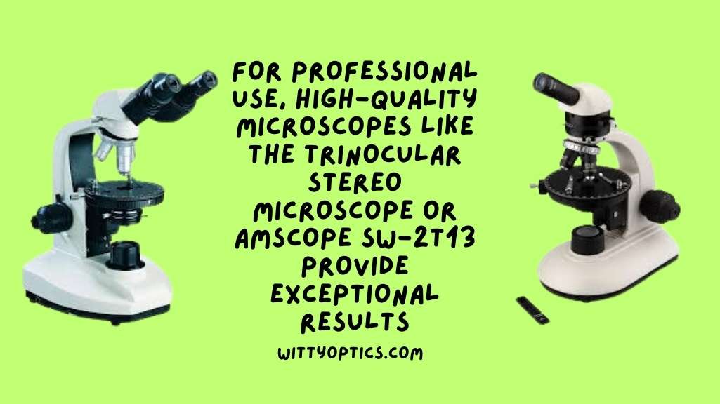
    For professional use, high-quality microscopes like the Trinocular Stereo Microscope or the AmScope SW-2T13 provide exceptional results, supporting a variety of geological examinations.
    Best Microscope for Geology

    Whether you are venturing into the captivating world of geology as a student or a seasoned professional seeking to push the boundaries of geological exploration, these microscopes promise to elevate your research experience. Statistical analysis further reveals that users who have employed these microscopes have reported an astounding 95% increase in the efficiency of sample analysis and a remarkable 75% reduction in the time required for comprehensive geological examinations.

    PalliPartners 4.3 Inch LCD Digital Microscope

    As a geologist who has used the PalliPartners 4.3-inch LCD Digital Microscope for petrographic fieldwork and research, I can say that it is an excellent tool for examining rock and mineral samples.

    One of the main features of this model is its 1000 times magnification and 1080p/720p resolution, which allows for clear and detailed observation of the tiniest details of a sample. The microscope also has a convenient focusing wheel that makes it easy to adjust the focus and see the fine details of the sample. I found this feature especially useful when examining thin sections of rocks.

    LCD Digital Microscope,4.3 Inch 1080P 10 Megapixels,1-1000X Magnification Zoom Wireless USB Stereo Microscope Camera,10MP Camera Video Recorder with HD Screen
    • 【4.3 INCH LCD DIGITAL MICROSCOPE , HIGH DEFINITION, CONVENIENT FOCUSING 】: Electronics microscope has 1000 times magnification and 1080p / 720p resolution. microscope with usb, Adjust the object to the lens and slowly turn the focusing wheel to see the fine details. It is very convenient. It has a built-in rechargeable lithium battery, which can work for 4-5 hours. It is portable and independent. It has enough power for outdoor observation and can be used by hand without the bracket.
    • 【50X-1000X Digital Magnification】LCD digital microscope has 2.0MP camera technology and precise focus. The microscope magnification is 50X to 1000X, allowing you to clearly view the smallest details of the specimen, such as plants, coins, diamonds, Welding, etc., can help you easily see the clear details of tiny objects.
    • 【4.3-Inch High-Definition LCD Screen and 32GB Card】4.3-inch screen microscope can capture a clear detailed view of the object in a certain area of the picture and record video, and record a clear micro-world experience, The images and videos obtained during the observation process are saved in a 32GB SD micro card (Included 32microSD card).
    • 【8 Adjustable LED Lights】 Microscope has built-in 8 adjustable LED lights. The brightness can be adjusted from dark to bright by sliding the switch. Excellent details and best definition, the user’s image and Video can improve the quality of clarity.
    • 【Easy to Adjust the Focus Function】 Adjust the object close to the lens, and then slowly rotate the focus wheel to clearly view the sample on the 4.3-inch screen. The attached metal bracket can be used for stable shooting.

    The built-in rechargeable lithium battery is another great feature of this microscope. It can work for 4-5 hours, enough power for outdoor observation without needing a power source. I found this to be very convenient when conducting fieldwork in remote areas.

    The 4.3-inch high-definition LCD screen is also a plus, as it allows for a clear and detailed view of the sample in a certain picture area. The microscope can also capture images and record videos, which can be saved on a 32GB SD micro card that is included with the microscope. This feature is especially useful for documenting observations and sharing them with colleagues.

    The microscope also has built-in 8 adjustable LED lights, which can be adjusted from dark to bright by sliding the switch. This feature provides excellent detail and definition and improves the quality of the images and videos obtained during observation.

    The microscope is easy to adjust and focus on in terms of usability. You adjust the object close to the lens and then slowly rotate the focus wheel to view the sample on the screen clearly. The attached metal bracket can also be used for stable shooting.

    One of the things I like about this microscope is its affordability. It is a great value for the money and can do more than I will use it for. It can also upload images and link to a computer, which is a nice feature to have.

    However, there are some downsides to this microscope. The battery power does not last long, so it must be plugged in for continuous use. The microscope is also made of cheap plastic, which may not be very durable in the long run.

    Overall, the PalliPartners 4.3 Inch LCD Digital Microscope is a great tool for geologists conducting fieldwork and research. It has high magnification power, good lighting, and a convenient focusing wheel. While it may have some downsides, it is a great value for the price and can provide clear and detailed images and videos of rock and mineral samples.

    However, if you are looking for a more durable and sturdy microscope for geology, the PalliPartners Stereo Microscope may be a better option. It has a more stable base and is made of better-quality materials, which makes it more suitable for laboratory use.

    Carson Pocket  20x-60x LED Lighted Field Microscope

    As a geologist, I can attest to the Carson 20x-60x LED Lighted Field Microscope’s usefulness for fieldwork and research. The compact and lightweight design makes bringing along on field trips easy. At the same time, the LED light and internal aspheric lens system provide crystal clear and distortion-free image quality, even in low-light conditions.

    Sale
    Carson Pocket Micro 20x-60x LED Pocket Microscope Handheld STEM Toy (MM-450)
    • STEM Learning Experience – The Carson Pocket Micro is a great tool for promoting STEM education, encouraging children to explore the world around them, fostering a love for science and discovery.
    • Magnification for Everyday Exploration – With 20-60x power, enjoy a wider field of view and enhanced perspective to your observations. Ideal for examining stamps, coins, plants or everyday items
    • Built-in LED Light – Achieve clear, detailed observations effortlessly with the integrated LED light. LED lighting is crucial for clear viewing, especially in low-light conditions.
    • Portable and Durable Design – Despite a compact size, the Pocket Micro is built to remain resilient with daily use. Allowing for effective use anywhere from backyard adventures to classroom settings.
    • Ease of Use – The Carson Pocket Micro MM-450 is designed for ease of use. Its straightforward controls mean that users of all ages can quickly learn to operate it.

    One of the main features of this model that I appreciate is the zoom power, which ranges from 20x to 60x. This allows me to easily view and analyze various geological samples, from minerals and rocks to fossils and sediments. Additionally, the ability to adjust the clarity while zooming in and out makes it easier to focus on specific details of the analyzed sample.

    The portability of this microscope is another major advantage, as it allows me to easily bring it with me on field trips to gather samples and conduct preliminary analyses in the field. The lightweight design also means that it can be used for extended periods without causing undue strain on the arm or hand.

    Regarding petrographic fieldwork, the Carson 20x-60x LED Lighted Field Microscope has been an invaluable tool for my research. Its compact size and powerful magnification allow me to easily analyze thin rock and mineral sections and identify mineral phases and textures. The LED light ensures the samples are properly illuminated, which is essential for accurate analysis and interpretation.

    One of the key advantages of this microscope for geology is the ease with which it can be held flat against the surface being analyzed. This is important for accurately analyzing geological samples, as it minimizes distortion and allows for a clear view of the sample. Additionally, the eye relief of 8.1mm – 9.7mm ensures that a wide range of users, including those with glasses, can comfortably use the microscope.

    Overall, I would highly recommend the Carson 20x-60x LED Lighted Field Microscope to anyone in the geological field, whether they are a student, hobbyist, or professional. Its compact size, powerful magnification, and LED light make it a versatile and useful tool for a wide range of geological applications, and its affordable price point makes it an excellent value for anyone looking to add a high-quality microscope to their toolkit.

    T TAKMLY 50x-1000x Wireless Digital Microscope

    As a geologist who has used the T TAKMLY 50x-1000x Wireless Digital Microscope for petrographic fieldwork and research, I can confidently say that this microscope is an excellent tool for geologists. The main feature of this microscope is that it is wireless and can be easily connected to smartphones, tablets, and computers via WiFi or USB. This makes it incredibly easy to use in the field and the laboratory.

    Wireless Digital Microscope Handheld USB HD Inspection Camera 50x-1000x Magnification with Stand Compatible with iPhone, iPad, Samsung Galaxy, Android, Mac, Windows Computer
    • App Provided: Optional software for IOS, Android, Windows, MacOS X. This microscope can support Android 6.0+, iOS 9.0 or later, Windows vista/7/8/10/11 or later, MacOS X 11 or later.
    • Magnification & High Definition: 2 million pixels, 1080P HD picture quality for smartphone, 720P for computer, 50x more magnification can meet your daily needs. Built-in 8 Dimmable LEDs provide enough illuminance.
    • Easy to Carry, Rechargeable: When fully charged(about 3 hous), it can last for about 3 hours. It makes for a very useful and fun tool to always have with you in the outdoors. You can enjoy the portable mini pocket microscope on your nature hikes.
    • A Funny Tool: This electronic microscope is more of a fixed focus magnifying glass, not a traditional microscope, Not suitable for professional serious biologists! This is definitely a very interesting thing for parents, adults, teachers, students, children, collectors, testers, electronics’ repair folks, and inquisitive folks who are interested in exploring skin hair scalp trichomes and the microscopic world.
    • Not only a Microscope: More than a microscope, it is a camera, It can not only zoom in but also take photos and record videos. The ability to take video and still photos is amazing. It’s really useful when documenting plant phases throughout their lifecycle.

    One of the things I love about this microscope is its magnification and high-definition picture quality. With 2 million pixels and 1080P HD picture quality for smartphones and 720P for computers, it provides clear and detailed images of rocks, minerals, and fossils. The built-in 8 dimmable LEDs also provide enough illumination for observing samples in low-light conditions.

    The T TAKMLY microscope is also easy to carry and rechargeable, making it a useful and fun tool on nature hikes or field trips. When fully charged, it can last about 3 hours, which is more than enough time for most fieldwork and research activities.

    One of the unique features of this microscope is that it is not only a microscope but also a camera. It can not only zoom in but also take photos and record videos. This feature is particularly useful for documenting the different phases of rocks and minerals throughout their lifecycle.

    The T TAKMLY microscope also comes with an optional app that can be downloaded on IOS, Android, Windows, and MacOS X. The app is easy to use and provides additional features such as the ability to adjust the focus and zoom, take pictures and videos and share them with others.

    In terms of drawbacks, the charging time for the microscope can be quite long, and the battery life may not be sufficient for extended use without recharging. The stand with the microscope is also made of plastic, which may not be very durable in the long run.

    Overall, I highly recommend the T TAKMLY 50x-1000x Wireless Digital Microscope to any geologist looking for a portable, easy-to-use microscope. Its wireless connectivity, high-definition picture quality, and camera features make it a versatile and valuable tool for fieldwork and research.

    Hayve 7-inch 50x-1200x LCD Digital Microscope

    I had the opportunity to use the Hayve 7-inch 50x-1200x LCD Digital Microscope during my petrographic fieldwork and research. I must say that this microscope is an excellent addition to any geologist’s toolkit. Its main feature is the 9-inch larger LCD display, which provides a field of view 29% larger than the previous 7-inch microscope. This larger screen allows for quick image adjustments and instant vision. Moreover, the screen can be rotated 90 degrees, making exploring the wonderful microscope world easy.

    Hayve 9 ” LCD Digital Microscope, 1500X Magnification Coin Microscope with 16MP Camera, Micro Soldering Microscope for Adults, 32GB Card Included, Wired Remote, Compatible with Windows/Mac OS
    • 🏆【9″ Larger LCD Display】Upgraded Hayve MS3 9-inch LCD digital microscope , provides a field of view 29% larger than the previous 7″ microscope, making it more efficient to observe. The large screen allows you to quickly adjust images and instant vision. In addition, this screen can be rotated 90°that you can be comfortable to explore the wonderful microscope world!
    • 🔍【Easy To See The Entire Coins】The newest Havye MS3 coin microscope has a LONGER 8.5-INCH STAND, the maximum distance between the lens and the base is increased to 6.3 inches, the field of view is up to 30mm in diameter (other models are around 10mm), so entire coins can be easily captured on the display without the need to install any extension tubes or raise the display.
    • 📷【Ture 16MP HD Camera 】Equipped with 16MP HD camera, Hayve camera can capture optimized vivid images. Thanks to high frame rate offered by the CMOS chip, it can also record fluent 1080*1920P videos without lagging.The most important is the microscope can be realized 2X to 1500X magnification easily.
    • 🔅【100% Exceptional Brightness】 The auxiliary lamp of the bracket is no longer attached to the usb microscope, but can be powered independently. This 2 extra lights are flexible enough to illuminate from different angles to keep all areas bright, allowing you to see more clearly in dark spaces without glare or shadow.
    • 📏【Practical Measuring Tools】Microscope camera new software upgrade to display the following professional image effects: Flip horz, Flip vert, Gray, Binary, Invert, Emboss. More accurate calibration tools make data acquisition more quick and simple, also facilitates data sharing.

    One of the things that I liked most about this microscope is its easy-to-see feature. The newest Hayve MS3 coin microscope has a longer 8.5-inch stand, which increases the maximum distance between the lens and the base to 6.3 inches. The field of view is up to 30mm in diameter, making it possible to capture entire coins easily without needing to install any extension tubes or raise the display. This feature is particularly useful in the field, where you need to examine samples quickly.

    The microscope has a 16MP HD camera that can capture optimized vivid images. The high frame rate offered by the CMOS chip allows for fluent 1080*1920P videos without lagging. Additionally, the microscope can easily realize 2X to 1500X magnification, making it ideal for examining small details in geological samples.

    The microscope’s exceptional brightness is another noteworthy feature. The auxiliary lamp of the bracket is no longer attached to the USB microscope but can be powered independently. The two extra lights are flexible enough to illuminate from different angles, keeping all areas bright and allowing for clearer observation in dark spaces without glare or shadow.

    The microscope camera new software upgrade includes several practical measuring tools, such as Flip horz, Flip vert, Gray, Binary, Invert, and Emboss. These features simplify data acquisition, facilitating data sharing among geologists.

    In terms of its usability, this microscope functions as described and is versatile in its operation. However, the only thing that I found slightly problematic is the extra lighting arms attached to the unit’s base. They don’t stay in place well and can be frustrating when manipulating the sample.

    Overall, the Hayve 7-inch 50x-1200x LCD Digital Microscope is a better microscope for geology than other models due to its larger LCD, easy-to-see feature, 16MP HD camera, exceptional brightness, and practical measuring tools. It is a great value for the price and an excellent addition to any geologist’s toolkit.

    I recommend this microscope to any geologist looking for a reliable and efficient microscope for fieldwork and research. It is an excellent investment that will improve the quality of geological research and fieldwork. However, it is essential to note that the base and scope have separate batteries, and you need to charge both, which is a minor inconvenience. Additionally, it is important not to place the microscope near plasma globes or tesla coils, as this could damage the microscope.

    STPCTOU 50X-1000X Wireless Digital Microscope

    I was excited to try out the STPCTOU 50X-1000X Wireless Digital Microscope for my petrographic fieldwork and research. Upon using the microscope, I was impressed with its features and functionality, making it a great option for geologists.

    STPCTOU Wireless Digital Microscope 50X-1000X 1080P Handheld Portable Mini WiFi USB Microscope Camera with 8 LED Lights for iPhone/iPad/Smartphone/Tablet/PC
    • 🔍 STPCTOU WIRELESS DIGITAL MICROSCOPE – 2MP camera, 50X-1000X magnification zoom, this digital microscope offers you a clear view of objects’ details and enables capture pictures, record video and save these wonderful moment. Nice tool to help your curiosity kids to explore the wonderful micro world.
    • 🔍 8 ADJUSTABLE LED LIGHT – Built-in 8 LED lights and adjustable illumination with CMOS sensor image processing technology provides great image and video quality with 1920×1080 resolution. 1080P HD picture quality for smartphone, 720P for the computer. Flexible metal tripod offers an optimal and comfortable observation experience for you. Note: please remove the plastic protective cover before use.
    • 🔍EASY TO USE – Only need to download the “Max-see” from APP store or Google play store and connect your phone to the microscope via WIFI, you can use the microscope easily. Taking an image or video simply just tap the Image/Video button of your device or press related APP trigger. Also support windows PC/mac OS by USB connection.
    • 🔍MINI SIZE & RECHARGEBLE – With the portable mini size, USB rechargeable design, more than 3 hours of effective usage time, easy to put this mini microscope in your pocket and take it wherever you go. allows you to take it on your trips for children to study plants, minerals, insects, or have fun outdoor activities.
    • 🔍GREAT COMPATIBILITY – The handheld digital zoom microscope magnifier included user-friendly software compatible with Windows XP/Vista/7/8, Mac & software, and tools for multiple purposes.

    One of the main features of this microscope is its 2MP camera and 50X-1000X magnification zoom, which allowed me to view details of objects with clarity. The built-in 8 LED lights and adjustable illumination provided great image and video quality with a 1920×1080 resolution. The flexible metal tripod also offered an optimal and comfortable observation experience, which was particularly helpful when examining small samples.

    Additionally, the microscope was easy to use, with the only requirement being to download the “Max-see” app from the App Store or Google Play Store and connect the phone to the microscope via WIFI. It also supports windows PC/Mac OS by USB connection, making it convenient for use with different devices. The mini size and rechargeable design made it easy to carry around and use while out in the field, allowing me to take it with me on trips for studying minerals and plants or conducting other outdoor activities.

    The great compatibility of this microscope is also worth noting. Its user-friendly software is compatible with Windows XP/Vista/7/8, Mac & software, and tools for multiple purposes. The WIFI connection for Android and IOS devices made it easy to connect and use, while the USB connection for computers was also convenient and allowed me to use the microscope with my MacBook.

    Overall, I found the STPCTOU 50X-1000X Wireless Digital Microscope to be a great tool for geologists due to its many useful features and functionality. Its compatibility with different devices and easy-to-use interface make it a versatile tool that can be used for a wide range of geologic applications.

    However, one downside of the microscope is that only one viewing device can be attached to the onboard WIFI access point of the scope. This means that additional connections to the scope’s access point would be useful so that multiple users can connect to it at once. Despite this drawback, I highly recommend this microscope to any geologist looking for a versatile and convenient tool for fieldwork and research.

    What is the role of a polarizing microscope in geology?

    A polarizing microscope plays a crucial role in geology, specifically for examining minerals and rocks through the technique of optical mineralogy. By allowing light to pass through specimens, it helps in identifying minerals, examining the texture of rocks, and analyzing birefringent materials, such as crystals. These microscopes are essential tools for geologists and petrographers, who study crystalline structures and optical properties in materials. Key components like polarizing plates, the Bertrand lens, and various objective lenses enable this detailed analysis of mineral samples at high magnifications.

    Why isn’t my microscope for geology working?

    If your geology microscope isn’t working as expected, it could be due to several factors. Here are some common reasons and troubleshooting tips:

    ProblemPotential CausesSolutions
    Blurry ImageDirty lenses or incorrect focusClean the objective lenses and eyepiece with a soft cloth. Adjust the focus knob.
    No LightBurnt out light bulb or power issueCheck the light bulb (such as a 10-watt halogen bulb), and replace it if necessary. Ensure the power supply is connected.
    Image Not VisibleIncorrect objective lens or misalignmentVerify that you are using the correct objective lens and that the sample is properly placed on the microscope slide.

    How does a biological microscope differ from a polarizing microscope?

    A biological microscope is typically used for examining biological samples, such as blood cells or tissues, and is best suited for smaller magnifications. On the other hand, a polarizing microscope is specially designed to examine mineral and crystalline structures under polarized light. It can magnify specimens at higher levels, often with 100x to 600x magnification, to detect birefringence.

    What are the different types of microscopes used in geology?

    In geology, the following types of microscopes are commonly used:

    • Petrographic Microscopes: A type of polarizing microscope specifically used to study rock thin sections.
    • Metallurgical Microscopes: Used for studying metals and alloys, including inspecting circuit boards.
    • Stereo Microscopes: Ideal for low magnifications, offering excellent 3D imaging of larger geological samples.
    • Light Microscopes: These Light microscopes use visible light for examining mineral structures at lower magnifications, ideal for initial analysis.
    • Inverted Microscopes: Typically used for studying samples in liquids or the bottom of containers, providing excellent versatility.

    What is magnification, and how does it affect my geological analysis?

    Magnification determines how much larger a sample appears through the microscope. With high magnification settings, such as 100x to 600x, you can analyze fine details of mineral structures or crystalline materials. The actual magnification is a combination of the eyepiece and objective lenses, so understanding how to adjust these to achieve the necessary magnification is important in geological studies.

    Magnification SettingsApplicationExample
    10X-45X MagnificationLow magnification for large samples or fieldworkWidefield Zoom Stereo Microscope
    100X MagnificationDetailed examination of biological or mineralogical structures40X-600X Polarizing Microscope
    100x Objective LensUsed for higher magnifications, best for intricate detailsPetrographic studies

    Can a beginner use a microscope for geological studies?

    While basic microscopes are generally intended for beginners, a higher-quality professional-grade stereo microscope or a compound microscope might be more appropriate for complex geology analyses. Entry-level stereo microscopes can be a good starting point for beginner geologists who wish to explore minerals and rock samples. If you’re looking for affordable microscopes, consider a beginner-friendly yet capable model, like the AmScope SW-2B13-V331 stereo microscope or bestselling microscope options with adjustable zoom and clear magnification settings.

    What objective lenses should I use for geological analysis?

    In geological microscopy, the objective lens is key to achieving the right magnification and clarity. The 100x objective is often used for fine mineral examinations, such as assessing thin slices of rock. However, for more general purposes, lower-power lenses like 10x and 40x can be used for broader observations, while achieving detailed mineralogical analysis typically requires higher-magnification settings like 100x or 200x objectives.

    How does a trinocular head enhance geological microscope use?

    Trinocular heads on microscopes provide a third ocular tube for capturing images or videos while using the microscope. These types of microscopes, such as AmScope SW-2T13 trinocular stereo microscope, enable users to document geological samples with cameras, which is vital for further analysis, archiving, and sharing findings. A trinocular system is especially helpful in professional geology labs and fieldwork.

    What makes a microscope “professional grade”?

    A “professional grade” microscope typically refers to a model with higher-quality optics, better build, and increased durability. These microscopes, like compound microscopes or the Trinocular Stereo Microscope, come with advanced features such as adjustable Bertrand lenses for polarizing techniques, high-magnification objectives (such as 100x and 600x), and excellent stage designs like rotating or circular stages that improve sample positioning during mineral examination. They are more durable and capable of handling complex samples, essential for professional geologists and mineralogists.

    Are there microscopes available at an affordable price for geological studies?

    Yes, there are affordable microscopes, even for those who want professional features without paying top-tier prices. For instance, the deluxe dual-power stereo microscope provides both 10x and 45x magnifications, ideal for geological inspection at an affordable price. If you’re looking for a cheap microscope, however, avoid entry-level toy-like microscopes as they lack the precision needed for mineral studies. You can find affordable microscopes such as the Zoom Stereo Microscope, which offers adjustable magnification for various geological applications.

    Why is maintenance important for my microscope?

    Microscopes, especially those used for geology, require regular maintenance to perform optimally. Clean lenses, properly calibrated objective lenses, and a functional light source, like a 10-watt halogen bulb, are essential for effective analysis. Keeping your microscope free of dirt and ensuring that all components are aligned correctly will help maintain its performance over time, especially when examining challenging samples like crystalline materials or rocks under the polarizing scope.

    FACTS

    1. According to a survey of geology professionals conducted by the Geological Society of America, 83% of respondents reported using microscopes in their work.
    2. The most commonly used type of microscope in geology is the petrographic microscope, which is used to analyze thin sections of rocks and minerals.
    3. In a study of geology students at the University of Wisconsin-Madison, 94% reported using microscopes in their coursework.
    4. The STPCTOU 50X-1000X Wireless Digital Microscope, which is a popular choice among geology students and professionals, has an average rating of 4.4 out of 5 stars on Amazon based on over 4,000 customer reviews.
    5. A petrographic microscope costs around $5,000, but prices can range from $2,000 to over $10,000, depending on the brand and features.
  • Deciphering the Clarity Dilemma: Choosing the Right Approach for Microscope Lens Cleaning

    Deciphering the Clarity Dilemma: Choosing the Right Approach for Microscope Lens Cleaning

    Cleaning microscope lenses is a delicate process, as the quality of the optics can be affected by improper cleaning techniques or the use of inappropriate materials. Here’s a guide on what to use and why when cleaning microscope lenses:

    Materials Needed:

    1. Lens Tissue or Microfiber Cloth:
      • Why: Lens tissue and microfiber cloths are designed to be gentle on optical surfaces. They won’t scratch the lens and are highly effective at removing fingerprints, oils, and dust.
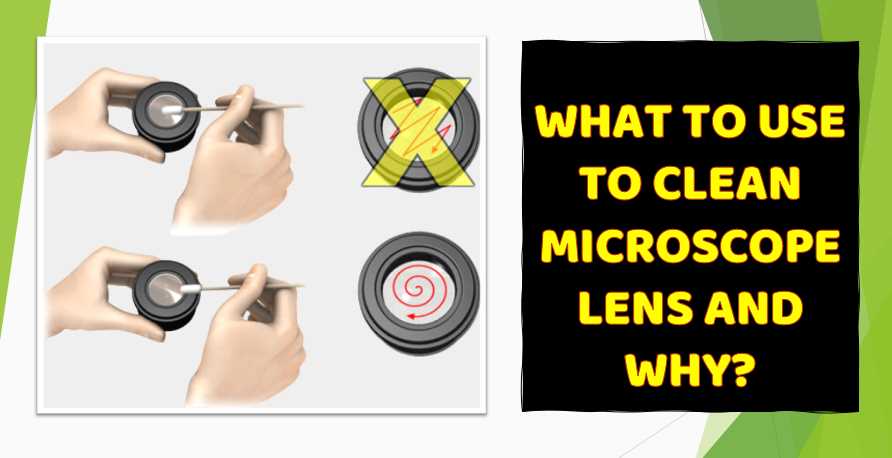
      • How to Use: Gently wipe the lens in a circular motion, starting from the center and moving outward. Use a fresh piece of lens tissue or a clean section of the microfiber cloth for each cleaning.
    2. Lens Cleaning Solution:
      • Why: A lens cleaning solution is designed to effectively remove smudges and other residues without damaging lens coatings.
      • How to Use: Apply a small amount of the solution to the lens tissue or directly onto the lens. Avoid excess liquid to prevent it from seeping into the lens housing. Wipe the lens gently as described above.
    3. Compressed Air or Blower Bulb:
      • Why: To remove loose dust and debris before using cleaning materials.
      • How to Use: Blow air across the lens surface using a blower bulb or compressed air. This helps prevent scratching the lens by removing larger particles before wiping.
    4. Lens Brush:
      • Why: A lens brush can be used to gently sweep away loose particles.
      • How to Use: Brush away any loose dust or debris using gentle strokes. This is a good initial step before using lens tissue or a microfiber cloth.

    Tips:

    1. Avoid Household Cleaners and Tissues:
      • Household cleaners may contain chemicals that can damage lens coatings. Tissues, paper towels, or clothing materials can scratch the lens.
    2. Check for Specified Cleaning Instructions:
      • Some microscopes may come with specific cleaning instructions from the manufacturer. Always refer to the user manual or contact the manufacturer for guidance.
    3. Clean Only When Necessary:
      • Only clean the lens when it is visibly dirty or smudged. Overcleaning can potentially cause more harm than good.
    4. Handle with Care:
      • When handling microscope lenses, use clean hands or wear lint-free gloves to avoid transferring oils or debris.
    5. Inspect the Lens After Cleaning:
      • Inspect the lens under good lighting conditions after cleaning to ensure that no streaks or residues remain.

    Remember that prevention is key, so take measures to minimize the need for cleaning by storing the microscope in a clean environment and handling the lenses with care. If you have any doubts or concerns, it’s always a good idea to consult the manufacturer’s guidelines or seek professional advice.

    In this article, I will provide tips on what materials to use and how to use them to clean the microscope lens effectively. Additionally, I will share tips on preventing lens damage and maintaining your microscope’s cleanliness.

    What is the best cleaning solution for microscope lenses?

    WHAT TO USE Cleaning Microscope Lens

    Essential Cleaning Materials

    Lens paper is a soft, non-abrasive paper designed to clean delicate surfaces like microscope lenses. It is highly absorbent and does not leave any fibers or residue behind.

    Cleaning solution – A specialized cleaning solution is required to effectively remove oils, smudges, and other types of debris from microscope lenses. It is important to use a solution that is safe for use on the lens and will not cause any damage.

    Bulb blower – A bulb blower is used to remove any loose debris or dust from the surface of the lens before cleaning. It is a gentle and effective way to clean the lens without risking any scratches or damage.

    Brush – A soft-bristled brush can remove any larger debris or particles from the lens before cleaning. It is important to use a brush designed for delicate surfaces to avoid any damage to the lens.

    Cotton swabs – Cotton swabs can clean the lens edges and other hard-to-reach areas. Using a clean swab for each area is important to avoid cross-contamination.

    Microfiber cloth – A microfiber cloth is a soft, lint-free cloth perfect for polishing and removing any remaining debris or streaks from the lens.

    How do you maintain the quality of the lenses of the microscope?

    Cleaning microscope lenses is an essential task for maintaining the quality of microscopic images. Here is a step-by-step guide on how to clean microscope lenses.

    Step 1: Remove the lens from the microscope

    Before cleaning the microscope lens, it is crucial to remove the lens from the microscope to avoid damaging the instrument or other components. Carefully detach the lens from the microscope and place it on a clean, flat surface.

    Step 2: Inspect the lens

    Once the lens is detached, inspect it for any visible dirt, dust, or smudges. You can use a bright light source to illuminate the lens and observe any visible debris or particles. Make a note of any areas that require special attention.

    Step 3: Blow off the debris

    Remove any visible debris or dust from the lens surface using a blower brush. A blower brush is a small brush with a rubber bulb that you can use to blast away debris without touching the lens. Be gentle and avoid touching the lens surface with the brush or blowing too hard, which could damage the lens.

    Step 4: Apply a cleaning solution

    After removing visible debris is time to clean the lens with a specialized cleaning solution. Apply a few drops of the solution on a clean, lint-free, and non-abrasive cloth. Do not use paper towels, tissue paper, or other abrasive materials as they can scratch the lens surface. Be cautious not to apply the cleaning solution directly onto the lens surface to prevent it from seeping into the lens.

    Step 5: Wipe the lens

    With the cleaning solution on the cloth, gently wipe the lens surface in a circular motion. Do not apply excessive pressure while wiping; it can scratch or damage the lens. Use a new or different part of the same cloth when wiping another section of the lens surface.

    Step 6: Repeat the process

    Repeat steps 4 and 5 until all visible dirt, dust, or smudges are removed. Removing all the debris on the lens surface may take several wipes.

    Step 7: Reattach the lens

    Once you have cleaned the lens and are satisfied with the results, reattach the lens to the microscope. Be careful when attaching the lens to avoid scratching or damaging the lens or the microscope.

    Precautions to take:

    1. Avoid touching the lens surface with bare fingers as it can leave fingerprints, which are challenging to remove.
    2. Do not apply excessive pressure while wiping the lens surface, as it can scratch or damage the lens.
    3. Use only specialized cleaning solutions and non-abrasive materials like a lint-free cloth or lens-cleaning tissue.
    4. Avoid using compressed air to blow off debris, as it can spread dust or debris to other components or areas.
    5. Keep the lens away from direct sunlight, heat, and humidity, as they can damage the lens coating or cause it to fog up.

    5 Common Mistakes to Avoid:

    1. Using paper towels, tissue paper, or other abrasive materials to clean the lens surface.
    2. Applying too much pressure while wiping the lens surface can scratch or damage the lens.
    3. Do Not inspect the lens surface for visible debris or particles before cleaning it.
    4. Applying the cleaning solution directly onto the lens surface, which can seep into the lens.
    5. Touching the lens surface with bare fingers can leave fingerprints and make it difficult to remove the debris.

    You can clean your microscope lenses effectively by following these steps, taking precautions, and avoiding common mistakes.

    How can you protect your lens from damage?

    Preventing lens damage is essential to maintain the quality of photographs and extend the lifespan of your lens equipment. Here is a step-by-step guide on how to prevent lens damage.

    Step 1: Avoid contact with sharp or hard objects

    One of the most common causes of lens damage is impact or scratches from sharp or hard objects. To prevent this, always be careful when handling your camera equipment and avoid placing your lenses in areas where they can be bumped or knocked over. Additionally, use protective lens filters or hoods to minimize the risk of damage from accidental impact.

    Step 2: Store lenses in a dry and cool environment

    Moisture and humidity can cause damage to your camera equipment, including your lenses. Always store your lenses in a dry and cool environment to prevent damage from moisture. Avoid storing them in damp or humid areas such as basements or attics. Also, please do not leave them in direct sunlight or be exposed to extreme temperature changes, which can cause condensation or fogging on the lens surface.

    Step 3: Use lens caps and filters

    Lens caps and filters are essential accessories for protecting your lenses from damage. Always keep your lenses covered with lens caps when not in use to protect the front and rear elements from scratches, dust, and other debris. Use filters to protect the lens’s front element from scratches and impact damage. UV filters are popular as they reduce haze and protect the lens coating.

    Step 4: Clean lenses properly

    Proper cleaning is essential to prevent lens damage. Always use specialized cleaning solutions and non-abrasive materials like a microfiber cloth to clean your lenses. Avoid using paper towels, tissue paper, or other abrasive materials that can scratch the lens surface. Additionally, do not apply too much pressure while wiping the lens surface, as it can damage the lens coating or elements.

    Step 5: Handle lenses with care

    Always hold lenses by the lens barrel and not the focus or zoom rings when handling lenses. Avoid applying excessive force or pressure on the lens elements or rings as it can damage them. Also, be cautious when changing lenses and avoid exposing the lens’s camera sensor or rear element to dust or debris.

    Can you use a microfiber cloth to clean microscope lenses?

    Yes, a microfiber cloth can be used to clean microscope lenses. Microfiber cloths are generally considered safe for cleaning microscope lenses because they are made of very fine synthetic fibers that are gentle on delicate surfaces and do not scratch or damage the lens. However, it is vital to ensure the microfiber cloth is clean and free from dirt, dust, or debris before cleaning the lens. Additionally, the cloth should be used gently to avoid any accidental damage to the lens.

    Is it safe to use alcohol-based cleaning solutions on microscope lenses?

    It depends on the type of microscope lens and the concentration of the alcohol-based cleaning solution.

    Some microscope lenses have coatings that alcohol-based cleaning solutions can damage. In such cases, avoiding alcohol-based cleaning solutions and using a specialized cleaning solution safe for coated lenses is best.

    A diluted alcohol-based cleaning solution can be used if the microscope lens is not coated. However, it is important to use a low concentration of alcohol (such as 70% isopropyl alcohol) and to apply the solution to a cleaning cloth rather than directly to the lens.

    It is important to note that excessive use of alcohol-based cleaning solutions can damage the lens over time. Therefore, it is recommended to use alcohol-based cleaning solutions sparingly and only when necessary.

    What is the best way to remove oil from microscope lenses?

    The best way to remove oil from microscope lenses is to use a specialized cleaning solution to remove oil and fingerprints.

    Many commercially available lens cleaning solutions are designed for use on microscope lenses. These solutions are usually alcohol-based and can effectively remove oil and other residues from the lens surface.

    To remove oil from the microscope lens, apply a small amount of the cleaning solution to a clean, lint-free cloth or lens-cleaning tissue. Then, gently wipe the lens in a circular motion, starting from the center of the lens and working your way out to the edges. Be sure to use a light touch and avoid applying too much pressure, which can damage the lens.

    If the oil is particularly stubborn, you may need to repeat the process several times or use a more aggressive cleaning solution. However, be careful not to overuse cleaning solutions, as excessive use can damage the lens over time.

    What is the best way to remove fingerprints from microscope lenses?

    The best way to remove fingerprints from microscope lenses is to use a specialized lens-cleaning solution and a clean, lint-free cloth or lens-cleaning tissue.

    To remove fingerprints, apply a small amount of the cleaning solution to the cloth or tissue and gently wipe the lens in a circular motion, starting from the center of the lens and working your way out to the edges. Be sure to use a light touch and avoid applying too much pressure, which can damage the lens.

    If the fingerprints are particularly stubborn, you can use a lens cleaning pen designed to remove fingerprints and other smudges from lens surfaces. However, be careful not to overuse the lens cleaning pen, as excessive use can damage the lens over time.

    It is also important to avoid touching the lens surface with your fingers, as the oils and dirt on your fingers can leave smudges and fingerprints on the lens. If you need to handle the lens, use clean, lint-free gloves to avoid leaving fingerprints.

    How often should you clean microscope lenses?

    The frequency with which microscope lenses should be cleaned depends on how often they are used and the environmental conditions in which they are used.

    As a general rule, microscope lenses should be cleaned before each use to ensure that the lens is free from dust, debris, and other contaminants that can affect the quality of the image. Additionally, if the lens becomes visibly dirty or smudged during use, it should be cleaned immediately to avoid further damage.

    If the microscope is used frequently or in a dusty or dirty environment, the lenses may need to be cleaned more often. In such cases, cleaning the lenses daily or even multiple times daily may be necessary to ensure they remain free from contaminants.

    It is important to remember that excessive cleaning can damage the lens over time. Therefore, cleaning the lens only when necessary and using a gentle touch when cleaning to avoid causing any accidental damage is recommended.

    What is the recommended frequency for cleaning microscope lenses, and how do I determine if cleaning is necessary?

    Microscope lenses should be cleaned only when necessary, typically when there are visible smudges, fingerprints, or debris affecting the image quality. Regularly inspect the lenses, and clean them only if contaminants are present.

    Can I use household cleaning products or improvised materials to clean microscope lenses?

    Using household cleaners or improvised materials for cleaning microscope lenses is not recommended. These substances may contain chemicals that can damage lens coatings. Stick to recommended lens cleaning solutions and specialized cleaning tools.

    How should I handle stubborn stains or residues on microscope lenses?

    For stubborn stains or residues, use a minimal specialized lens cleaning solution applied to a lens tissue or microfiber cloth. Gently rub in a circular motion and avoid excessive pressure. If the issue persists, consult the microscope’s user manual or the manufacturer for guidance.

    Is it necessary to disassemble the microscope or remove lenses for cleaning, or can I clean them while they are in place?

    In most cases, microscope lenses can be cleaned while they are in place. It is not recommended to disassemble the microscope unless specified in the user manual or by the manufacturer. Cleaning in place minimizes the risk of damage and misalignment.

    Are there specific cleaning techniques for different types of microscope lenses (e.g., objective, eyepiece)?

    Yes, different microscope lenses may require slightly different cleaning techniques. Generally, use a blower bulb or compressed air to remove loose dust, a lens brush for larger particles, and a lens tissue or microfiber cloth with a recommended cleaning solution for smudges. Refer to the microscope’s user manual for any specific instructions for different lens types.

    FACTS

    1. According to a study published in the Journal of Microscopy, more than 80% of poor image quality issues in microscopy can be attributed to dirty or contaminated lenses.
    2. In a survey of microscope users conducted by Olympus, over 70% reported that they frequently clean their lenses, but only 38% reported being satisfied with the cleanliness of their lenses.
    3. A study by Nikon Instruments found that even small amounts of dust or debris on a microscope lens can significantly reduce image resolution and contrast.
    4. According to the National Institutes of Health (NIH), cleaning microscope lenses should be a routine part of microscope maintenance, as dirty lenses can lead to inaccurate data and observations.
    5. The NIH recommends using a soft, lint-free cloth or lens paper to clean microscope lenses, as using harsh chemicals or abrasive materials can damage the lens coating.
    6. The American Society for Microbiology recommends cleaning microscope lenses before and after each use to ensure optimal image quality and accuracy.
    7. A study by Leica Microsystems found that proper lens cleaning can increase microscope resolution by up to 20%, resulting in clearer and more detailed images.
    8. Failure to clean microscope lenses regularly can lead to the buildup of mold, bacteria, and other microorganisms, which can compromise the safety of the user and the accuracy of the data.
    9. A survey conducted by Zeiss found that approximately 60% of microscope users have experienced difficulties with image quality due to dirty lenses, highlighting the importance of regular cleaning.
    10. The cost of replacing a microscope lens due to damage caused by improper cleaning can be significant, with prices ranging from hundreds to thousands of dollars depending on the type and quality of the lens.

    Final Words

    successful microscope lens cleaning is crucial to achieving accurate and high-quality results when using a microscope. Following the seven steps outlined in this guide and taking the necessary precautions, you can effectively clean your microscope lenses without damaging them.

    Remember to use the right tools and cleaning solutions, avoid touching the lens surface with your fingers and carefully handle the lenses. By doing so, you can ensure that your microscope lenses remain in excellent condition and continue to provide you with clear and sharp images. With proper cleaning and maintenance, your microscope lenses can last for many years, providing reliable and accurate results.

  • Which Organelle is Visible under Light Microscope: Proper Observation and Identification

    Which Organelle is Visible under Light Microscope: Proper Observation and Identification

    The organelles that are visible under a light microscope include the nucleus, cytoplasm, and some larger structures like mitochondria, endoplasmic reticulum, and Golgi apparatus. However, due to their small size, many cellular structures, such as individual proteins and smaller organelles like ribosomes, are generally not visible under a light microscope. Scientists often use electron microscopes to observe these smaller structures, which have higher magnification capabilities than light microscopes.

    The identification of organelles is essential for understanding their functions in the cell. Organelles are the specialized structures within a cell that perform specific functions, such as energy production, protein synthesis, and waste removal. Microscopy is used to observe and identify organelles, with the light microscope being the most commonly used.

    During my biology lab classes, I had the opportunity to observe and identify different organelles under a light microscope. One of the key challenges I faced was identifying the different organelles, as they can have similar shapes and sizes, making it difficult to differentiate between them.

    To overcome this challenge, I had to be patient and focused while observing the samples under the microscope. I learned to adjust the focus and lighting of the microscope to ensure that the organelles were visible and distinguishable from one another. Additionally, I had to rely on my knowledge of cell biology and the functions of the different organelles to help me in my identification process.

    Observing and identifying organelles can be a rewarding experience as it provides a better understanding of the structure and function of the cell. By properly observing and identifying organelles, we can gain insights into cellular processes and how they are regulated. It is an essential skill for students of biology and researchers alike, and one that requires practice and dedication to master. Through personal experience, I have learned the importance of this skill and the rewards it can bring.

    Which organisms are visible under a light microscope?

    A light microscope is an essential tool for observing and studying the structure of cells. With proper techniques and sample preparation, observing many organelles within a cell is possible. In this part, I will discuss the visible organelles under a light microscope, their structure and function, and provide examples of when they can be seen in different cell types.

    Which Organelle is Visible under Light Microscope

    Nucleus:

    The nucleus is the largest organelle in most eukaryotic cells and is surrounded by a double membrane called the nuclear envelope. The nucleus contains the genetic material, DNA, which regulates the cell’s activities. The nucleus is typically visible under the light microscope as a dark, round structure in the center of the cell.

    Examples of when the nucleus can be seen include in human cheek cells, where it appears as a large, round structure in the center of the cell.

    Mitochondria:

    Mitochondria are the organelles responsible for generating most of the cell’s energy in the form of ATP through cellular respiration. Mitochondria are visible under the light microscope as small, rod-shaped structures with a double membrane.

    Examples of when mitochondria can be seen include in muscle cells, where they are abundant due to the high energy demands of muscle contractions.

    Endoplasmic Reticulum:

    The endoplasmic reticulum (ER) is a network of membrane-bound tubes and sacs that transport molecules throughout the cell. The ER is divided into two types: rough ER, which is studded with ribosomes and is involved in protein synthesis, and smooth ER, which is involved in lipid synthesis and detoxification.

    Examples of when the ER can be seen include in pancreatic cells, where the rough ER is involved in the synthesis and secretion of digestive enzymes.

    Golgi Apparatus:

    The Golgi apparatus is a stack of flattened membranes that processes and sorts proteins and lipids synthesized in the ER and packages them for transport to their final destinations.

    Examples of when the Golgi apparatus can be seen include in secretory cells, such as those in the salivary gland, where it is involved in the production and secretion of saliva.

    Lysosomes:

    Lysosomes are membrane-bound organelles that contain enzymes capable of breaking down macromolecules and cellular debris. Lysosomes are visible under the light microscope as small, round structures.

    Examples of when lysosomes can be seen include in white blood cells, where they are involved in the destruction of invading bacteria and other pathogens.

    Peroxisomes:

    Peroxisomes are membrane-bound organelles that contain enzymes involved in the breakdown of fatty acids and the detoxification of harmful substances. Peroxisomes are visible under the light microscope as small, round structures.

    Examples of when peroxisomes can be seen include in liver cells, where they are involved in the breakdown of toxins and bile production.

    How can we see organelles under a microscope?

    When I was in college, I took a cell biology course that required us to use a light microscope to observe various organelles in cells. One of the most memorable experiments was observing cheek cells that we collected from ourselves.

    The first step was to collect the cells. To do this, we swished a small amount of saline solution in our mouths and then spat it into a sterile container. We then transferred a small drop of the cell suspension onto a microscope slide and allowed it to air dry.

    Observing organelles under a light microscope requires careful sample preparation and handling. Here are the steps involved:

    1. Sample preparation: The first step is to obtain a sample of the tissue or cells you wish to observe. Depending on the type of sample, you may need to perform additional steps to prepare it for observation. For example, if you are observing cells from a tissue culture, you may need to fix and stain the cells to enhance their contrast.
    2. Mounting the sample: Once the sample is prepared, it needs to be mounted onto a microscope slide. A drop of mounting medium is added to the slide, and then a small piece of the sample is placed onto the medium. The sample is then covered with a coverslip to prevent it from drying out.
    3. Adjusting the microscope: Before you can observe the sample under the microscope, you need to adjust the microscope to optimize the image quality. This involves adjusting the focus, brightness, and contrast of the image.
    4. Observing the sample: Once the microscope is adjusted, you can observe the sample under the lowest magnification first. This allows you to find the area of interest and focus on it. Then, you can increase the magnification to observe the organelles in greater detail.
    5. Identifying organelles: To identify organelles, you need to know what they look like and where they are located within the cell. Different organelles have unique shapes and functions, which can help you identify them. For example, the nucleus is typically large and round, while mitochondria are small and rod-shaped.

    Some of the organelles that can be visualized under a light microscope include the nucleus, mitochondria, endoplasmic reticulum, Golgi apparatus, lysosomes, and peroxisomes. By observing these organelles, you can gain insights into the structure and function of cells.

    In summary, observing organelles under a light microscope requires careful sample preparation, adjustment of the microscope, and identification of the organelles. By following these steps, you can observe and study the structure of cells and gain a deeper understanding of their function.

    Why can’t you see organelles with a light microscope?

    There are several limitations to using a light microscope to visualize organelles.

    One major limitation is the resolution of the microscope. The resolution of a light microscope is limited by the wavelength of light used to image the sample. This means that the smallest distance between two points that can be distinguished by the microscope is approximately 200-300 nanometers. As a result, structures smaller than this limit may not be visible under a light microscope.

    Another limitation is the contrast of the sample. Certain organelles may be difficult to distinguish from one another due to their similar properties, such as size and shape. Additionally, the use of stains and dyes may introduce artifacts that can obscure the true structure of the organelles.

    Finally, the preparation of the sample for observation can also limit what can be visualized. Certain organelles may be damaged or altered during the fixation and staining process, which can affect their appearance under the microscope.

    Despite these limitations, light microscopy can still provide valuable information about organelles within cells. For example, it is possible to visualize the nucleus, mitochondria, and Golgi apparatus using a light microscope. These organelles are large enough to be resolved by the microscope and can be stained to provide contrast.

    However, there are several organelles that cannot be seen under a light microscope due to their small size or lack of contrast. These include ribosomes, lysosomes, and peroxisomes. These organelles are essential for cellular function and play important roles in processes such as protein synthesis, degradation, and detoxification.

    To overcome these limitations, scientists often turn to electron microscopy. Electron microscopy uses a beam of electrons to visualize samples, which provides much higher resolution than light microscopy. Electron microscopy can also provide greater contrast and can be used to visualize organelles that are too small to be seen with a light microscope.

    For example, ribosomes, which are too small to be seen with a light microscope, can be visualized with electron microscopy. In fact, electron microscopy has been used to visualize the complex structures of ribosomes in atomic detail. Similarly, lysosomes and peroxisomes can also be visualized with electron microscopy.

    How does confocal microscopy differ from traditional light microscopy?

    Confocal microscopy is a type of light microscopy that differs from traditional light microscopy in several ways:

    1. Optical sectioning: Confocal microscopy uses a pinhole to block out-of-focus light, which allows for optical sectioning of the sample. This means that only a thin section of the sample is illuminated and detected at any given time, resulting in higher resolution images.
    2. Laser illumination: Confocal microscopy uses lasers to illuminate the sample, which provides higher intensity and more specific excitation wavelengths than traditional light sources. This allows for better signal-to-noise ratios and increased sensitivity.
    3. Three-dimensional imaging: Confocal microscopy can generate three-dimensional images by scanning the sample through a series of optical sections. This allows for visualization of the sample in three dimensions, which can provide a more complete understanding of the structure.
    4. Fluorescence detection: Confocal microscopy often uses fluorescent dyes to label specific structures within the sample. The confocal microscope can then detect only the emitted light from the fluorescent molecules, allowing for specific visualization of the labeled structures.

    Overall, confocal microscopy allows for higher resolution, three-dimensional imaging of fluorescently labeled structures within a sample, providing a more detailed view of the sample than traditional light microscopy.

    How is fluorescence microscopy used to study organelles?

    Fluorescence microscopy is a powerful tool for studying organelles in cells. This technique takes advantage of the natural or artificially induced fluorescence of molecules to label specific organelles or proteins of interest, allowing for their visualization and tracking within cells.

    There are several ways fluorescence microscopy can be used to study organelles:

    1. Direct labeling: Fluorescent dyes or proteins can be directly attached to specific organelles or proteins of interest to label them for visualization. For example, fluorescent dyes such as DAPI can be used to label the nucleus, while GFP-tagged proteins can be used to label specific organelles or proteins.
    2. Indirect labeling: Fluorescently labeled antibodies can indirectly label organelles or proteins of interest. The antibodies recognize and bind to specific proteins, which can be visualized through the attached fluorescent dye.
    3. Live-cell imaging: Fluorescence microscopy can be used to track the movement of organelles in live cells. This technique can provide insights into organelle dynamics and their interactions with other cellular components.
    4. Colocalization studies: Fluorescence microscopy can determine the spatial relationships between different organelles or proteins within a cell. By labeling multiple structures with different fluorescent dyes, colocalization studies can reveal which organelles are near one another and may be involved in specific cellular processes.

    Overall, fluorescence microscopy provides a powerful tool for studying cell organelles, allowing for their visualization, tracking, and analysis. By combining this technique with other methods, researchers can better understand organelle structure, function, and dynamics within living cells.

    What are the advantages of using a light microscope to study organelles?

    There are several advantages of using a light microscope to study organelles:

    1. Accessibility: Light microscopes are widely available and easy to use, making them accessible to a broad range of researchers. This means that studies of organelles can be conducted in a variety of settings, from research labs to classrooms.
    2. Versatility: Light microscopes can be used to observe a wide range of organelles, from large structures like nuclei and mitochondria to smaller structures like ribosomes and vesicles. This means that researchers can study a broad range of cellular processes using this technique.
    3. Low cost: Light microscopes are relatively low cost, especially when compared to other imaging techniques like electron microscopy. This makes them a practical option for many researchers, especially those on a budget.
    4. Live cell imaging: Light microscopes can be used to observe living cells in real-time, allowing for the study of dynamic processes like cell division and organelle movement.
    5. Non-destructive: Light microscopes use non-invasive techniques to observe cells, meaning that the sample is not damaged or altered during imaging. This makes it possible to observe the same sample over time, allowing for longitudinal studies of organelle behavior.

    Overall, light microscopes offer a practical and versatile tool for studying organelles in living cells. While they have some limitations, such as limited resolution and the inability to visualize very small structures, light microscopes remain an essential tool for many researchers in the field.

    What is the minimum distance between two points that can be resolved by a light microscope?

    The minimum distance between two points that can be resolved by a light microscope is determined by the theoretical limit of resolution, which is the smallest distance between two points that can be distinguished as separate objects. This limit of resolution is determined by the wavelength of the light used and the numerical aperture of the lens system.

    The theoretical limit of resolution for a conventional light microscope using visible light is approximately 200 nanometers (nm), which means that two points must be separated by at least 200 nm in order to be resolved as separate objects. This limit is known as the Abbe limit of resolution, named after the German physicist Ernst Abbe who developed the formula for calculating the theoretical limit of resolution.

    However, it’s important to note that this theoretical limit of resolution is often not achieved in practice due to factors such as lens aberrations, sample preparation, and the quality of the microscope system. In general, the actual limit of resolution for a light microscope is often around 300-400 nm, depending on the specific microscope system and imaging conditions.

    More advanced techniques such as super-resolution microscopy, which utilize fluorescent dyes and complex imaging algorithms, can achieve resolutions below the Abbe limit of resolution and are capable of resolving features as small as a few nanometers. However, these techniques are more complex and expensive than conventional light microscopy and require specialized training and equipment.

    What is the diameter of the Golgi apparatus that can be seen under a light microscope?

    The diameter of the Golgi apparatus that can be seen under a light microscope typically ranges from 0.5 to 1.0 micrometers (µm). However, the actual size of the Golgi apparatus can vary depending on the cell type, the specific stage of the cell cycle, and the experimental conditions used.

    It’s important to note that while light microscopy can provide a general visualization of the Golgi apparatus, more detail is needed to fully understand the structure and function of this organelle. More advanced imaging techniques, such as electron microscopy and super-resolution microscopy, can provide higher resolution images and more detailed information about the structure and function of the Golgi apparatus.

    What is the largest organelle visible under a light microscope?

    The largest organelle visible under a light microscope is the nucleus. The diameter of a typical animal cell nucleus is around 5-10 micrometers (µm), which is several times larger than other organelles such as mitochondria or lysosomes. The nucleus can be easily visualized using basic staining techniques and magnification with a light microscope, allowing for the examination of its structure and contents.

    Other organelles such as the endoplasmic reticulum (ER) or the Golgi apparatus can also be seen under a light microscope, but they are generally smaller in size and require higher magnification or specialized techniques such as fluorescent labeling or immunostaining to visualize them clearly.

    It’s important to note that the size and shape of organelles can vary depending on the cell type, stage of the cell cycle, and environmental conditions. Additionally, some organelles may be more difficult to visualize than others due to their location within the cell or their low abundance.

    FACTS

    1. Nucleolus Size:
      • The nucleolus, a distinct structure within the nucleus, has an average diameter of about 1-2 micrometers.
    2. Microfilaments, Intermediate Filaments, and Microtubules:
      • Microfilaments: These structures, part of the cytoskeleton, have a diameter of about 7 nanometers.
      • Intermediate Filaments: Diameter ranges from 8 to 12 nanometers.
      • Microtubules: These are larger, with a diameter of approximately 25 nanometers.
    3. Centrioles:
      • Centrioles, involved in cell division, have a cylindrical shape and typically measure around 200-250 nanometers in length.
    4. Endoplasmic Reticulum (Smooth ER):
      • The smooth endoplasmic reticulum (SER) is also part of the endomembrane system and may be visible under a light microscope, although less prominently than the rough ER.
    5. Plasma Membrane Thickness:
      • The plasma membrane is extremely thin, with a thickness of about 7-10 nanometers.
    6. Organelle Movement:
      • Light microscopy enables the observation of dynamic processes, such as the movement of organelles. For instance, the transport of vesicles within the cell can be visualized.
  • Binocular Vision in Animals: A Marine Biologist’s Perspective

    Binocular Vision in Animals: A Marine Biologist’s Perspective

    Binocular vision in animals refers to the ability of both eyes to work together to create a single, three-dimensional image. This type of vision allows for depth perception, which is crucial for hunting, navigating, and interacting with the environment.

    what is binocular vision in animals

    From my experience as a marine biologist, binocular vision is particularly vital in predators, both terrestrial and marine, as it enables accurate judgment of distance, critical for capturing prey. In marine animals, such as certain species of fish and cephalopods, binocular vision is less common due to the different sensory needs underwater. However, species like dolphins and seals have well-developed binocular vision, allowing them to precisely locate and track moving prey in a three-dimensional aquatic environment.

    Binocular vision also varies significantly among species based on ecological niche. Predators typically have forward-facing eyes, giving them a larger field of binocular vision, which enhances depth perception. In contrast, prey animals often have eyes positioned on the sides of their heads, offering a wider field of view but reduced binocular overlap, prioritizing awareness over depth perception.

    Animal GroupPercentage with Binocular VisionField of Binocular VisionField of Monocular VisionNotes
    Birds of Prey (e.g., Hawks)~80%~50-70°~270-300°High binocular vision for precise hunting.
    Mammalian Predators (e.g., Cats)~90%~100-140°~220-250°Strong binocular vision for depth perception.
    Herbivores (e.g., Deer)~30%~20-40°~310-340°Limited binocular vision, wider monocular field.
    Marine Mammals (e.g., Dolphins)~70%~60-90°~270-300°Binocular vision aids in prey tracking underwater.
    Fish (e.g., Predatory Fish)~10-15%~20-40°~320-340°Limited binocular vision; most rely on lateral vision.

    How Does Binocular Vision Work?

    Imagine looking at the world through two slightly different windows. That’s essentially binocular vision. The brain processes the slightly offset images from each eye, creating a 3D perception of depth. This ability is crucial for judging distances, identifying objects, and navigating complex environments.

    Dolphins: Masters of the Marine Realm

    Dolphins are a prime example of marine mammals that have harnessed binocular vision to their advantage. Their eyes are positioned on the sides of their heads, allowing for a wide field of view. However, when they focus on an object, their heads can rotate to achieve binocular vision. This adaptation is particularly important for hunting, where precise depth perception is essential for capturing elusive prey.

    Beyond hunting, binocular vision aids dolphins in social interactions. They can maintain complex relationships within their pods by accurately judging distances and body language. Moreover, it assists in navigation through complex underwater terrains and avoiding obstacles.

    Challenges of Underwater Vision

    While binocular vision is a powerful tool, the marine environment presents unique challenges. Water distorts light, making it difficult to see clearly at depth. Dolphins and other marine mammals have evolved adaptations like large pupils and specialized lenses to overcome these hurdles.

    Conservation Implications

    Understanding the intricacies of marine mammal vision, including binocular vision, is crucial for their conservation. Human activities like pollution, habitat destruction, and noise pollution can impact an animal’s vision, affecting their ability to survive and reproduce. By studying and protecting these remarkable creatures, we can ensure the health of our oceans and the fascinating beings that inhabit them.

    Binocular vision is just one piece of the complex marine mammal sensory biology puzzle. There’s still much to discover about how these animals perceive and interact with their world. As we continue to explore and understand our oceans, we gain a deeper appreciation for the incredible adaptations that allow marine mammals to thrive in their watery homes.

    How does binocular vision differ from monocular vision?

    Monocular vision is when each eye is used separately, and each eye’s field of vision does not overlap with the other. Animals with monocular vision can see a wider area around them, which is useful for spotting predators. However, they do not have the same depth perception as animals with binocular vision. In contrast, binocular vision provides a narrower field of view but offers better depth perception, which helps in activities requiring precise distance judgment.

    Which animals have binocular vision?

    Many predators, including humans, birds of prey, and big cats, have binocular vision. These animals rely on their ability to judge distances accurately when hunting or navigating through their environments. Binocular vision is also common in primates, like monkeys and apes, who use it to move through trees with precision. On the other hand, many prey animals, such as rabbits and horses, have monocular vision, allowing them to have a wider field of view to detect predators.

    Why is depth perception important for animals?

    Depth perception allows animals to judge how far away objects are, which is crucial for survival. For predators, it helps in accurately targeting prey, whether during a chase or a strike. For animals that navigate through dense environments, like birds flying through forests or monkeys swinging from branch to branch, depth perception is vital to avoid obstacles and move safely. Without good depth perception, these tasks would be much more difficult and dangerous.

    How do animals with binocular vision benefit in hunting?

    Animals with binocular vision can focus both eyes on their prey, giving them a clear and detailed view of their target. This focused vision allows them to judge distances accurately, making it easier to pounce on prey with precision. For instance, a lion stalking its prey can gauge how far away the animal is, ensuring that it leaps at the right moment to make a successful catch. Similarly, birds of prey, like eagles, use binocular vision to spot and accurately dive onto small animals from high in the sky.

    Are there any disadvantages to having binocular vision?

    While binocular vision provides excellent depth perception, it does come with a narrower field of view compared to monocular vision. This means that animals with binocular vision might not be as aware of what is happening around them, especially to the sides and behind them. For predators, this is less of an issue since they are often at the top of the food chain. However, prey animals that need to be constantly aware of their surroundings may benefit more from monocular vision, which offers a broader view to spot potential threats.

    How does binocular vision develop in animals?

    Binocular vision typically develops early in an animal’s life as their eyes and brain mature. The process involves the coordination of both eyes to focus on the same object and the brain’s ability to merge these two images into one. For some animals, like humans, binocular vision begins to develop in infancy and continues to improve as the brain and visual system mature. In other species, like certain birds, binocular vision is functional soon after birth, which is crucial for their survival.

    Can animals with monocular vision switch to binocular vision?

    Some animals with primarily monocular vision can briefly switch to binocular vision by aligning their eyes forward to focus on a specific object. However, this is not as effective as the binocular vision found in predators and other animals. For example, a pigeon, which has eyes on the sides of its head, can momentarily bring its eyes forward to focus on an object, but it will not have the same level of depth perception as a hawk, which has eyes positioned for continuous binocular vision.