
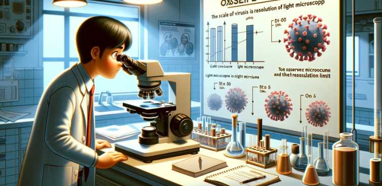
No, a light microscope cannot typically see viruses due to their small size, which is below the resolution limit of light microscopy.
Light microscopes use visible light to magnify and visualize specimens. However, they have a resolution limit imposed by the wavelength of light, making it challenging to observe objects smaller than the wavelength of light itself. Viruses are generally much smaller than the wavelength of visible light, making them invisible under a standard light microscope.
| Microscope Type | Light Microscope | Electron Microscope |
|---|---|---|
| Resolution Limit | Limited by the wavelength of visible light (around 200 nanometers) | Electron beams have much shorter wavelengths (0.005 nanometers or less) |
| Viruses Visibility | Typically cannot see viruses due to their small size | Electron microscopes can visualize viruses and other subcellular structures |
Overview Of Light Microscopes
Light microscopes, or optical or compound microscopes, are widely used in various scientific fields to observe small objects or organisms not visible to the naked eye. These microscopes use visible light to illuminate the specimen, allowing for detailed observation and analysis. Here are the key components and features of light microscopes:
-
Objective Lenses:
- Light microscopes have multiple objective lenses with different magnification levels. Users can switch between these lenses to achieve varying levels of magnification.
-
Eyepiece (Ocular Lens):
- The eyepiece further magnifies the image produced by the objective lens. Typically, it provides additional magnification, such as 10x.
-
Stage and Stage Clips:
- The stage is where the specimen is placed for observation. Stage clips secure the specimen in place.
-
Illumination Source:
- Light microscopes use a light source, often located beneath the stage, to illuminate the specimen. This transmitted light passes through the specimen for observation.
-
Condenser Lens:
- The condenser lens focuses the light onto the specimen, enhancing contrast and brightness.
-
Diaphragm:
- The diaphragm controls the amount of light reaching the specimen. Adjusting the diaphragm helps optimize the image quality.
-
Coarse and Fine Adjustment Knobs:
- These knobs enable precise focusing. The coarse adjustment is used for initial focusing, while the fine adjustment provides finer focus.
-
Body Tube:
- The body tube holds the eyepiece and connects it to the objective lenses, maintaining the proper distance between them.
-
Base and Arm:
- The base supports the microscope, while the arm connects the base to the body tube. These components provide stability and support.
Table: Components of a Light Microscope
| Component | Function |
|---|---|
| Objective Lenses | Provide various levels of magnification |
| Eyepiece (Ocular Lens) | Further magnifies the image produced by the objective lens |
| Stage and Stage Clips | Platform for placing and securing the specimen |
| Illumination Source | Provides light to illuminate the specimen |
| Condenser Lens | Focuses light onto the specimen for enhanced contrast |
| Diaphragm | Controls the amount of light reaching the specimen |
| Coarse and Fine Adjustment Knobs | Enable focusing for optimal image quality |
| Body Tube | Holds the eyepiece and connects it to the objective lenses |
| Base and Arm | Provide stability and support for the microscope |
Characteristics Of Viruses
| Characteristic | Description |
|---|---|
| 1. Non-cellular | Lack cellular structure |
| 2. Genetic Material | Contains DNA or RNA |
| 3. Host Dependency | Relies on host cells for replication and processes |
| 4. Lack Metabolic Machinery | No cellular machinery for metabolic activities |
| 5. Inert Outside Host | Inactive outside a host |
| 6. Structural Diversity | Varied shapes, including helical, icosahedral, and complex |
| 7. Protein Coat (Capsid) | Capsid surrounds genetic material |
| 8. Envelope Presence | Some viruses have a lipid envelope derived from host cell |
| 9. Specific Host Range | Shows specificity in infecting particular hosts |
| 10. Infection Mechanism | Uses host cell machinery for replication |
Advancements In Microscopy Techniques
Microscopy techniques have evolved significantly, offering unprecedented insights into the world of the small. Super-resolution microscopy, such as STED, surpasses the diffraction limit, providing detailed images at the nanoscale. Cryo-EM preserves specimens in a frozen state, allowing for high-resolution imaging of delicate biological structures. Light sheet microscopy reduces phototoxicity and enables 3D imaging by illuminating thin sections of a specimen. Single-molecule microscopy allows the visualization of individual molecules, aiding in the study of molecular dynamics. Correlative microscopy integrates various techniques for a comprehensive analysis of specimens.
Atomic force microscopy (AFM) probes surfaces at the atomic level, offering high-resolution imaging of non-conductive samples. Multi-photon microscopy enhances tissue penetration by exciting fluorophores with longer-wavelength photons. Digital holographic microscopy records holograms for 3D imaging without traditional lenses. Expansion microscopy swells biological samples, improving resolution for detailed imaging.
| Microscopy Technique | Description |
|---|---|
| Super-Resolution Microscopy | Overcomes diffraction limits, enabling nanoscale imaging. |
| Cryo-Electron Microscopy | Preserves specimens in a frozen state for high-resolution imaging of biological structures. |
| Light Sheet Microscopy | Illuminates thin sections, reducing phototoxicity and enabling 3D imaging. |
| Single-Molecule Microscopy | Visualizes individual molecules, facilitating the study of molecular dynamics. |
| Correlative Microscopy | Integrates multiple techniques for comprehensive specimen analysis. |
| Stimulated Emission Depletion | Uses focused laser beams to enhance resolution beyond traditional limits. |
| Atomic Force Microscopy | Probes surfaces at the atomic level, allowing high-resolution imaging of non-conductive samples. |
| Multi-Photon Microscopy | Excites fluorophores with longer-wavelength photons, enabling deeper tissue penetration. |
| Digital Holographic Microscopy | Records holograms for 3D imaging without traditional lenses. |
| Expansion Microscopy | Swells biological samples, enhancing resolution for detailed imaging. |
Current Research And Findings
Recent advancements in imaging technology have enabled researchers to gain unprecedented insights into the interactions between viruses and host cells. Advanced microscopy techniques have allowed scientists to visually capture the behavior of viruses at a level of detail that was previously unattainable. These technological breakthroughs have significantly impacted virus research, providing new opportunities to study viral replication, infection mechanisms, and the host immune response.
Researchers can now visualize the intricate interactions between viruses and host cells, shedding light on the molecular and cellular processes involved. The ability to observe viral behavior in real time has not only deepened our understanding of viral pathogenesis, but has also paved the way for the development of novel antiviral therapies. By leveraging advanced imaging, scientists are gradually unraveling the complex dynamics of viral infections, offering valuable insights that may ultimately lead to the discovery of more effective treatments.
Challenges And Future Perspectives
The ability of light microscopes to visualize viruses has long been a challenge due to the limitations imposed by the wavelengths of visible light. However, recent advancements have provided hope for overcoming these barriers and expanding our understanding of viruses through light microscopy techniques.
Overcoming the limitations of light microscopy involves innovative approaches such as enhancing resolution and sensitivity. One potential breakthrough lies in the use of super-resolution microscopy techniques, such as stimulated emission depletion (STED) microscopy and structured illumination microscopy (SIM). These techniques allow for visualization of viral structures at a higher level of detail.
Another approach involves the incorporation of fluorescent labeling techniques to specifically target viral components, enabling their visualization within infected cells. This technique, known as immunofluorescence, allows researchers to track and study the behavior of viruses in real-time.
| Breakthrough | Description |
|---|---|
| Enhanced Resolution Microscopy | Super-resolution microscopy techniques, such as STED and SIM, enable visualization of viral structures with higher detail. |
| Fluorescent Labeling | Immunofluorescence allows for targeted visualization of viral components within infected cells, facilitating real-time tracking and study of virus behavior. |
These advancements in light microscopy have the potential to revolutionize our understanding of viruses and improve our ability to combat viral diseases. By overcoming the limitations of traditional light microscopy, researchers can delve deeper into the complex world of viruses and uncover new insights that may pave the way for innovative treatments and preventative measures.
Can a standard light microscope detect viruses?
Using a standard light microscope, it is challenging to directly visualize viruses due to their small size. Most viruses fall below the resolution limit of light microscopes, which typically ranges from 200 to 300 nanometers. To put this into perspective, many viruses are much smaller, making them difficult to observe using conventional light microscopy.
| Microscope Type | Resolution Limit |
|---|---|
| Light Microscope | 200-300 nm |
| Virus Size | < 200 nm |
Are there specific techniques to enhance virus visibility under a light microscope?
Yes, there are specialized staining techniques that can improve the contrast and visibility of viruses under a light microscope. By using dyes or fluorescent tags that selectively bind to viral components, researchers can enhance the details and make viruses more discernible. However, even with these techniques, there are limits to the size of viruses that can be effectively visualized.
| Technique | Purpose |
|---|---|
| Staining | Enhances contrast for better visibility |
| Fluorescent Labeling | Attaches fluorescent tags for detection |
What is the smallest virus observable with a light microscope?
The smallest viruses that can be observed using a light microscope are typically in the range of 50-100 nanometers. However, this requires advanced microscopy techniques and often the use of contrast-enhancing methods such as electron microscopy for better clarity.
| Minimum Observable Size |
|---|
| 50-100 nm |
How does a light microscope compare to an electron microscope for virus observation?
Light microscopes use visible light to illuminate specimens, while electron microscopes use electron beams. Electron microscopes have much higher resolution capabilities, allowing the observation of smaller structures, including viruses. While light microscopes have their limitations, electron microscopes are more suitable for studying viruses at the molecular level.
| Microscope Type | Resolution Limit |
|---|---|
| Light Microscope | 200-300 nm |
| Electron Microscope | Sub-nanometer resolution |
Can light microscopes be used for preliminary virus identification?
Light microscopes can play a role in preliminary virus identification, especially in clinical settings. While they may not provide detailed views of viral structures, they can aid in the observation of infected cells and help identify characteristic changes associated with viral infections. However, for precise virus characterization, advanced techniques like PCR and electron microscopy are often necessary.
| Role in Identification | Limitations |
|---|---|
| Preliminary Observation | Limited structural details of the virus |
| Identification Aid | Supplementary to advanced characterization |
What are the challenges of using a light microscope to study viruses?
The main challenges include the limited resolution of light microscopes, making it difficult to visualize viruses below a certain size threshold. Additionally, viruses often require specific staining or labeling techniques for improved visibility, and even with these methods, detailed structural information may remain elusive.
| Challenges | Mitigation Strategies |
|---|---|
| Limited Resolution | Complementary use of electron microscopy |
| Staining Requirement | Utilization of advanced staining and labeling techniques |
| Incomplete Structural Information | Integration with other characterization methods |
Conclusion
The capabilities of a light microscope in detecting viruses are limited due to their small size. While it can capture larger structures and some cells, viruses often fall below the microscope’s resolution limits. To visualize viruses, more advanced tools like electron microscopes are necessary for accurate observation and study.
Books and Textbooks:
- Explore textbooks on advanced microscopy techniques, such as “Handbook of Biological Confocal Microscopy” edited by James Pawley or “Principles of Fluorescence Spectroscopy” by Joseph R. Lakowicz.

Fahim Foysal is a well-known expert in the field of binoculars, with a passion for exploring the great outdoors and observing nature up close. With years of experience in the field, Fahim has honed his skills as a binocular user and has become a go-to resource for those seeking advice on choosing the right binoculars for their needs.
Fahim’s love for the natural world began during his time at The Millennium Stars School and College and BIAM Laboratory School, where he spent much of his free time exploring the outdoors and observing the wildlife around him. This passion for nature led him to pursue a degree in Fine Arts from the University of Dhaka, where he gained a deep understanding of the importance of observation and attention to detail.
Throughout his career, Fahim has used his expertise in binoculars to help others discover the beauty of the natural world. His extensive knowledge of binocular technology and optics has made him a trusted advisor for amateur and professional wildlife observers alike. Whether you’re looking to spot rare birds or observe animals in their natural habitats, Fahim can help you choose the perfect binoculars for your needs. With his guidance, you’ll be able to explore the outdoors with a newfound appreciation for the beauty of the natural world.
Table of Contents
Pingback: Through the Lens of Progress: Advancing Scientific Frontiers with Laboratory Microscopes
Pingback: Are Chloroplasts Visible Under a Light Microscope? Unveiled Secrets!