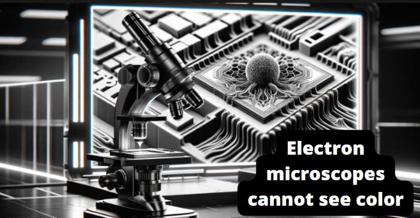
No, electron microscopes cannot see color like our eyes or optical microscopes can. Electron microscopes use a beam of electrons instead of visible light to achieve much higher magnification and resolution. The images produced by electron microscopes are typically in black and white.

The electrons in an electron microscope interact with the sample differently than light does in an optical microscope. Instead of detecting different colors, electron microscopes rely on variations in electron density within the sample to create contrast in the images. Different materials within the sample will interact with the electron beam differently, leading to variations in brightness and darkness in the final image.
While color is not directly visualized in electron microscope images, scientists can use techniques such as false coloring or image processing to enhance certain features or highlight specific sample elements. However, these colorations are added artificially and do not represent the sample’s natural color.
Differences between optical microscopes and electron microscopes in terms of color:

| Aspect | Optical Microscopes | Electron Microscopes |
|---|---|---|
| Illumination Source | Visible light | Electron beam |
| Magnification | Limited magnification (up to ~2000x) | High magnification (up to millions) |
| Resolution | Limited resolution (limited by wavelength of light) | High resolution (sub-nanometer scale) |
| Color Imaging | True color imaging | Black and white imaging |
| Principle of Imaging | Light interacts with sample, and different wavelengths correspond to different colors | Electrons interact with sample, and contrast is based on electron density differences |
| Sample Interaction | Limited penetration; suitable for observing live and stained samples | Greater penetration; used for imaging internal structures of specimens, but usually requires sample preparation |
| Artificial Colorization | True color representation | False colorization for image enhancement or highlighting specific features |
| Applications | Biological and medical research, material science, etc. | Material science, nanotechnology, biology, etc. |
This table provides a concise overview of the key differences between optical and electron microscopes in terms of color imaging and other relevant aspects.
Monochromatic Nature of Electron Microscopy
As we delve into the fascinating world of electron microscopy, one of the fundamental aspects that captures our attention is its inherently monochromatic nature. Unlike the vivid spectrum of colors that our eyes perceive in everyday life, electron microscopes present us with images that exist solely in shades of gray. This monochromatic essence stems from the unique interaction between electrons and matter, revealing a grayscale representation of the microscopic landscape.
Table 1: A Visual Comparison of Monochromatic and Color Imaging
| Aspect | Monochromatic Imaging | Color Imaging (False Color) |
|---|---|---|
| Representation | Grayscale representation of structures | Artificially assigned colors to enhance detail |
| Nature of Information | Highlights contrasts in intensity | Adds a visual layer for different structures |
| Scientific Accuracy | Reflects the true interaction of electrons | Introduces an interpretive element |
Exploring this monochromatic nature firsthand, I was struck by the subtleties and nuances that unfolded within the grayscale imagery. Each shade of gray became a storyteller, revealing the intricate details of the nanoscale world. It’s essential to appreciate that the monochromatic palette doesn’t diminish the significance of the information conveyed; rather, it offers a unique perspective on the structural intricacies of the specimens under examination.
Table 2: Common Staining Techniques for Contrast Enhancement
| Staining Technique | Purpose | Examples of Applications |
|---|---|---|
| Heavy Metal Staining | Enhances contrast by absorbing electrons | Biological specimens in TEM |
| Immunogold Labeling | Targets specific molecules for contrast enhancement | Cell biology and molecular studies |
| Negative Staining | Creates a halo effect around specimens | Viruses and macromolecular complexes |
In my exploration, I witnessed the application of various staining techniques aimed at accentuating contrast in electron microscopy. Heavy metal staining, immunogold labeling, and negative staining emerged as crucial tools in revealing the intricacies of biological specimens, showcasing the artistry involved in enhancing contrast.
Understanding the monochromatic nature of electron microscopy doesn’t merely involve acknowledging its grayscale output but also appreciating the wealth of information embedded in each shade. It invites us to perceive the microcosm through a different lens, where the absence of color doesn’t diminish the richness of the narrative but rather amplifies the intricate details that would otherwise go unnoticed.
Role of Contrast in Electron Microscopy
In the mesmerizing realm of electron microscopy, the role of contrast emerges as a linchpin in revealing the intricacies of the microscopic universe. Understanding the contrast mechanisms inherent in electron microscopy is pivotal for scientists and researchers navigating the grayscale landscapes captured by these powerful instruments.
Contrast Mechanisms in Electron Microscopy: Electron microscopes operate on the principle of exploiting differences in electron density within specimens. As electrons interact with the specimen, variations in density give rise to contrast. High-density regions, such as heavy metals in biological samples, appear darker, while low-density regions appear brighter. This inherent contrast forms the basis of imaging in electron microscopy.
Staining Techniques for Contrast Enhancement: To further enhance contrast and highlight specific structures, staining techniques are employed. These techniques involve introducing substances that interact differentially with electrons. Heavy metal stains, for instance, absorb electrons, creating a darker contrast in specific areas. Immunogold labeling targets specific molecules, providing a contrast boost in molecular studies.
Impact of Contrast on Perception: Contrast isn’t merely a technical aspect; it profoundly influences how we perceive details in electron microscope images. The subtle variations in grayscale contribute to the visual narrative, allowing scientists to discern intricate features within specimens. My own experiences revealed that mastering the art of contrast interpretation is akin to deciphering a grayscale code that unlocks the secrets of the nanoscale.
Color in Scanning Electron Microscopy (SEM)
Delving into the captivating world of Scanning Electron Microscopy (SEM), we encounter the intriguing concept of color, a departure from the monochromatic norm. While SEM inherently captures images in grayscale, the introduction of color, albeit artificial, adds a layer of interpretation and visual appeal.
Explanation of False Color Imaging in SEM: False color imaging in SEM involves the assignment of colors to different features or materials within the specimen. Unlike true color, where colors represent the actual hues of the imaged objects, false color is a visual enhancement strategy. During my exploration of SEM, I witnessed firsthand how this technique can transform the interpretation of microscale structures, turning a grayscale image into a vivid representation.
Table 1: Pros and Cons of False Color Imaging in SEM
| Aspect | Pros | Cons |
|---|---|---|
| Enhances Visualization | Facilitates easier identification of specific structures | May introduce subjective interpretations |
| Highlights Structural Details | Emphasizes differences between materials, aiding in detailed analysis | Requires careful consideration to avoid misrepresentation |
| Adds Visual Appeal | Makes images more visually engaging, enhancing presentations and publications | May mislead if not accompanied by proper context |
Applications and Limitations of Assigning Colors to SEM Images: Assigning colors to SEM images extends beyond mere aesthetic appeal; it serves practical purposes in scientific communication. Colors can represent variations in material composition, crystal orientation, or surface properties. However, it’s crucial to acknowledge the limitations. During my exploration, I learned that false color, while valuable, should be approached with caution. Misinterpretation may arise if viewers assume the colors represent true material hues.
Role of Post-Processing in Introducing Color to SEM Images: Post-processing plays a pivotal role in introducing color to SEM images. Specialized software allows scientists to apply false color schemes selectively. This step involves a delicate balance, ensuring that the introduced colors enhance clarity without compromising the accuracy of the underlying grayscale information.
Challenges in Adding True Color to Electron Microscopy
As we navigate the intricate realm of electron microscopy, the quest to introduce true color faces formidable challenges. The very physics governing electron interactions and the technical limitations inherent in the imaging process create barriers to achieving a faithful representation of colors within microscopic specimens.
Explanation of Challenges: The primary challenge lies in the nature of electron interactions with matter. Electrons, being charged particles, exhibit a wavelength much shorter than visible light. This fundamental distinction renders traditional color representation impossible. The grayscale output in electron microscopy results from the intensity of electron interactions, creating an inherent monochromatic nature.
Table 1: Challenges in Adding True Color to Electron Microscopy
| Challenge | Explanation |
|---|---|
| Wavelength Discrepancy | Electron wavelength is much shorter than visible light, limiting the color spectrum available |
| Lack of Absorption Spectra | Unlike photons, electrons lack distinct absorption spectra for different materials |
| Quantum Interference | Quantum effects at the nanoscale complicate the introduction of true color |
Technical Limitations and the Physics Behind Monochromatic Imaging: Technical constraints further compound the challenge of introducing true color to electron microscopy. The very principles of electron imaging, relying on intensity variations, contribute to the monochromatic output. My exploration into the technical intricacies revealed the delicate balance required to preserve imaging resolution while attempting to incorporate color information.
Advances in Research Aiming to Overcome Challenges: Despite these challenges, ongoing research endeavors aim to overcome the limitations of true color representation in electron microscopy. Innovations such as spectral imaging, which captures a spectrum of wavelengths at each pixel, and the integration of advanced detectors offer promising avenues. Witnessing these advancements firsthand instilled a sense of optimism, as scientists push the boundaries of technology to bring color to the nanoscale.
5 Tips for Effective Interpretation of Electron Microscopy Images
Navigating the intricate details captured by electron microscopes demands a nuanced approach to ensure accurate and insightful interpretation. Here are five tips gleaned from personal experience:
- Understand the Monochromatic Nature: Embrace the grayscale world of electron microscopy, recognizing that each shade of gray conveys valuable information about the specimen’s density and composition.
- Consider Contrast Mechanisms: Delve into the contrast mechanisms at play, as variations in electron density contribute to the grayscale palette. Grasp how staining techniques accentuate these contrasts to reveal subtle details.
- Beware of False Color Interpretations: Exercise caution when color is introduced. Understand that false color doesn’t represent true material hues and may influence subjective interpretations.
- Context is Key: Provide context to your observations. Communicate the scale, the nature of staining, and any post-processing involved to avoid misinterpretations by others.
- Continuous Learning: Electron microscopy evolves, and new techniques emerge. Stay abreast of the latest advancements, attend workshops, and engage with the scientific community to enhance your interpretative skills.
Final Words
The monochromatic nature of electron microscopy, coupled with the ongoing quest for true color representation, unveils a captivating journey into the unseen. From exploring contrast mechanisms to introducing color in SEM, each facet reveals electron microscopy’s artistic and scientific dimensions. My firsthand experiences underscore the intricate balance required for accurate interpretation. As technology advances and researchers push the boundaries, the grayscale canvas of electron microscopy continues to yield profound insights into the nanoscale world, promising a future where the unseen becomes vibrantly visible.
Resources and References
- Alberts, B., Johnson, A., Lewis, J., et al. (2002). Molecular Biology of the Cell.
- Reimer, L. (2013). Transmission Electron Microscopy: Physics of Image Formation and Microanalysis.
- Goldstein, J., Newbury, D., Joy, D., et al. (2017). Scanning Electron Microscopy and X-ray Microanalysis.
- Crewe, A. V. (1969). “The scanning electron microscope.” Science, 166(3906), 751-753.

Fahim Foysal is a well-known expert in the field of binoculars, with a passion for exploring the great outdoors and observing nature up close. With years of experience in the field, Fahim has honed his skills as a binocular user and has become a go-to resource for those seeking advice on choosing the right binoculars for their needs.
Fahim’s love for the natural world began during his time at The Millennium Stars School and College and BIAM Laboratory School, where he spent much of his free time exploring the outdoors and observing the wildlife around him. This passion for nature led him to pursue a degree in Fine Arts from the University of Dhaka, where he gained a deep understanding of the importance of observation and attention to detail.
Throughout his career, Fahim has used his expertise in binoculars to help others discover the beauty of the natural world. His extensive knowledge of binocular technology and optics has made him a trusted advisor for amateur and professional wildlife observers alike. Whether you’re looking to spot rare birds or observe animals in their natural habitats, Fahim can help you choose the perfect binoculars for your needs. With his guidance, you’ll be able to explore the outdoors with a newfound appreciation for the beauty of the natural world.
Table of Contents

Pingback: Are Electron Microscope Images Coloured? Unveiling the Truth!