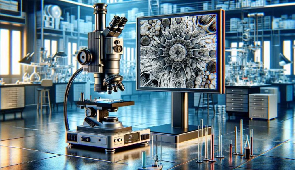
No, electron microscope images are not inherently colored. By default, these images are captured in black and white, reflecting the monochromatic nature of the electron signals used in imaging. The grayscale representation offers high contrast and detailed views of microscopic structures. However, scientists often apply color to these images through pseudocoloring techniques for specific purposes. Pseudocoloring helps highlight distinct features or materials within the specimen, aiding in the interpretation and communication of scientific findings. The choice of colors is strategic and does not signify inherent colors present in the specimens. While artistic renderings and false coloration techniques exist, the core of electron microscope imaging lies in its ability to provide precise, high-resolution, and uncolored representations of the microscopic world.
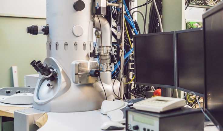
The Basics of Electron Microscopy
Delving into the microscopic universe, electron microscopy serves as a transformative tool, revealing intricacies beyond the reach of traditional optical microscopes. Understanding the fundamentals of electron microscopy is essential to appreciate its capabilities and applications.
Types of Electron Microscopes
Electron microscopes come in diverse types, each tailored to specific scientific needs. The three primary types are Transmission Electron Microscope (TEM), Scanning Electron Microscope (SEM), and Scanning Transmission Electron Microscope (STEM). Each type offers unique advantages, allowing scientists to explore different aspects of specimens in unprecedented detail.
Types of Electron Microscopes
| Electron Microscope Type | Characteristics |
|---|---|
| Transmission Electron Microscope (TEM) | Penetrates thin specimens for detailed internal structures |
| Scanning Electron Microscope (SEM) | Provides 3D surface images with excellent depth of field |
| Scanning Transmission Electron Microscope (STEM) | Combines features of TEM and SEM for versatility |
How Electron Microscopes Work

- Electron Beam Generation:
- Electron microscopes use electron guns to generate focused beams of electrons.
- Magnetic lenses focus these beams onto the specimen.
- Interaction with Specimens:
- When the electron beam strikes the specimen, interactions with atoms generate signals.
- Detection of Signals:
- Detectors capture signals like secondary electrons, backscattered electrons, and transmitted electrons.
- Image Formation:
- Captured signals are translated into high-resolution images, providing detailed views of specimen structures.
Characteristics of Electron Microscope Images

Characteristics of Electron Microscope Images
| Aspect | Description |
|---|---|
| High Resolution | Exceptional detail and clarity |
| Monochromatic Nature | Native images are black and white |
| Depth of Field | SEM provides excellent depth perception |
| Internal Structures | TEM reveals internal structures in fine detail |
Electron microscope images stand out for their high resolution, monochromatic default state, and the ability to unveil internal structures with precision.
Advantages and Limitations
- Advantages:
- High resolution for detailed imaging.
- Capability to explore internal structures.
- SEM excels in revealing fine surface details.
- Limitations:
- Meticulous sample preparation is essential.
- Vacuum conditions are required.
- Artefacts may be introduced during sample processing.
Innovations in Electron Microscopy
Continual innovations enhance electron microscopy’s capabilities:
- Cryo-Electron Microscopy (Cryo-EM):
- Allows imaging of biological specimens in their native state.
- Minimizes damage from sample preparation.
- Correlative Light and Electron Microscopy (CLEM):
- Integrates data from light and electron microscopy for comprehensive analysis.
- Bridges information across macro and micro scales.
Understanding these basics sets the stage for exploring the colorful and captivating world of electron microscope images, debunking misconceptions and appreciating the intersection of science and art at the microscopic level.
Black and White: The Native State of Electron Microscope Images
Contrary to popular belief, the native state of electron microscope images is monochromatic, devoid of the vibrant hues that often characterize other imaging techniques. Electron microscopy relies on the interaction of electron beams with specimens and the subsequent capture of signals emitted during this interaction. The result is a raw, high-resolution image that is inherently black and white.
Characteristics of Native Electron Microscope Images
| Aspect | Description |
|---|---|
| Monochromatic Nature | Lack of inherent color in native images |
| Electron Signal Interaction | Detectors capturing electron signals |
| Raw Representation | Unaltered depiction of specimen structures |
The monochromatic nature of these images arises from the fundamental principles of electron microscopy. Electron beams, focused onto the specimen, interact with the atoms present. Detectors capture the signals generated by this interaction, forming an image that faithfully represents the structural details without the introduction of artificial colors.
These native black and white images offer a stark beauty, emphasizing the intricate details of the microscopic world. The absence of color allows scientists to focus on the structural information, discerning the fine nuances within cells, materials, or any specimen under examination.
Appreciating the native state of electron microscope images is crucial for accurate interpretation and communication of scientific findings. While the absence of color might seem surprising to those new to the field, it underscores the precision and objectivity inherent in electron microscopy, where the focus is on revealing the true nature of microscopic structures. As we delve deeper into the world of electron microscopy, it becomes evident that the beauty lies not in the colors that adorn the images but in the rich tapestry of details that monochromatic images unveil.
Pseudocoloring in Electron Microscopy
In the realm of electron microscopy, the quest to unveil intricate details often extends beyond the limitations of black and white images. Pseudocoloring emerges as a powerful technique, enabling scientists to enhance visibility, highlight specific features, and convey additional information in electron microscope images.
Pseudocoloring involves the deliberate assignment of colors to different parts of an image, creating a visually enriched representation without altering the inherent grayscale nature of electron microscope images. This technique is particularly valuable when researchers seek to emphasize specific elements within a specimen.
The application of pseudocoloring is not arbitrary; instead, it adheres to scientific principles. By assigning distinct colors to different structures or materials, scientists can create a more intuitive visual interpretation of the microscopic world. For instance, in a biological sample, cell membranes may be pseudocolored in one hue, while organelles or other structures are represented in contrasting colors.
Pseudocoloring Techniques
| Technique | Purpose |
|---|---|
| Heatmap Pseudocoloring | Emphasizes variations in temperature or intensity |
| Rainbow Pseudocoloring | Enhances contrast by assigning colors across the spectrum |
| Grayscale with Color Highlights | Retains black and white base while accentuating specific details |
Heatmap pseudocoloring is often employed to visualize variations in temperature or intensity within a specimen, providing a gradient of colors that intuitively represent different levels. Rainbow pseudocoloring, on the other hand, enhances contrast by assigning colors across the visible spectrum, making subtle differences more apparent.
Pseudocoloring is not limited to scientific utility; it also serves an essential communicative role. By introducing colors, researchers can create images that are not only scientifically informative but also visually engaging. This aids in effective communication of complex findings to diverse audiences, from fellow scientists to students and the general public.
While pseudocoloring undeniably enhances the interpretability of electron microscope images, researchers must use it judiciously. The colors assigned should accurately reflect the scientific context, ensuring that the visual representation aligns with the underlying data. In navigating the microscopic landscape with pseudocoloring, scientists embark on a journey where art and science converge, bringing forth a richer understanding of the hidden world magnified by electron microscopy.
False Coloration and Artistic Rendering in Electron Microscopy
Beyond the realm of pseudocoloring, electron microscopy offers another layer of visual complexity through techniques such as false coloration and artistic rendering. These approaches elevate scientific images beyond mere representations, transforming them into captivating works that marry the precision of science with the aesthetics of art.
False Coloration:
False coloration involves the intentional application of colors to an image, departing from the natural grayscale representation. Unlike pseudocoloring, where colors are assigned based on scientific principles, false coloration allows for more subjective choices. This technique is commonly employed to emphasize specific details or highlight different elements within a specimen.
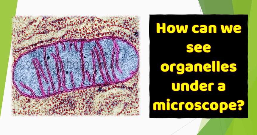
False coloration often serves to enhance contrast and clarity, making subtle structures more discernible to the human eye. For example, in biological samples, false coloration may be used to distinguish between different cell types or to highlight areas of interest such as cellular organelles.
Artistic Rendering:
Artistic rendering takes electron microscope images to a different realm, where scientific visualization meets creative expression. While the primary goal of scientific imaging is to accurately represent the specimen, artistic rendering allows for a departure from strict realism. This technique involves applying artistic elements such as shading, texture, and even unconventional colors to create visually striking representations.
Examples of Artistic Rendering:
- Neuronal Connections:
- False coloration is used to distinguish between neural pathways, creating a visually appealing representation of the intricate connections within the brain.
- Cellular Landscape:
- Artistic rendering transforms a typical cellular landscape into a visually engaging masterpiece, where scientific accuracy converges with creative expression.
Artistic rendering not only produces aesthetically pleasing images but also invites viewers to appreciate the beauty inherent in the microscopic world. It bridges the gap between science and art, making complex scientific concepts more accessible to a broader audience.
Both false coloration and artistic rendering contribute to the narrative of scientific discovery. They go beyond the mere documentation of structures and delve into the realm of visual storytelling. These techniques play a crucial role in communicating the wonder of the microscopic universe, fostering a deeper appreciation for the beauty concealed within the seemingly mundane world magnified by electron microscopy.
Factors Influencing Coloration in Electron Microscopy
The introduction of color to electron microscope images is a deliberate process influenced by various factors. Understanding these factors is crucial for accurately interpreting the visual information conveyed through the vibrant hues that adorn the microscopic world.
Specimen Composition:
The materials constituting the specimen play a pivotal role in influencing coloration. Different substances interact with electrons in distinctive ways, leading to variations in the colors observed. For instance, biological specimens may exhibit different colors based on the composition of cellular structures or the presence of specific molecules.
Imaging Techniques:
The choice of imaging techniques in electron microscopy can significantly impact color representation. Different techniques, such as transmission electron microscopy (TEM) and scanning electron microscopy (SEM), may produce varied color schemes. The interaction of electrons with specimens and the subsequent detection methods contribute to the diversity of colors observed in the final images.
Post-processing:
The coloration of electron microscope images is not solely determined during the imaging process but can be further modified during post-processing. Adjustments in contrast, brightness, and color balance can influence the final appearance of the image. Researchers often fine-tune these parameters to enhance the visual clarity and emphasize specific features within the specimen.
Factors Influencing Coloration
| Factor | Influence on Color Representation |
|---|---|
| Specimen Composition | Different materials may exhibit distinct colors |
| Imaging Techniques | Varied techniques may result in different color schemes |
| Post-processing | Adjustment of contrast and brightness impacts color |
Navigating the intricacies of coloration in electron microscopy requires a nuanced understanding of these factors. The interplay between specimen composition, imaging techniques, and post-processing intricacies contributes to the rich palette of colors that brings the microscopic world to life. As scientists continue to refine their methods, the resulting images become not only scientific tools but also visual masterpieces that capture the essence of the microscopic realm in vibrant detail.
Common Misconceptions about Color in Electron Microscopy
Misconceptions often surround the representation of color in electron microscope images, stemming from assumptions and unfamiliarity with the underlying principles. Let’s address and debunk some of these common misconceptions:
Debunked Misconceptions
| Misconception | Clarification |
|---|---|
| Inherent Color in Electron Images | Electron microscope images are naturally black and white |
| Unrestricted Pseudocoloring | Pseudocoloring is deliberate and guided by scientific principles |
- Inherent Color in Electron Images:
- Misconception: Assuming that electron microscope images inherently possess color.
- Clarification: Native electron microscope images are monochromatic, capturing the grayscale nuances of specimen structures. Color is introduced through deliberate techniques.
- Unrestricted Pseudocoloring:
- Misconception: Believing that pseudocoloring involves arbitrary assignment of colors.
- Clarification: Pseudocoloring follows scientific principles, with colors assigned purposefully to enhance visibility and convey specific information without distorting the underlying data.
Understanding these misconceptions is essential for appreciating the accuracy and intentionality involved in the coloration of electron microscope images. By dispelling these myths, we foster a more informed interpretation of the mesmerizing visuals that emerge from the microscopic world.
6 Tips for Interpreting Electron Microscope Images
Interpreting electron microscope images requires a nuanced approach to fully grasp the intricate details captured at the microscopic level. Here are six tips to enhance your understanding:
- Recognize Pseudocoloring:
- Be aware that colors introduced to electron microscope images through pseudocoloring are intentional and serve a specific purpose. Understanding this technique aids in accurate interpretation.
- Understand the Native State of Images:
- Acknowledge that native electron microscope images are monochromatic. This awareness helps in distinguishing between inherent features and those highlighted through coloration techniques.
- Context Matters:
- Consider the scientific context in which the images are presented. Colors may be used to emphasize specific structures or materials, providing vital clues to the researcher’s intended focus.
- Be Mindful of Artistic Rendering:
- Understand the distinction between scientifically accurate representation and artistic rendering. While the former is based on data, the latter introduces creative elements for visual appeal.
- Consider Specimen Composition:
- Different materials within a specimen may naturally exhibit distinct colors. Take into account the inherent properties of the materials being imaged for a more accurate interpretation.
- Stay Informed About Imaging Techniques:
- Different electron microscopy techniques may produce varied color schemes. Familiarize yourself with the imaging method used to capture the images, as this influences the visual outcome.
3 Major Challenges in Electron Microscopy Imaging
While electron microscopy has revolutionized our ability to visualize the microscopic world, it is not without its challenges. Three major hurdles persist in the field:
- Color Representation:
- The debate over standardized colorization in scientific imaging remains ongoing. Establishing consistent color representation is challenging, as different labs may employ varied methods, impacting the reproducibility and comparability of results.
- Technological Advancements:
- Despite continuous innovations, electron microscopy faces technological limitations. Improving spatial resolution, reducing specimen damage during imaging, and enhancing imaging speed are ongoing challenges that researchers strive to overcome.
- Standardization in Electron Microscopy:
- The field lacks standardized protocols for colorization and imaging procedures. The absence of uniform standards hinders collaboration and makes it challenging to establish a universal language for interpreting electron microscope images. Efforts to standardize practices are underway but remain a complex endeavor.
Do Electron Microscope Images Have Inherent Color?
Electron microscope images are inherently monochromatic. The default representation is black and white, devoid of inherent color. This monochromatic nature arises from the fundamental principles of electron microscopy, where electron beams interact with specimens to create high-resolution images captured in grayscale.
How are Colors Introduced to Electron Microscope Images?
Colors are introduced through deliberate processes such as pseudocoloring and false coloration. Pseudocoloring involves assigning colors to specific features, enhancing visibility without altering the native grayscale. False coloration, on the other hand, allows for more subjective color choices, often used to highlight structures or create visually engaging representations.
Is Pseudocoloring Arbitrary, or Does it Follow Scientific Principles?
Pseudocoloring is not arbitrary; it follows scientific principles. Colors are assigned purposefully to enhance the interpretation of specific features within a specimen. This intentional approach ensures that the coloration aligns with the scientific context and aids accurate interpretation.
Can Electron Microscope Images Be in True Color?
True color, as perceived by the human eye, is not present in electron microscope images. These images are formed by capturing electron signals, resulting in a monochromatic representation. The introduction of color is a deliberate and strategic process aimed at enhancing scientific interpretation.
What is the Difference Between Pseudocoloring and False Coloration?
Pseudocoloring and false coloration both involve introducing color to electron microscope images, but they differ in their approach. Pseudocoloring follows scientific principles, assigning colors deliberately, while false coloration allows for more subjective and artistic color choices to enhance contrast and visual appeal.
Are There Different Techniques for Pseudocoloring?
Various techniques are employed for pseudocoloring in electron microscopy. Heatmap pseudocoloring emphasizes variations in temperature or intensity. Rainbow pseudocoloring enhances contrast with a spectrum of colors, and grayscale with color highlights retains a black and white base while accentuating specific details. These techniques cater to different imaging needs and objectives.
Why Use Coloration in Electron Microscopy?
Coloration in electron microscopy serves multiple purposes. It enhances visibility, making specific features more discernible. It allows for the conveyance of additional information, aiding in the interpretation of complex structures. Moreover, coloration adds a visual dimension to microscopic images, making them more accessible and engaging for a broader audience.
What is the Role of Artistic Rendering in Electron Microscopy?
Artistic rendering in electron microscopy involves creative embellishments to scientific images. While scientific accuracy remains crucial, artistic rendering adds aesthetic elements, such as shading and texture, contributing to visually striking representations. This approach bridges the gap between science and art, making microscopic imagery more captivating.
Are Electron Microscope Images Ever in True Color?
True color, as perceived by the human eye, is not inherent in electron microscope images. The introduction of color is a deliberate process, whether through pseudocoloring, false coloration, or other techniques. This deliberate approach ensures that the colors used in the images serve a scientific purpose, aligning with the researcher’s objectives.
What Challenges Exist in Representing Color in Electron Microscopy?
Challenges in representing color in electron microscopy include debates on standardized colorization, technological limitations, and the lack of universal standards. Achieving consistency in color representation remains a complex task, requiring ongoing efforts to enhance imaging technology and establish standardized practices for accurate and reproducible results.
Final Words
As we conclude our journey into the world of electron microscope images, the truth becomes evident—electron microscope images are not inherently colored. Instead, deliberate processes such as pseudocoloring and artistic rendering bring these microscopic wonders to life. Armed with newfound knowledge, we can appreciate the marriage of science and art that defines the captivating images revealing the hidden beauty of the microscopic realm.
Resources and References
For those eager to delve deeper into the realm of electron microscopy, the following resources provide a wealth of knowledge and insight:
- “Principles of Electron Optics: Applied Geometrical Optics” by Peter W. Hawkes
- This comprehensive book delves into the principles of electron optics, providing a solid foundation for understanding the intricacies of electron microscopy.
- “Introduction to Electron Microscopy” by John J. Bozzola and Lonnie D. Russell
- An excellent introductory text covering the fundamentals of electron microscopy, from basic principles to advanced techniques.
- “Cryo-EM: A Unique Tool for the Visualization of Macromolecular Complexity” by Sriram Subramaniam
- This seminal article, published in Nature, explores the application of cryo-electron microscopy in visualizing macromolecular structures.
- “Current Trends in SPM Instrumentation for Nanoscale Imaging and Spectroscopy” by Andrea Schwartz and Enrico Gnecco
- A valuable resource on scanning probe microscopy, offering insights into nanoscale imaging and spectroscopy techniques.
These references, coupled with the firsthand experiences shared in this article, offer a comprehensive understanding of the intricacies surrounding the coloration of electron microscope images. Whether you are a novice or an experienced researcher, these resources provide valuable insights into the evolving field of electron microscopy.

Fahim Foysal is a well-known expert in the field of binoculars, with a passion for exploring the great outdoors and observing nature up close. With years of experience in the field, Fahim has honed his skills as a binocular user and has become a go-to resource for those seeking advice on choosing the right binoculars for their needs.
Fahim’s love for the natural world began during his time at The Millennium Stars School and College and BIAM Laboratory School, where he spent much of his free time exploring the outdoors and observing the wildlife around him. This passion for nature led him to pursue a degree in Fine Arts from the University of Dhaka, where he gained a deep understanding of the importance of observation and attention to detail.
Throughout his career, Fahim has used his expertise in binoculars to help others discover the beauty of the natural world. His extensive knowledge of binocular technology and optics has made him a trusted advisor for amateur and professional wildlife observers alike. Whether you’re looking to spot rare birds or observe animals in their natural habitats, Fahim can help you choose the perfect binoculars for your needs. With his guidance, you’ll be able to explore the outdoors with a newfound appreciation for the beauty of the natural world.
Table of Contents

Pingback: Can Electron Microscopes See Color? Unraveling the Monochromatic Mystery of Electron Microscopy