Microscopes are indispensable tools for scientists and researchers in various fields, from biology to materials science. As a researcher, I have gathered information on the most commonly used microscopes based on research and user experience. Having encountered and analyzed many information about these microscopes, I can introduce the five most commonly used microscopes: Compound, Stereo, Digital, Electron, and Confocal.
In this introduction, I will share insights into each microscope’s features, applications, and benefits based on my analysis and experience. Whether you are a novice or an expert in microscopy, learning about these microscopes’ unique strengths and limitations will aid in selecting the best one for your research needs.
| Image | Product | Detail | Price |
|---|---|---|---|
 | Carson MicroBrite Plus 60x-120x LED Lighted Pocket Microscope |
| See on Amazon |
 | Elikliv LCD Digital Coin Microscope |
| See on Amazon |
 | AmScope M150 Series Portable Compound Microscope |
| See on Amazon |
 | PalliPartners Compound Microscope for Adults & Kids |
| See on Amazon |
 | Skybasic 50X-1000X Magnification WiFi Portable Handheld Microscopes |
| See on Amazon |

Skybasic 50X-1000X Microscope
It is a great tool when you are in the lab, field, or on an adventure. This microscope is ideal for schools, laboratories, and even home use. It can be used for animal observation, plant inspection, and other small objects. It is perfect for educational purposes such as biology, chemistry, astronomy, etc.
Doctors can also use it to check the condition of the patients etc. With its portable design and USB interface support, this microscope gives you brilliant image quality. You can take it anywhere, and it will provide you with 100% satisfaction in both your professional & personal life.
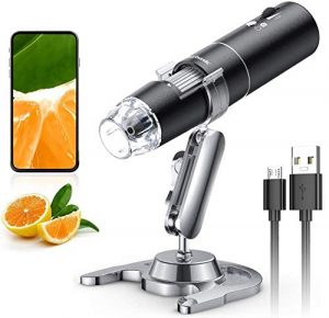
Ultra-High resolution: The lens of this microscope is made of high-quality glass, and the magnification is adjustable to 50X, 100X, 200X, 400X, and 1000X. With this microscope, you can take clear photos or videos of your specimens.
1.5″ LCD Screen: The large LCD screen displays clear images from edge to edge. Three brightness levels are available for you to choose from low brightness (yellow), medium brightness (orange), and high brightness (white).
Adjustable LED Light: You can adjust the brightness and contrast of the light for a clearer view. The microscope has a white LED light source that can be changed from dark to bright. It provides harsh brightness for most applications. And the light is free from electromagnetic radiation, so it is safe for children and adults.
HD USB Microscope Camera Compatible with iPhone, Android, iPad, Windows Mac Computer: Allow you to take high-quality pictures and videos via USB cable.
Continuous use for 3 hours – The battery lasts up to 3 hours if you use it continuously, which means you can enjoy your microscope for a long time without worrying about charging it again.
Excellent Compatibility – Supporting Android and IOS systems allows you to enjoy the fun of microscopy with your friends or families anytime wherallowse.
One-year replacement service – If you have any problem with any time and anywhere after purchase, we will replace it for free.
Multi-Function: The microscope can be used for viewing, photographing, recording, etc. With the adjustable stand, you can make the microscope at any angle you like in a very convenient way; With the big eyepiece, it is easy to see details of the object through it.
TOMLOV DM9 7′ LCD Digital Microscope
This model is the most commonly used microscope by professionals and students. We have this model in our lab. It has great clarity and brightness. The stand is also perfect. It’s easy to adjust the microscope’s height, focus, and objective lens. Also, you can see it in color and black/viewing screen (the image is transmitted through USB), which helps you a lot when researching. The objectives are easily interchangeable with different magnification – 4X, 10X, 40X(S), 100X(S), 400(S), 1000X(S).
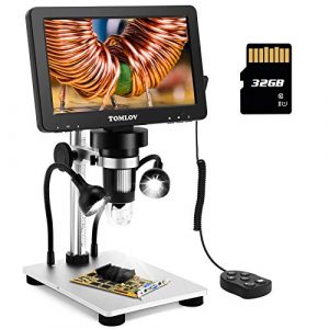
50X-1200X MAGNIFICATION: The microscope has a magnification range of 50X-1000X and the highest magnification of 1200X. You can observe the sample from top to bottom. TOMLOV offers a one-year warranty service. If there are any problems, please get in touch with them, and they will try their best to help you solve them.
7-INCH ROTATABLE FHD SCREEN: It comes with a 7-inch FHD screen, which can be rotated 90° left or right and is convenient for you to adjust the screen to your most desirable angle. The wired remote control makes it easy to capture images or videos by pressing a button on the remote—no need to use your phone or tablet to control the microscope.
12MP ULTRA-PRECISE FOCUSING MECHANISM: The microscope has a high-precision focusing mechanism that is smooth and accurate. You can focus the image precisely on the screen and capture high-quality images.
LED FILL LIGHT: The microscope comes with 4 LED lights, illuminating the object and making it stand out clearly on the screen. You can observe details of the specimen even in low light conditions.
PC VIEW & SD CARD READER: With the PC view function, you can connect to your computer via a USB cable and use software to record live videos and take photos directly onto your computer. The microscope adopts a solid metal framework with stable performance. The rubber foot on the stand is soft and durable, protecting your desk well.
Jiusion USB 2.0 Digital Microscope
This Jiusion USB 2.0 40-1000x is one of the best tools for anyone who needs a microscope to see things at a higher level! This microscope combines modern technology with the classic, hand-held device to give you a fantastic view of your favorite items. It can magnify objects up to 1000x while still keeping things very clear. You can use it on anything from food to hair strands, bugs, and more! It has eight bright LED lights that will illuminate your subject and make it easier to view!
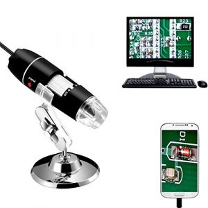
High quality: a high-quality USB microscope with a working distance of up to 4.7in, perfect for inspecting small parts, jewelry, coins, and more. This microscope is made of high-quality material that prevents it from breaking easily. It features a sturdy yet lightweight design that makes it easy to carry around when needed.
Wide application: it can be used for smartphones, laptops, computers, and tablets with a USB port. It’s compatible with most Windows, Mac, and Linux systems. AVI video output format can be used on Windows 7/8/10 and Mac OS X 10.4 or above to play the video directly on your computer.
Convenient focusing: two pairs of focusing lenses provide a large viewing area and an easy way to focus on your object. Simply use the two adjusting knobs to zoom in and out or adjust the focus as needed. You don’t need a power cord or external power supply, making it more convenient for your use.
Great for beginners: It comes with an aluminum stand, making it easier to steady your microscope. The frame also makes it easy to set up on any flat surface. This is an excellent tool for everyone, from kids to adults interested in exploring the world around them. It is a must-have tool for those who want to observe what they cannot see by the naked eye.
High definition: it’s equipped with eight built-in led lights that help you see your object in low-light conditions. It also has a 640 x 480 resolution that provides clear images. The metal stand makes it easy to adjust the microscope angle and rotate 360 degrees.
Light Microscopes: Shedding Light on the Microscopic World
Light microscopes, often the first encounter with the microscopic world for students and scientists alike, have played a pivotal role in advancing our understanding of biology and medicine. This section provides an in-depth look into the history, evolution, components, types, applications, and the advantages and limitations of light microscopes.
Brief History and Evolution
Light microscopy has a rich history, dating back to the late 16th century when pioneers like Hans Janssen and Zacharias Janssen created the first compound microscope. The development continued through the centuries, with notable contributions from Anton van Leeuwenhoek, who achieved magnifications up to 300 times. In the 19th and 20th centuries, advancements in lens technology and the introduction of phase contrast and fluorescence microscopy further expanded the capabilities of light microscopes.
Basic Components and Functionality
Light microscopes operate on the principle of using visible light to magnify and illuminate specimens. Key components include:
- Objective Lens: The primary magnifying lens closest to the specimen.
- Eyepiece (Ocular Lens): The lens through which the observer views the magnified specimen.
- Illuminator: Light source that passes through the specimen.
- Stage: Platform holding the specimen.
- Focus Mechanism: Adjustments for sharp focusing.
The combination of these components enables the observer to visualize details not visible to the naked eye.
Types of Light Microscopes
Light microscopes come in various types, each designed for specific applications:
| Type | Description | Applications |
|---|---|---|
| Compound Microscopes | Utilize multiple lenses for high magnification | Cellular biology, histology |
| Stereo Microscopes | Provide three-dimensional views with lower magnification | Dissection, microsurgery |
Applications in Biology and Medicine
Light microscopes are indispensable tools in biological and medical research, enabling scientists to study:
- Cellular Structure: Revealing details of cell organelles and structures.
- Histology: Examining tissue sections for pathology and diagnostics.
- Microorganisms: Identifying and studying bacteria, fungi, and protozoa.
- Live Cell Imaging: Observing dynamic processes in living cells.
Advantages and Limitations
Advantages:
- Ease of Use: Light microscopes are user-friendly and require minimal training.
- Cost-Effective: Generally more affordable than advanced microscopy techniques.
- Versatility: Suitable for a wide range of biological and medical applications.
Limitations:
- Limited Resolution: Constrained by the wavelength of visible light, limiting resolution.
- Magnification Limits: Typically lower magnification compared to electron microscopes.
- Specimen Requirements: Samples may require staining, affecting their natural state.
In conclusion, light microscopes have been instrumental in unraveling the mysteries of the microscopic world. From their humble beginnings to the sophisticated instruments of today, these microscopes continue to be essential tools in biology and medicine, offering a balance between accessibility and capability for researchers and educators alike.
Electron Microscopes: Peering Deeper into the Nano Realm
Introduction to Electron Microscopy
In the vast landscape of microscopy, electron microscopes stand as powerful tools capable of revealing details at the nanoscale. Unlike light microscopes that use visible light, electron microscopes employ electron beams to achieve unprecedented resolution, offering insights into the intricacies of materials and biological specimens.
Transmission Electron Microscopes (TEM)
Working Principle: Transmission Electron Microscopes function based on the interaction of electrons with the specimen. Electrons pass through the ultra-thin specimen, and the resulting transmitted electrons are used to form an image. The wavelength of electrons is much shorter than that of visible light, allowing for significantly higher resolution.
Applications in Material Science: TEM is extensively employed in material science for detailed analysis of structures at the atomic and molecular levels. It enables researchers to examine the composition and crystallography of materials, making it invaluable in the development of advanced materials and nanotechnology.
Scanning Electron Microscopes (SEM)
3D Imaging Capabilities: Scanning Electron Microscopes, on the other hand, operate by scanning a focused beam of electrons across the specimen’s surface. The interaction between the electrons and the specimen generates signals that are used to create a three-dimensional image. This provides a depth of field not achievable with traditional light microscopes.
Geological and Biological Applications: SEM finds applications in diverse fields, including geology and biology. In geology, it aids in the examination of mineral surfaces and the study of geological formations. In biology, SEM is used to visualize the surface structures of cells and tissues, providing detailed information about the topography.
Comparison with Light Microscopes
The comparison between electron microscopes and light microscopes highlights the superiority of electron microscopy in terms of resolution and magnification.
| Criteria | Electron Microscopes | Light Microscopes |
|---|---|---|
| Resolution | Nanometer scale | Micrometer scale |
| Magnification | Higher magnification (up to 50 million times) | Limited magnification (usually up to 2,000 times) |
| Wavelength | Shorter wavelength of electrons | Longer wavelength of visible light |
| Depth of Field | Greater depth of field | Shallow depth of field |
Technological Advancements in Electron Microscopy
Continual advancements in electron microscopy technology have expanded its capabilities and usability.
- Cryo-Electron Microscopy (Cryo-EM): This technique involves freezing biological samples, preserving their natural state for examination. It has revolutionized the study of biological macromolecules, including proteins and viruses.
- Environmental Electron Microscopy: Designed to operate in controlled environments, this technology allows researchers to study materials and biological samples under specific conditions, such as varying temperatures or gas atmospheres.
- In-situ Electron Microscopy: Enabling real-time observations of dynamic processes, in-situ electron microscopy provides insights into changes at the nanoscale as they occur.
In conclusion, electron microscopes, encompassing both TEM and SEM, have significantly advanced our ability to explore the nano realm. From unraveling the atomic structures of materials to providing three-dimensional insights into biological specimens, electron microscopy continues to be an indispensable tool for researchers across various scientific disciplines. The ongoing technological innovations in this field promise even greater revelations in the microscopic world.
Fluorescence Microscopy: Illuminating the Invisible
Principle of Fluorescence Microscopy
Fluorescence microscopy is a powerful imaging technique that exploits the natural fluorescence of certain substances. The basic principle involves light absorption by fluorophores, followed by their re-emission at longer wavelengths. This emitted fluorescence is then captured to create detailed images of the specimen.
Types of Fluorescence Microscopes
Fluorescence microscopes come in various types, each catering to specific imaging needs.
- Confocal Microscopes:
- Working Principle: Use of pinholes to eliminate out-of-focus light, enabling sharper images.
- Applications: High-resolution imaging of biological specimens, live cell imaging.
- Two-Photon Microscopes:
- Working Principle: Simultaneous absorption of two photons to excite fluorophores, reducing photodamage.
- Applications: Deep tissue imaging, neuroscience research.
Applications in Cell Biology and Biochemistry
Fluorescence microscopy has revolutionized the fields of cell biology and biochemistry, offering unique advantages in visualizing and understanding cellular processes.
- Cellular Dynamics: Real-time observation of dynamic processes within living cells.
- Protein Localization: Labeling specific proteins with fluorophores for precise localization studies.
- Cellular Markers: Tracking cellular structures and organelles with fluorescent markers.
Pros and Cons of Fluorescence Microscopy
Pros:
- High Sensitivity: Fluorescence microscopy is highly sensitive, allowing the detection of low concentrations of fluorophores.
- Selective Labeling: Specific structures or molecules can be targeted and labeled with fluorescent dyes.
- Live Cell Imaging: Ideal for studying dynamic processes in living cells over time.
Cons:
- Photobleaching: Prolonged exposure to light can lead to the fading of fluorescence over time.
- Phototoxicity: High-intensity illumination may cause damage to living cells or specimens.
- Cost and Complexity: Advanced fluorescence microscopy setups can be expensive and require expertise.
In conclusion, fluorescence microscopy has become an indispensable tool in the biological sciences, providing researchers with the ability to illuminate and observe the invisible intricacies of cellular and molecular structures. The diverse types of fluorescence microscopes cater to different research needs, allowing for a range of applications in cell biology, biochemistry, and beyond. Despite some limitations, the advantages of fluorescence microscopy continue to drive innovation and enhance our understanding of the microscopic world.
Atomic Force Microscopes: Feeling the Microscopic Terrain
Introduction to Atomic Force Microscopy (AFM)
Atomic Force Microscopy (AFM) stands out as a powerful technique in the realm of microscopy, offering a unique approach to imaging at the nanoscale. Unlike conventional microscopy methods that rely on optics or electron beams, AFM utilizes a physical probe to “feel” the surface of a sample, providing detailed information about its topography.
Working Principle
The core principle of AFM lies in the interaction between a sharp tip at the end of a flexible cantilever and the sample surface. As the tip scans the surface, the cantilever deflection is measured. This information is then used to generate a three-dimensional map of the sample’s surface, revealing features at the atomic and molecular levels.
Applications in Nanotechnology and Surface Analysis
AFM finds extensive applications in various scientific domains with a particular emphasis on nanotechnology and surface analysis.
| Application | Description |
|---|---|
| Nanotechnology Research | Characterizing nanomaterials, studying nanoparticles, and manipulating individual atoms or molecules. |
| Material Science | Analyzing surface properties, studying thin films, and investigating the mechanical properties of materials. |
| Biology and Life Sciences | Imaging biological samples, studying DNA, proteins, and cell structures with high resolution. |
| Surface Chemistry | Probing chemical interactions at the nanoscale, understanding surface roughness, and analyzing molecular structures. |
Advantages and Limitations
Advantages:
- High Resolution: AFM can achieve sub-nanometer resolution, allowing the visualization of individual atoms.
- Versatility: Applicable to a wide range of materials, from biological specimens to solid surfaces.
- Non-destructive: AFM is a non-destructive technique, preserving the integrity of samples during imaging.
Limitations:
- Speed: Imaging with AFM can be time-consuming compared to other microscopy methods.
- Complexity: The instrument setup and operation can be complex, requiring specialized training.
- Sample Limitations: Samples must be relatively flat, and imaging liquids can be challenging.
In conclusion, Atomic Force Microscopy has proven to be an invaluable tool in various scientific disciplines, offering a tactile approach to imaging at the nanoscale. Its ability to provide high-resolution topographical information and study a diverse range of materials positions AFM as a key player in nanotechnology, material science, and life sciences research. Despite its limitations, the unique advantages of AFM make it an indispensable tool for researchers exploring the microscopic terrain of the nanoworld.
Other Specialized Microscopes: Niche Tools for Specific Needs
Specialized microscopes cater to unique research requirements, offering advanced capabilities for specific applications.
| Microscope Type | Applications | Key Features |
|---|---|---|
| Scanning Tunneling Microscopes (STM) | Nanoscience | Visualizing individual atoms and manipulating surfaces at the atomic level. |
| Phase-Contrast Microscopes | Live Cell Imaging | Enhancing contrast in transparent specimens without the need for staining. |
| Darkfield Microscopes | Observing Unstained Specimens | Illuminating specimens against a dark background, revealing fine details. |
| Polarizing Microscopes | Geological and Material Analysis | Analyzing birefringent materials and studying the optical properties of crystals. |
Scanning Tunneling Microscopes (STM): Used extensively in nanoscience, STMs employ a sharp tip to scan surfaces at the atomic level, providing detailed information about individual atoms and their arrangement.
Phase-Contrast Microscopes: Ideal for live cell imaging, phase-contrast microscopy enhances contrast in transparent specimens by exploiting differences in refractive index, allowing for detailed observation without the need for staining.
Darkfield Microscopes: By illuminating specimens against a dark background, darkfield microscopy enhances contrast, revealing fine structures and details in unstained specimens that might be otherwise challenging to observe.
Polarizing Microscopes: Specifically designed for geological and material analysis, polarizing microscopes analyze birefringent materials, providing insights into crystal structures and optical properties.
In conclusion, these specialized microscopes serve as indispensable tools in various scientific disciplines, addressing specific research needs that may not be adequately met by conventional microscopy techniques. From the manipulation of individual atoms to the detailed analysis of crystal structures, these niche tools contribute to the advancement of knowledge across diverse fields.
Recent Technological Advancements in Microscopy
Recent technological advancements have propelled microscopy to new heights, breaking traditional barriers and expanding the capabilities of these powerful tools.
| Advancement | Description | Key Features |
|---|---|---|
| Super-Resolution Microscopy | Breaking the Optical Limit | Achieving resolutions beyond the diffraction limit of light, enabling visualization of structures at the molecular and even atomic levels. |
| Correlative Microscopy | Combining Multiple Techniques | Integrating various microscopy techniques, such as light and electron microscopy, to provide a more comprehensive understanding of specimens. |
| Artificial Intelligence Integration | Enhancing Image Analysis | Incorporating artificial intelligence for automated image analysis, improving accuracy, and enabling faster processing of large datasets. |
Super-Resolution Microscopy: Overcoming the limitations imposed by the diffraction of light, super-resolution microscopy techniques, including STED (Stimulated Emission Depletion) and PALM (Photoactivated Localization Microscopy), enable researchers to delve into details at the nanoscale, unraveling structures that were once beyond the reach of conventional microscopy.
Correlative Microscopy: By combining multiple techniques, correlative microscopy allows researchers to leverage the strengths of each method. For example, combining light microscopy for live-cell imaging with electron microscopy for high-resolution structural details provides a more comprehensive view of biological specimens.
Artificial Intelligence Integration: The integration of artificial intelligence (AI) in microscopy has revolutionized image analysis. AI algorithms can quickly and accurately process vast amounts of microscopy data, aiding in the identification of patterns, anomalies, and complex structures.
These advancements not only push the boundaries of what is observable but also streamline the workflow for researchers, making microscopy more accessible and efficient. As technology continues to advance, these recent innovations pave the way for even more exciting developments in the field of microscopy.
Future Trends in Microscopy: Pushing the Boundaries
The realm of microscopy is continually evolving, with researchers pushing the boundaries of what is possible. This section delves into the future trends of microscopy, exploring nanoscale imaging breakthroughs, advancements in in vivo imaging, and the integration of microscopy with other analytical techniques.
Nanoscale Imaging Breakthroughs
Nanoscale imaging has been a focal point in microscopy research, driven by the need to visualize structures at unprecedented resolutions. Recent breakthroughs have opened new frontiers, allowing scientists to explore the intricate details of biological, material, and chemical samples at the nanometer scale.
| Breakthrough | Technique | Resolution Achieved | Applications |
|---|---|---|---|
| Cryo-Electron Microscopy (Cryo-EM) | Utilizing cryogenic temperatures to image frozen samples | Sub-nanometer | Structural biology, drug discovery |
| 3D Structured Illumination Microscopy (3D-SIM) | Enhancing lateral resolution in fluorescence microscopy | ~100 nanometers | Cellular and subcellular imaging |
| Stimulated Emission Depletion Microscopy (STED) | Implementing stimulated emission to reduce the focal spot | <50 nanometers | Super-resolution imaging of cellular structures |
These breakthroughs are revolutionizing our understanding of the nanoworld, enabling researchers to explore biological processes and materials with unprecedented detail.
Advancements in In Vivo Imaging
In vivo imaging, the visualization of biological processes within living organisms, is a crucial aspect of medical and biological research. Advancements in this area are providing researchers with real-time insights into dynamic processes, aiding in the development of therapies and the understanding of diseases.
| Advancement | Technique | Key Features | Applications |
|---|---|---|---|
| Light Sheet Fluorescence Microscopy (LSFM) | Illuminating a thin section of the sample to reduce phototoxicity | High-speed imaging, minimal damage to living tissues | Developmental biology, neuroscience |
| Multiphoton Microscopy | Using infrared light for deeper penetration in tissues | 3D imaging at depth, reduced photodamage | Imaging of live tissues, neuroscience |
| In Vivo Magnetic Resonance Imaging (MRI) | Non-invasive imaging using strong magnetic fields | Soft tissue visualization, functional imaging | Clinical diagnostics, neuroscience |
These advancements are transforming our ability to study living organisms at the cellular and molecular levels, providing a deeper understanding of physiological processes.
Integration with Other Analytical Techniques
The future of microscopy lies not only in improving imaging capabilities but also in integrating microscopy with other analytical techniques. This synergy enhances the overall understanding of samples by combining the strengths of different methods.
| Integration | Techniques | Synergistic Benefits | Applications |
|---|---|---|---|
| Correlative Microscopy | Combining light and electron microscopy for comprehensive imaging | Ultrastructural and functional information | Cell biology, materials science |
| Mass Spectrometry Imaging (MSI) | Linking microscopy with mass spectrometry for chemical analysis | Spatially resolved molecular information | Metabolomics, drug development |
| Raman Imaging | Integrating microscopy with Raman spectroscopy | Molecular identification and localization | Materials analysis, pharmaceuticals |
The integration of microscopy with other analytical techniques enhances the depth of information obtained from a sample, providing a more comprehensive understanding of its structure and composition.
In conclusion, the future of microscopy is marked by groundbreaking advancements in nanoscale imaging, in vivo visualization, and the seamless integration of microscopy with other analytical techniques. These developments not only push the boundaries of scientific exploration but also open new avenues for discoveries in fields ranging from medicine to materials science. As technology continues to advance, researchers can anticipate even more powerful tools to unravel the mysteries of the microscopic world.
Which structure is best observed using a compound light microscope?
Several different structures can be observed using a compound light microscope; the most famous form is the cell. Cells can be kept in various states, including mitosis, meiosis, and fertilization. Additionally, cells can be examined for signs of disease or damage.
One of the great things about compound light microscopy is that it can provide a high-resolution image that is easy to navigate. Additionally, it is versatile and can be used to identify both plant and animal cells. If you want to pursue a career in science or medicine, a compound light microscope is an essential tool in your arsenal.
How to use the Edu science microscope?
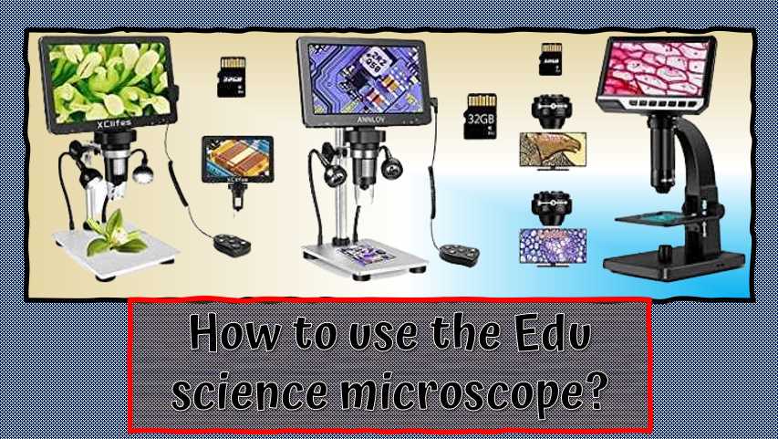
To use an Edu science microscope, first, choose the magnification you want. Then, adjust the lens to get the best view. Finally, focus the microscope using the crosshairs and the buttons on the side to move the object around. If you need to correct, simply press the reset button. If you want to take a picture, press the picture button and select the file type you want. You can also save your images to your computer using a USB connection.
How to focus a microscope using a high-power lens?
There are a few methods for focusing a microscope using a high-power lens. One method is the cross-hairs method, which involves using the cross-hairs to focus the lens on an object. Another method is the zone method, which focuses the le, and this is on an object’s specific area.
The last method is the sliding method. This involves moving the lens along with the object and then focusing it.
What is the most common lab microscope in schools?
There are various types of lab microscopes, but the most common is the binocular microscope. Binocular microscopes allow two people to view the same object simultaneously, making it ideal for class experiments. They are also less expensive than other microscopes and can be easily moved from one location to another.
Another common type of microscope is the compound microscope, which is used to view large specimens such as cells or tissues. It is a stationary microscope requiring a space of at least 30x30x50 mm. The third type of microscope is the scanning electron microscope, which images large objects such as crystals or metals. It requires a high-voltage power supply and is usually only used in research laboratories.
What kind of microscope is most commonly used globally?
The most commonly used microscope is the fluorescence microscope, which uses fluorescent dyes to visualize cells and tissues. It is a versatile tool that can be used for various applications, including medical research, biology, and chemistry. It is also a cost-effective option that can be used in the laboratory and home.
What microscope is commonly used in a healthcare setting?
The most common microscope used in healthcare is the light microscope,d it, and it, and it, and it, and it, and it, and it, and it, and it, and it is an,d it is used to view cells, tissues, and organs in detail. Additionally, this microscope can be used to diagnose and treat diseases.
Which microscope is commonly used in science classes?
Some of the most commonly used microscopes in science classes include the light, electron, and scanning electron microscopes. While all three of these microscopes are incredibly useful, the light microscope is often the one that is used in elementary and middle school science classes. This microscope uses light to view objects, and as a result, it is easy to use and is generally portable. It is also affordable, making it a good choice for classrooms with a limited budget.
The electron microscope is used to view the minute details of objects, and it is often used in research labs. It has a higher price tag but offers vast magnification capabilities and is used to study materials that are too delicate for the other two microscopes. The scanning electron microscope is used to image objects with high resolution and is often used in research labs and for medical diagnosis.
Which microscope is most commonly used in crime elaborates?
Various microscopes are used in crime laboratories; a scanning electron microscope is the most commonly used ureteroscope. This microscope can image structures at an extremely high resolution, making it a valuable tool for determining the composition and structure of objects. It is also used to identify chemical and biological stains.
Another ordinary microscope used in crime laboratories is the light microscope. This microscope can see small objects and image them using a beam of light. It is perfect for examining biological specimens, such as bacteria and viruses. Additionally, the light microscope can be used for fingerprinting and DNA analysis.
The last microscope used in crime laboratories is the stereomicroscope. This microscope can provide three-dimensional images of objects and is beneficial for examining biological specimens, such as cells and tissues. It can also be used to identify blood stains and body fluids.
Final Words:
Microscopes come in different magnifications and are convenient for a variety of purposes. Some models come with a rotatable screen that helps you get a clear view of the object you’re examining. Additionally, some microscopes offer wireless capabilities that make them easier to use.
Thank you for reading our blog post on the most commonly used microscopes. In it, we outlined the different features that make a microscope a powerful tool for research or simply studying tiny organisms. We also included a list of the best microscopes for convenience, magnification, and Rotatable screens.
SupposeSupposeSupposeSupposeSuppose you are in the market for a microscope and can’t decide which one to choose. In that case, I recommend checking out the Skybasic 50X-1000X Magnification wireless digital microscope, as it is the most commonly used model! W hope you have found this helpful article. Please comment and let us know what you want to see in future posts.

I am an enthusiastic student of optics, so I may be biased when I say that optics is one of the most critical fields. It doesn’t matter what type of optics you are talking about – optics for astronomy, medicine, engineering, or pleasure – all types are essential.
Table of Contents

Pingback: What Is the Best Way to Focus on a Microscope?: A Beginner's Guide
Pingback: 5 ways to Improve Microscope Resolution: Video Guide Explained
Pingback: What Does Skeletal Muscle Look Like under Microscope: A Microscopic Marvel
Pingback: Are Electron Microscope Images Coloured? Unveiling the Truth!
Pingback: Are Microscope Objectives Interchangeable?