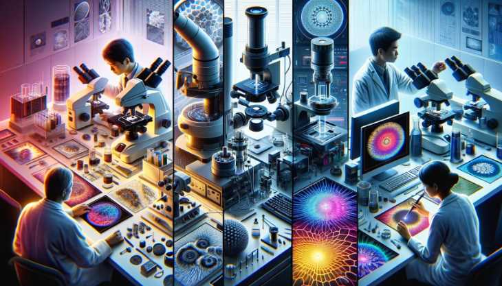
We all know that magnifying glasses allow us to see more clearly, but what does this mean? A magnification of 100x can make something about 5cm tall look like it’s about 40cm tall. And a microscope lets you see objects that are even smaller than that!
The best way to understand the science behind a microscope is by looking at the mechanism that makes it work. Microscopes and their use are interesting and complex topics, and this article explores an in-depth guide to how a microscope works.

Before exploring how a microscope functions, we must first address the available types.
Types Of Microscopes:
Overview of Different Microscope Types
Microscopes are instrumental in magnifying the invisible, with various types tailored to specific applications. This overview provides a glimpse into the diverse world of microscopes, each wielding its unique capabilities.
Light Microscopes
Introduction: As a novice explorer of the microscopic, the journey often begins with light microscopes. These instruments utilize visible light to illuminate specimens, allowing for a detailed examination of biological samples and materials.
Applications:
- Invaluable in Biology: Light microscopes are workhorses in biology, enabling scientists to study the intricate structures of cells, tissues, and microorganisms.
- Material Science Insight: Their simplicity makes them a go-to tool in material science, aiding in the analysis of material composition and structure.
Advantages:
- Accessibility: Light microscopes are widely accessible, making them essential for educational settings and routine laboratory work.
- Real-time Observation: Ideal for observing living specimens in real-time, providing a dynamic view of biological processes.
Electron Microscopes
Paradigm Shift: Venturing further into the microscopic frontier, the transition to electron microscopes marks a paradigm shift. These instruments employ beams of electrons instead of visible light, propelling microscopy into the nanoscale.
Nanoscale Revelation:
- Unparalleled Resolution: Electron microscopes unveil intricate details at the nanoscale, offering unprecedented resolution for studying atomic and molecular structures.
- Nanotechnology Marvel: Revolutionizing fields like nanotechnology, electron microscopes play a pivotal role in designing and analyzing nanomaterials.
Applications:
- Materials Science Precision: In materials science, electron microscopes facilitate precise characterization of materials, contributing to advancements in semiconductor technology and metallurgy.
- Biological Frontiers: In biology, electron microscopes delve into the ultrastructure of cells, revealing details beyond the capability of light microscopes.
In navigating the world of microscopes, understanding the nuances of light and electron microscopes is paramount. Each type, with its distinctive features, contributes uniquely to scientific exploration, offering a glimpse into realms previously hidden from our sight.
Fundamental Principles of Microscopy
Microscopy, a science of discovery, is anchored in fundamental principles that govern the magnification and resolution of microscopic images. In this exploration, we delve into these core principles, understanding the mechanisms that bring the unseen into focus.
Magnification
Magnification is the bedrock of microscopy, enabling us to explore the microscopic world with enhanced clarity. The process of magnification involves increasing the apparent size of an object, allowing for a detailed examination beyond the limits of our natural vision.
Key Components of Magnification:
| Component | Description |
|---|---|
| Objective Lens | Primary magnifying lens near the specimen. |
| Eyepiece | Further magnification through the eyepiece. |
| Total Magnification | Calculated by multiplying objective and eyepiece magnifications. |
Significance of Magnification: Understanding magnification is pivotal. As an amateur microscopist, I discovered that higher magnification reveals finer details, providing a more comprehensive view of the specimen’s structure.
Resolution
Resolution, the microscope’s ability to distinguish between two closely spaced objects, is equally crucial. While magnification brings objects closer, resolution ensures that these objects are distinct entities rather than a blur.
Factors Influencing Resolution:
| Factor | Description |
|---|---|
| Wavelength of Light/Electrons | Smaller wavelength allows higher resolution. |
| Numerical Aperture | Determines the resolving power of the objective lens. |
Importance of Resolution: Resolution became a fascinating aspect of my journey. I realized that even with high magnification, poor resolution could compromise the clarity of the observed details. Achieving optimal resolution is vital for a comprehensive understanding of the microscopic world.
Parts of a Microscope and their Functions
Microscopes are complex instruments, weaving together various components to unlock the mysteries of the microscopic world. Understanding the interplay between these components is essential for harnessing the full potential of these scientific marvels.
Optical Microscopes
Optical microscopes, often the starting point in microscopy, employ visible light to illuminate and magnify specimens. As I delved into the intricacies, I discovered the synergy of components that form the backbone of optical microscopy.
Key Components:
| Component | Description |
|---|---|
| Objective Lens | Primary lens near the specimen, providing magnification. |
| Eyepiece | Further magnifies the image for observation. |
| Condenser | Focuses and directs light onto the specimen. |
| Diaphragm | Regulates the amount of light reaching the specimen. |
Functionality:
- Objective Lens:
- Varied Magnification: Different objective lenses offer varying levels of magnification.
- Numerical Aperture: Influences resolution and light-gathering ability.
- Eyepiece:
- Additional Magnification: Multiplies the magnification obtained from the objective lens.
- Observation Interface: Where the observer views the magnified specimen.
- Condenser:
- Light Focus: Concentrates light onto the specimen for optimal illumination.
- Aperture Control: Adjusts the size of the aperture to regulate light intensity.
- Diaphragm:
- Light Regulation: Controls the amount of light passing through the specimen.
- Enhances Contrast: Adjustments aid in improving contrast for better visibility.
Electron Microscopes
Electron microscopes, representing a technological leap, utilize beams of electrons for imaging, achieving unparalleled resolution at the nanoscale. My exploration into electron microscopy unveiled a realm of advanced components.
Key Components:
| Component | Description |
|---|---|
| Electron Gun | Emits a focused beam of electrons. |
| Magnetic Lenses | Focus and steer the electron beam. |
| Detector Systems | Capture and convert electrons into an image. |
Functionality:
- Electron Gun:
- Electron Emission: Generates a focused beam of electrons.
- Beam Control: Directs electrons towards the specimen.
- Magnetic Lenses:
- Focusing Electrons: Magnetic fields focus and steer the electron beam.
- Precision Control: Achieves high-resolution imaging at the nanoscale.
- Detector Systems:
- Electron Detection: Capture electrons transmitted through or scattered by the specimen.
- Image Conversion: Converts electron signals into a visible image.
Navigating the components of microscopes is akin to deciphering a symphony of precision. Whether in the realm of optical or electron microscopy, each component plays a unique role, contributing to the clarity and detail of the microscopic images that captivate scientific minds.
How does a Microscope work?
How Microscopes Produce Images
The production of microscopic images is a complex yet fascinating process, involving intricate mechanisms that transform minuscule details into visible wonders. Understanding the nuances of this process is key to appreciating the beauty and depth of microscopic exploration.
Light Microscopes
Light microscopes, relying on visible light to illuminate specimens, orchestrate a symphony of components to produce clear and magnified images. My journey through the workings of light microscopy revealed the elegance behind the creation of these images.
Key Processes:
| Process | Description |
|---|---|
| Illumination | Light source provides illumination for the specimen. |
| Lenses | Objective and eyepiece lenses magnify and focus the image. |
| Image Formation | Interplay of lenses creates a magnified and observable image. |
Illumination and Lenses:
- Illumination:
- Light Source: Provides a steady stream of light to illuminate the specimen.
- Condenser Lens: Focuses and directs light onto the specimen for optimal visibility.
- Lenses:
- Objective Lens: Magnifies the specimen, capturing details for observation.
- Eyepiece Lens: Further magnifies the image for the observer.
- Image Formation:
- The coordinated action of lenses produces a magnified and focused image for observation.
- Adjustments to focus and magnification contribute to image clarity.
Electron Microscopes
In the realm of electron microscopy, the process takes a quantum leap into the nanoscale, where beams of electrons replace visible light. This shift introduces an intricate dance of electrons to create images with unprecedented resolution.
Key Processes:
| Process | Description |
|---|---|
| Electron Beam Generation | Electron gun emits a focused beam of electrons. |
| Electron Interaction | Electron beam interacts with the specimen, revealing details. |
| Image Detection | Detector systems capture electron signals and convert them into an image. |
Electron Beam and Interaction:
- Electron Beam Generation:
- Electron Gun: Emits a focused and controlled beam of electrons.
- Magnetic Lenses: Focus and steer the electron beam with precision.
- Electron Interaction:
- The electron beam interacts with the specimen, revealing intricate details at the nanoscale.
- Scattering and transmission of electrons provide information about the specimen’s structure.
- Image Detection:
- Detector systems capture the transmitted or scattered electrons.
- Conversion of electron signals into a visible image completes the imaging process.
In both light and electron microscopes, the production of images is a carefully choreographed process, where the manipulation of light or electrons transforms the invisible into the visible, allowing us to explore the hidden realms of the microscopic universe.
5 Tips for Maximizing Your Microscope Experience
Microscope use is an art that combines precision and technique to unlock the mysteries of the microscopic world. Whether you are a novice or seasoned microscopist, these tips will enhance your microscopy experience, ensuring optimal results.
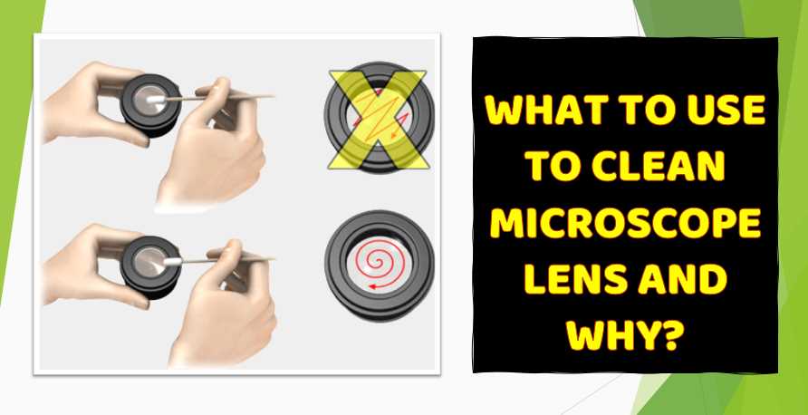
1. Proper Handling and Care
Microscopes are delicate instruments requiring careful handling. Always use both hands when carrying or adjusting the microscope to prevent damage. Regular cleaning, especially of lenses, ensures clear and accurate imaging. Proper storage in a dust-free environment protects the instrument’s longevity.
2. Optimizing Illumination and Focus
Adjusting illumination and focus is an art that significantly impacts image quality. Start with low light and gradually increase to prevent specimen damage. Use fine and coarse adjustments for precise focusing, ensuring a crisp and clear image for observation.
3. Choosing the Right Objective Lens
Selecting the appropriate objective lens is pivotal for obtaining the desired magnification and resolution. Begin with low magnification for an overall view, then progressively move to higher magnifications for detailed observations. Always clean lenses before switching objectives.
4. Utilizing Immersion Oil Effectively
In certain microscopy techniques, immersion oil enhances resolution. When using oil immersion objectives, apply a small amount of oil to the specimen slide. Ensure the oil has the same refractive index as the lens for optimal imaging and wipe it off carefully after use to prevent contamination.
5. Record Keeping and Documentation
Maintaining a detailed record of observations is crucial for research and analysis. Use a microscope camera or smartphone attachment to capture images. Annotate findings, including magnification levels and specimen details. This not only aids in data analysis but also serves as a valuable reference for future studies.
Implementing these tips transforms microscope use into a refined skill, enhancing the quality and longevity of your microscope and ensuring a rewarding exploration of the microscopic realm.
How do Digital microscopes work?

Components of Digital Microscopes
| Component | Description |
|---|---|
| Digital Camera | Integrated camera captures images of the specimen. The camera resolution impacts the clarity of the digital image. |
| Objective Lens | Functions similarly to traditional microscopes, providing magnification and focusing capabilities. The choice of the objective lens influences the overall optical performance. |
| Illumination Source | Light source, often LED, provides consistent illumination for the specimen. Some digital microscopes offer adjustable illumination settings for optimizing image quality. |
| Digital Image Sensor | Converts light information into digital signals. The type and quality of the image sensor influence the final image’s resolution and sensitivity. |
| Processor and Software | The processor manages image processing tasks, and software applications handle image capture, storage, and analysis. Advanced software allows users to enhance and manipulate captured images. |
| Display Screen | Digital microscopes feature built-in screens or connect to external monitors. The display screen allows real-time observation and analysis of specimens, eliminating the need for traditional eyepieces. |
Functionality of Digital Microscopes
- Image Capture and Processing:
- The digital camera captures high-resolution images of the specimen.
- The digital image sensor converts the captured light information into digital signals.
- Image Display and Analysis:
- The processed digital image is displayed on the microscope’s built-in screen or an external monitor.
- Digital microscopes often feature advanced software for real-time analysis, annotation, and manipulation of captured images.
- Storage and Sharing:
- Captured images are stored digitally, eliminating the need for physical slides.
- Digital microscopes enable easy sharing of images through electronic means, facilitating collaboration and remote analysis.
- Adjustable Illumination:
- LED illumination sources provide consistent and adjustable lighting for optimal specimen visibility.
- Users can control illumination sett
How does an electron microscope work?
Electron microscopes are devices that use a beam of electrons to illuminate an object and then capture images of the object.
The electron beam is shot at the sample and can be manipulated by changing its trajectory with magnetic lenses or a position sensor. The light scattering by the electrons causes interference patterns on a detector used to reconstruct an image.
What is the importance of lenses in a microscope?
The lens is an essential part of a microscope and helps to focus light from the object being observed onto the detector. The lens also affects how much light the instrument collects and how much magnification can be achieved.
A good quality microscope will have two lenses, one for illuminating light and collecting it (also called an objective). In most cases, this objective is fixed to the body of the microscope and not removable. This means that when using a microscope with interchangeable objectives, one must first remove this set objective before switching to another one on the other side of the body or in another compartment.
What is fluorescence microscopy?
Fluorescence microscopy is a type of microscopy that uses a laser to excite fluorescent molecules, which emit light in response. This allows the fluorescence microscope to visualize cells and tissues in real time or over long periods.
The advantage of fluorescence microscopy is that it provides high-resolution images with minimal phototoxicity and damage to living tissue. It also offers 3D imaging, which can be used for many purposes, such as cancer diagnosis, studying cellular behavior, or determining cell function.
It would be best to use this technique to have a fluorophore and the appropriate wavelength laser light source.
Do microscopes need electricity?
Yes, microscopes need electricity, and this is because they are electronic devices that use electric signals to operate and observe things at the nanometer scale.
Microscopes use light or electron beams to illuminate objects and then use the light reflected from these objects to form an image on a monitor.
How does magnification work in a microscope?
Magnification is the process of making something appear larger. You can do that by either increasing the size of an object or reducing the distance between a subject and an observer.
Magnification can be achieved in a microscope by moving one or more lenses to different focal lengths.
When looking through a microscope, there are two ways that magnification can occur:
1) The light from your sample enters the microscope through a condenser lens, reducing its numerical aperture (NA). The objective lens then moves closer to the model to collect more light. In this case, you will see an increase in magnification because more light is coming into your field of view and less going out.
2) The second way that magnification occurs in microscopes is by using a tube lens instead of a condenser lens. Tube lenses work like telephoto lenses when placed at the front end of your microscope tube.
What do dead cells look like under a microscope?
Dead cells look like faded, dry structures that have lost shape and are no longer alive.
Under a microscope, dead cells can be seen to lack a nucleus and other cellular organelles. They also show signs of cell death, such as dendrites or spines, which generally help transfer healthy cells’ electrical signals.
What does it mean if a micrograph is false-colored?
When an image is falsely colored, it means that different colors have been assigned to other regions in the image to be easier for the viewer to identify them.
Micrographs are pictures of microscopic structures that are obtained by using a microscope. The images obtained from these micrographs can be false-colored to help the viewers visualize certain features and systems.
How does a microscope use light?
A microscope uses light in two ways:
1. To illuminate the specimen under observation and allow it to be seen.
2. To collect images of the specimen and provide a digital record of its structure.
What does the diaphragm do on a microscope?
The diaphragm is a thin membrane that controls the amount of light passing through an optical system. It is a simple mechanical device used in microscopy and other fields, including astronomy.
The diaphragm is attached to the objective lens on most microscopes and can be moved from side to side. It is used with both low-power (low magnification) and high-power (high magnification) objectives.
How far can a microscope zoom in?
According to Wikipedia, microscopes with a minimum magnification of 100x have been invented, and they’re capable of zooming into an object that is only 0.5mm wide (1/20th of an inch).
There are two main ways to measure the distance between two objects.
The first is with a ruler. You can use this method by holding a ruler on top of your object and measuring the length in millimeters or inches, then multiplying that number by 100 to find out how far away it is from you.
The second way is to put your eye at one end of the microscope lens and measure the size of your image on the other side. That will give you an idea of how far away your object is.
What is the iris diaphragm on a microscope?
The iris diaphragm is a type of aperture stop in the lens of a microscope. It limits the amount of light that enters the objective lens and can be set to any desired level by adjusting its size or location.
What can you see with a 250x microscope?
A 250x microscope is an optical microscope that can magnify up to 250 times its original size. This microscope type is used to view small specimens, such as cells or other minute objects.
If you want to see something with a 250x microscope, you must use the lens adapter on the objective lens and attach it to your camera to capture images from your perspective.
What is the smallest object ever seen through a microscope?
The smallest object ever seen through a microscope is an atom.
A micrograph is a photograph of an object that is magnified frequently to be seen clearly. It’s called “micro.” Because the image has been made with a microscope, making it look like a miniature copy of what’s in front of the lens.
An electron microscope can show objects as small as molecules and atoms.
Final words:
Microscopes are used to magnify objects to make them visible. There are various types of microscopes, and each one has its purpose. In this blog post, we will be discussing the difference between a compound microscope and a stereo microscope. If you would like to learn more about these two types of microscopes, please take some time to read through the article below!
If you’re a student or have been studying science for some time, you may already know how a microscope works. But if you’re new to this field, it’s probably worth knowing what’s going on under the hood. This blog post should help you understand the essential parts and their functions for magnification and light paths. What is your opinion about magnifications?
Let us know in the comments below!
Resources and References
For those eager to delve further into the captivating world of microscopy, a wealth of resources awaits. Books such as “Microscopy: A Very Short Introduction” by Terence Allen and “Introduction to Light Microscopy” by Randy O. Wayne provide comprehensive insights. Online platforms like MicroscopyU offer interactive tutorials and reference materials. Academic journals such as the “Journal of Microscopy” and “Microscopy Research and Technique” provide in-depth research articles. This article serves as a stepping stone, and these resources offer a continuous journey, providing nuanced perspectives and expanding your understanding of microscopy’s intricacies.

I am an enthusiastic student of optics, so I may be biased when I say that optics is one of the most critical fields. It doesn’t matter what type of optics you are talking about – optics for astronomy, medicine, engineering, or pleasure – all types are essential.
Table of Contents

Pingback: 5 Best Microscope For Soil Biology: Reviews With Expert Recommendation
Pingback: 5 Best Labomed Dental Microscope Review: A Complete Buying Guide 2022
Pingback: Lumagny MP7545 Lighted Microscope Review: Instructions Explained
Pingback: 5 Best Vanguard Microscope Review: A Complete Buying Guide 2022
Pingback: 5 Best Microscope For Laboratory: Reviews With Buying Guide in 2022
Pingback: 5 Best Microscope For Photomicrography: A Complete Buying Guide with Comparison
Pingback: 5 Best Chinese Microscopes: A Complete Buying Guide in 2022
Pingback: 5 Best Most Commonly Used Microscopes We Tested in the Last 2 weeks: Comparison With Buying Guide
Pingback: 5 Best LCD Microscope We Tested Recently: Comparison With Buying Guide
Pingback: What you Need to Know About the Latest Vanguard Microscope - Watch Video Now!
Pingback: 6 Reasons Why You Need Microscope For Parasites: What It Is and How To Use It (Video Guide)
Pingback: We Tested Wireless Digital Microscopes to See If They Work: Including a Comparison with Video Guide
Pingback: 5 Best Mobile Phone Microscope: Comparison with Buying Guide in 2022
Pingback: 5 Best USB Microscopes for Educational and Professional Use
Pingback: Seeing the Unseen: 5 Best Advanced Microscopes for Visualizing Atoms and Molecules
Pingback: Beyond the Visible: Exploring the Microscopic World with a Powerful Light Microscope
Pingback: How To Choose the Most Magnifying Microscope: An Infographic Guide With Buying Guide
Pingback: One Eye Binoculars: What Are They and How Do They Work?