Parasites are a big problem for many people, especially those who live in developing countries. Chronic infection by parasites can lead to serious health problems, including an increased risk of death. Fortunately, there are some simple and easy ways to treat and get rid of these parasites. One of the most effective ways is to use a microscope.
Are you looking for a microscope to see parasites or other tiny organisms? If so, you are at the right place! In this blog, we will be discussing the microscopes for parasites with complete buying guides. So, whether you are a student, researcher, or just interested in biology, read on to learn more about the benefits of using a microscope for parasites.
| Image | Product | Detail | Price |
|---|---|---|---|
 | Carson MicroBrite Plus 60x-120x LED Lighted Pocket Microscope |
| See on Amazon |
 | Elikliv LCD Digital Coin Microscope |
| See on Amazon |
 | AmScope M150 Series Portable Compound Microscope |
| See on Amazon |
 | PalliPartners Compound Microscope for Adults & Kids |
| See on Amazon |
 | Skybasic 50X-1000X Magnification WiFi Portable Handheld Microscopes |
| See on Amazon |

Types of Microscopes for Parasite Detection: Navigating the Microscopic Landscape
In the realm of parasitology, the choice of microscope is pivotal to unlock the secrets concealed within the microscopic dimensions of parasites. Different types of microscopes offer unique advantages, catering to the diverse needs of researchers and clinicians. Let’s explore these microscopes in detail.
1. Light Microscopes (LM)
Principles of Operation: Light microscopes, with their fundamental principles of using visible light to illuminate specimens, are the workhorses of the microscopic world. These microscopes are excellent for identifying larger parasites, providing a broad overview of their morphology.
Applications: Light microscopes find their application in the examination of blood smears, fecal samples, and tissue sections. They are particularly useful for identifying parasites that are larger in size, such as adult helminths and certain stages of protozoa.
2. Electron Microscopes (EM)
Principles of Operation: Electron microscopes utilize a beam of electrons instead of light, allowing for much higher magnification and resolution compared to light microscopes. This makes them invaluable for studying microscopic parasites in great detail.
Applications: Electron microscopes are employed in the examination of ultra-small parasites, such as certain stages of protozoa and minute helminth eggs. They provide high-resolution images, revealing intricate structures that are otherwise invisible under light microscopy.
3. Fluorescence Microscopes
Principles of Operation: Fluorescence microscopes use specific wavelengths of light to excite fluorescent dyes, allowing for the visualization of specific structures within parasites. This technique enhances contrast and facilitates the identification of particular components.
Applications: Fluorescence microscopy is particularly useful in highlighting specific organelles or structures within parasites. It is commonly used in the study of protozoa and certain stages of helminths, providing insights into their biology and behavior.
Microscope Types Comparison Table
| Microscope Type | Principles of Operation | Magnification | Resolution | Applications |
|---|---|---|---|---|
| Light Microscopes | Use visible light | Low to high | Limited | Identification of larger parasites |
| Electron Microscopes | Use electron beams | Very high | Very high | Detailed examination of ultra-small parasites |
| Fluorescence Microscopes | Use fluorescent dyes | Moderate to high | Moderate to high | Highlighting specific structures in parasites |

Notable Microscopic Techniques for Parasite Identification: Unveiling the Secrets
In the intricate realm of parasitology, employing the right microscopic techniques is pivotal for accurate identification and detailed understanding of parasites. Several techniques stand out for their efficacy in revealing the nuances of parasite morphology and behavior. Let’s explore these notable microscopic techniques that have become indispensable tools in the field.
1. Staining Methods
Giemsa Staining: Giemsa staining is a versatile technique widely used in parasitology. It imparts a purple-blue color to parasite structures, enhancing contrast and making them more visible. This staining method is particularly effective for blood smears and thin tissue sections.
Gram Staining: While primarily known for bacterial identification, Gram staining can be adapted for certain parasites. It helps differentiate between different types of microorganisms and highlights specific structures within parasites.
Ziehl-Neelsen Staining: This acid-fast staining method is instrumental in identifying mycobacteria, including the causative agents of diseases like tuberculosis and leprosy. It is particularly useful for detecting acid-fast parasites in clinical samples.
2. Immunofluorescence
Immunofluorescence is a sophisticated technique that relies on specific antibodies to bind to particular antigens within parasites. These antibodies are labeled with fluorescent dyes, allowing for the visualization of specific structures or components. This technique is highly sensitive and is commonly used in the diagnosis of parasitic infections.
Applications in Parasite Identification:

- Highlighting Specific Structures: Immunofluorescence can be tailored to target specific structures within parasites, aiding in their identification.
- Clinical Diagnostics: The technique plays a crucial role in clinical settings, where rapid and accurate identification of parasites is essential for patient treatment.
These notable microscopic techniques offer a diverse toolkit for researchers and diagnosticians. Whether through traditional staining methods, which provide enhanced contrast and reveal morphology, or advanced immunofluorescence, which enables specific and targeted identification, these techniques contribute significantly to unraveling the secrets of parasites under the microscope.
Tips for Effective Microscopic Examination of Parasites: Navigating Precision
Mastering the art of microscopic examination is fundamental in the realm of parasitology. The following tips serve as a guide to ensure accuracy and efficacy in the observation of parasites under the microscope.
1. Sample Preparation Mastery:
- Diligently follow standardized protocols for sample preparation to avoid artifacts.
- Ensure thin and even smears for optimal visibility of parasites.
2. Focusing and Alignment Techniques:
- Calibrate the microscope regularly to maintain accurate focus.
- Practice the fine art of focusing, adjusting for variations in sample thickness.
3. Quality Control Protocols:
- Regularly maintain and calibrate the microscope to ensure optimal performance.
- Monitor and control environmental factors, such as temperature and humidity, to prevent interference with observations.
4. Illumination Optimization:
- Adjust illumination techniques based on the type of parasite and staining method.
- Experiment with different lighting conditions to enhance contrast and visibility.
5. Patience in Observation:
- Take the time to thoroughly observe and scan each field of view.
- Exercise patience, especially when dealing with challenging or subtle specimens.
6. Utilize Digital Imaging:
- Embrace digital imaging capabilities for archiving and analysis.
- Leverage imaging software to document and share findings efficiently.
These practical tips, derived from hands-on experience, form a holistic approach to effective microscopic examination. As one navigates the microscopic landscape, the fusion of precision, patience, and technological tools becomes the compass, ensuring that the unseen world of parasites unfolds with clarity and accuracy.
AmScope M102C-PB10 40X-1000X Microscope
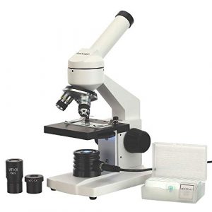
Looking for a microscope that is both affordable and has a wide range of features? Look no further than the AmScope M102C-PB10. This microscope is perfect for students and professionals who need a high-quality microscope that is easy to use. Plus, its range of 40X-1000X makes it ideal for examining samples of all sizes.

- Monocular offers five magnification powers up to 1000X
- Single lens condenser with disc diaphragm
- LED Illumination powered by wall-power
- Sturdy all-metal framework and all optical glass lenses
- 5 blank and 5-prepared slide set included
I am happy with my new microscope! I was worried that it might be too easy to set up, but it wasn’t. The images are apparent and crisp, and the single-lens condenser is perfect. The included 5-prepared slides were great to practice focusing on, and the monocular provided good information and guidance when I subsequently purchased a microscope camera. I’m thrilled with my purchase and would recommend it to other users.
It was a straightforward process regarding the microscope setup, which is excellent as there were so many steps, and I didn’t want anything to confuse me. The image quality right away is top-level seeing, far better than any previous equipment I have used for this purpose! So clear that even on my little 10-inch HD tv, you can see individual particles from 175 miles away from Earth with enough detail to discern what the drop is made of or even know if it has any salt on it!
OMAX – M837ZL-C100U 40X-2500X Microscope
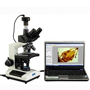
Looking for a microscope that can do it all? Look no further than the OMAX M837ZL-C100U microscope. This microscope boasts an intuitive and easy-to-use control panel, an 8x digital zoom, a bright and vivid display, and a built-in digital camera. With all these features, you’ll be able to explore specimens in greater detail and make more accurate diagnoses. So what are you waiting for? Check out the OMAX LED Microscope today!

- included_components : Full Size Lab Digital Trinocular Compound LED Microscope
This is a fantastic microscope! The size is perfect for smaller labs and has a translation range of 3in x 2in. I also like that it comes with a black palm rest on the base and metal mechanical components. The illumination is transmitted (lower), so you can use this microscope in any light condition.
Additionally, the power supply is compatible worldwide (the US and Canada) and includes a 100V~240V 50 /60Hz range adapter. That alone makes this stand out from others!
I am very disappointed that the included sample does not match what is advertised in detail on the website for this product. This may be a result of translation or just some misunderstanding. Still, it would have been nice to have what was advertised as pictured on OMAX’s site when you open up the box for your microscope–it kills the mystery! (I don’t blame OMAX for this, merely my observation and opinion.)
One other thing that is a little disappointing is the switch to move between high-definition images. Sure there’s an efficient lockout button, but it doesn’t feel like you have much tactility when manipulating the control. That can be overcome with mounting brackets made of balsa wood or learning how anatomy works by gutting flies as fast as possible!
My overall impression of the OMAX model is that it’s a well-made (steel construction), compact product with great features and capabilities. I highly recommend this unit for those who want to study small creatures in their field or for use as an insect trapping device.
MAXLAPTER 100X-2000X Microscope
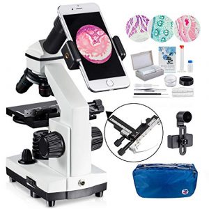
If you’re looking for a microscope that can take crisp images and is easy to use, then the MAXLAPTER 100X-2000X is the perfect option. This microscope lets you easily see clear pictures of small objects and details. Its compact design makes it easy to take with you wherever you go. So what are you waiting for? Check out the MAXLAPTER microscope below to learn more about this fantastic equipment!

- 🔬【𝐄𝐱𝐜𝐞𝐩𝐭𝐢𝐨𝐧𝐚𝐥 𝐂𝐥𝐚𝐫𝐢𝐭𝐲 𝐚𝐧𝐝 𝐌𝐚𝐠𝐧𝐢𝐟𝐢𝐜𝐚𝐭𝐢𝐨𝐧】- Experience unparalleled detail with this advanced monocular microscope, featuring a built-in 2x fully multi-coated (FMC) multiplier lens and a high-quality WF25x eyepiece. With objective lenses of 4x, 10x, and 40x and a 360-degree rotatable head, enjoy magnification from 100x to 2000x for an up-close look at the microscopic world.
- 🔬【𝐏𝐫𝐞𝐜𝐢𝐬𝐢𝐨𝐧 𝐅𝐨𝐜𝐮𝐬𝐢𝐧𝐠】 - Crafted with a robust metal frame and dual coarse and fine focusing knobs, this microscope ensures precise adjustments. Achieve sharp, crystal-clear images every time, perfect for both educational and professional use.
- 🔬【𝐀𝐝𝐯𝐚𝐧𝐜𝐞𝐝 𝐋𝐄𝐃 𝐈𝐥𝐥𝐮𝐦𝐢𝐧𝐚𝐭𝐢𝐨𝐧】- Illuminate your specimens with the top and bottom LED lighting of this microscope, equipped with a 0.65 NA single-lens condenser and disc diaphragm. Whether at home, school, or in a lab, get consistent, brightfield lighting for optimal viewing at any time.
- 🔬【𝐂𝐨𝐦𝐩𝐫𝐞𝐡𝐞𝐧𝐬𝐢𝐯𝐞 𝐀𝐜𝐜𝐞𝐬𝐬𝐨𝐫𝐲 𝐊𝐢𝐭】 - This set includes everything needed for immediate exploration: a smartphone adapter, wire camera shutter, durable carrying bag, operational accessories, and 15 prepared slides. It's all designed to enhance your microscopic journey, right out of the box.
- 🔬 【𝐕𝐞𝐫𝐬𝐚𝐭𝐢𝐥𝐞 𝐄𝐝𝐮𝐜𝐚𝐭𝐢𝐨𝐧𝐚𝐥 𝐓𝐨𝐨𝐥】 - From budding students to amateur scientists, this 100X-2000X biological microscope is an invaluable learning tool. It's ideal for classroom labs, home education, or personal exploration, providing a hands-on educational experience that inspires discovery across all ages.
If you are looking for a high-quality microscope that offers 2000x resolution and is perfect for school or home education, look no further than the MAXLAPTER microscope with a mechanical platform slide set. This microscope will give you the quality eyewear you need to see everything clearly with its outstanding lenses and phone adapter.
Additionally, a built-in light makes viewing slides easy and allows you to see what is under the slide. With a lens made of high-quality Japanese polycarbonate, this microscope will give you excellent optical and mechanical properties that enhance your viewing experience. Also included are two automatic lenses that slide on the microscope stage for quick and easy adjustment of focus depth.
My family has been using the MAXLAPTER 100X-2000X microscope for about a year, and we love it! The moving range is perfect for our needs as it can go up to 40×60 mm. I am also delighted with the clarity of this microscope’s images, especially when using the magnification levels of 100x, 200x, 250x, 500x, 400x, and 1000x.
The LED illumination is also a huge plus and makes it easier to observe the sample without an uncomfortable amount of light shining. Another great thing about this microscope is that you can adjust both sides up or down to help keep from getting any error in your viewing process, which we have needed before when receiving samples for education purposes!
Overall, I recommend investing in one if you want a reasonable selling price and quality components.
Swift SW200DL Compound Monocular Microscope
Do you want to view an object at a very close range with high accuracy? The Swift SW200DL is the perfect tool if your answer is yes. This tool is highly versatile and can be used for various applications such as medical examinations, research, and teaching. Its simple and user-friendly design is perfect for anyone who wants to get the most out of their microscope. Read on to find out more about this powerful compound monocular microscope!
I am writing this product review based on my recent purchase of the Swift Compound Microscope SW200DL. I was looking for a microscope to use at home, and after doing some research, I decided on the SW200DL.

- 【MAGNIFICATION SETTING】Available magnification settings of 40X, 100X, 250X, 400X, and 1000X through aberration-correcting 4X, 10X, and 40X glass objectives with wide-field 10X and 25X eyepieces
- 【Dual lllumination】Dual illumination system allows you to examine both transparent and solid specimens; cool LED lights protect eyesight and live specimens
- 【Excellent material】Rugged design with metal arm and base, carrying handle, and cordless capability make this compound microscope a practical pick for field experiments
- 【Suitable】Designed forDesigned for students/kids/adults/beginner/amateur scientist/hobbyists, perfect for science fairs, at home experiments, and school lab experiments
- 【Features】Fully rotatable monocular head for easy shared use or one-on-one instruction in the classroom. Our customer service is ready at all the time, please contact us if you have any problems
This model is very lightweight and portable; it took only a few minutes to set up using the included instructions. The 40x-1000x magnification capability is excellent for viewing small objects like insects and other specimens.
The SW200DL has a durable design that comfortably fits the hand, allowing for comfortable use. As I started to take some photos of delicate objects under glass slides, I realized that I needed to purchase two synthetic 1-micron grade filters (1mm nominal size) for my camera flash unit’s settings. It will adequately light up the object on film, giving me much better image quality than natural daylight illumination.
I also found that the 35mm lens provided better image quality, especially for photographing insects with their bodies covered in mucus or oils, than using a 60x objective and 100x eyepiece (40× on this microscope).
The Swift SW200DL is perfect for young people, students, and hobbyists who want to make observations at home.
With 40X-1000X magnification available through aberration-correcting 4X, 10X, and 40X glass objectives with wide-field 10X and 25X eyepieces, this microscope has excellent material for examining both transparent and solid specimens.
The dual illumination system allows you to examine solid objects through the objectives and transparent specimens like plant leaves, flowers, or insects. Cool LED lights protect eyesight for both you and live models!
OMAX 40X-2000X LED Microscope
When it comes to viewing microscopy specimens, you need to be able to see small details. That’s where an excellent binocular microscope comes in handy. These microscopes are perfect for examining small objects and are also known as compound microscopes. These microscopes use two lenses to give you a broad and deep view of your specimen. In this section, we will look at the OMAX 40X-2000X LED microscope. We will discuss its features, pros, and cons, and finally, give you our verdict on whether or not this is the suitable microscope for you.
If you’re in the market for a versatile and high-quality microscope, I highly recommend the OMAX 40X-2000X. This microscope has a revolving quadruple nosepiece, double layer mechanical stage with scales, size of 4-1/2inchx 4-15/16inch (115mm x 125mm), translation range of 2-13/16inch x 1 -3/16inch (70mm x 30mm).

- Magnification: The OMAX microscope boasts 4 achromatic DIN objectives and offers 8 levels of high-resolution magnification from 40X to 2000X
- Advanced Illumination: Equipped with variable intensity LED light, this compound microscope ensures clear visualization of microscope slides
- Adjustable Viewing: Features a binocular viewing head with adjustable interpupillary distance and diopter, providing comfortable and precise viewing
- Premium Construction: This microscope for adults includes a double layer mechanical stage with scale, coaxial coarse and fine focus knobs, and a sturdy metal frame
- About AmScope: We have the industry's leading collection of microscopes, microscopes cameras, accessories, and other related products
It sets itself apart by having a solid construction using a stain-resistant enamel finish with a 5-year warranty against manufacturing defects and my favorite features such as focus coaxial coarse and fine knobs on both sides to set the magnification, as well as built-in mechanical stage scale. They also have a replaceable capsule which is nice because sometimes, when microscope parts get older, they don’t guarantee the quality anymore.
Overall this microscope is excellent to use at home or in school and reasonably priced compared to many of its competitors. The only downside is that the colored filters are hard to remove from their holder, with less experience doing so than other people looking for an easy way out.
Case Studies and Applications: Illuminating Real-World Insights

In the dynamic field of parasitology, the application of microscopic techniques in real-world scenarios has profound implications for both diagnosis and research. Examining notable case studies provides valuable insights into the practical applications of these techniques, showcasing their impact on public health and scientific advancements.
1. Clinical Diagnosis
Case Study 1: Malaria Diagnosis Using Giemsa Staining
| Patient Profile | Method Employed | Key Findings |
|---|---|---|
| Symptomatic individual | Giemsa staining | Identification of Plasmodium species in blood |
| Rapid and accurate diagnosis | Facilitation of timely and targeted treatment |
2. Research and Surveillance
Case Study 2: Surveillance of Waterborne Parasites
| Research Objective | Microscopic Technique Employed | Key Outcomes |
|---|---|---|
| Monitoring water quality | Darkfield Microscopy | Detection of waterborne parasites in environmental samples |
| Identification of potential health risks | Facilitation of preventive measures and public health interventions |
What does the malaria parasite look like under a microscope?
Malaria is a deadly disease caused by the parasite Plasmodium falciparum. This parasite is shaped like a long, thin comma and lives in the red blood cells of its host. Under a microscope, malaria parasites can be seen as a colorful array of cells. They can move around and cause damage to the surrounding cells.
Although malaria is not easily treatable, early diagnosis and treatment are essential for the best possible outcome. Understanding how malaria parasites look under a microscope can help in making better decisions when it comes to treatment. Additionally, knowing the parasite can help develop new and more effective anti-malarial drugs.
FAQs
How to count malaria parasites under a microscope?
There are several ways to count malaria parasites under a microscope. Still, the most commonly used methods involve the use of hematoxylin and eosin (H&E) staining and Plasmodium falciparum (P. falciparum) enzyme-linked immunosorbent assay (ELISA). H&E staining is used to identify the presence of red blood cells that have been invaded by parasites, while ELISA is used to determine the number of parasites in a sample.
Counting malaria parasites under a microscope is essential in diagnosing and treating the disease. Knowing the number and location of parasites, you can develop a treatment plan that is most effective for you.
Can you see parasites in a light microscope?
Yes, parasites can be seen in a light microscope. However, because parasites are small and delicate, it is often difficult to see them. It is best to use higher magnification to get a better view. Parasites can be seen as small, round, dark spots with a characteristic “horn” or “nose.”
How to find parasites using a microscope?
Using a microscope to find parasites can be a handy tool in a physician’s diagnostic arsenal, as it can help identify and diagnose various health conditions. Parasites can be found in multiple places, including the digestive, respiratory, and urinary systems.
The most common parasites detected using a microscope are Giardia lamblia and Cryptosporidium parvum. Giardia is a parasite found in the intestines, and Cryptosporidium is a parasite found in the water and soil. These parasites can cause significant health complications if not diagnosed and treated properly.
By using a microscope, doctors can also identify other types of parasites, such as Toxoplasma gondii, which is associated with an increased risk of schizophrenia and other mental health issues, and Trichinella spiralis, which is associated with Chronic Gastrointestinal Illness (CGI).
How to identify chicken parasites under a microscope?
Under a microscope, chicken parasites can be easily identified as small, elongated creatures with a head and two pairs of legs. They are usually red but can also be green or black. The parasites live inside the chicken’s intestines and feed on the blood and other tissues in the intestines. They can cause severe health problems for the chicken, including weight loss, diarrhea, and anemia. If you suspect that your chicken has chicken parasites, it is best to get it checked out by a veterinarian.
How can you identify fecal parasites microscope?
There are a few ways to identify fecal parasites, including examining the stool for the presence of eggs, larvae, or adults, performing a caulicolous culture, or completing a microscope. Coproscope is a special microscope that can identify parasites by their morphology, and it is also the most reliable method for diagnosing helminthic infections.
How to measure parasite size with a Leica microscope?
Parasites are tiny creatures that can cause significant health problems. One of the best ways to measure parasite size is with a Leica microscope. This microscope can provide detailed images of parasites that can help diagnose and track the infection’s progress. Additionally, it can assist in determining the disease’s severity and the best course of treatment.
Before using the Leica microscope, make sure that you understand the principles of light and optics. Once you have set up the microscope and are ready to start collecting data, use a light source that emits short and long-wavelength light.
This process will enable you to see different types of parasites in detail. Next, adjust the focus and magnification to get a perfect view of the parasites. Finally, take notes on the other characteristics of the parasites and their location for future reference.
By using a Leica microscope, you can significantly contribute to the diagnosis and treatment of parasites.
What setting should my microscope be on to see parasites?
The best setting to see parasites would be under a light microscope, allowing you to know the organism more clearly and distinguish between different species. Under a standard microscope, parasites appear as dark dots or tiny black spots.
Benefits
A microscope can be a great way to identify and diagnose parasites, including those that might be difficult to see with the naked eye. Using a microscope, you can see the detail of parasites that would be otherwise invisible to the human eye. This process can help diagnose and treat parasites, as well as provide information that can be used in developing new antiparasitic drugs.
A microscope can help identify parasites, but it can also be used to study their life cycle. By examining the stages of parasite development, you can develop better anti-parasitic strategies. Additionally, a microscope can identify parasites in food and water sources. By doing this, you can prevent the spread of parasites and help to protect the public.
Microscopes are also a great way to teach students about the fundamentals of anatomy and physiology. Using a microscope, students can learn about the structure and function of cells and organs.
What are some of the challenges with using a Microscope for Parasites?
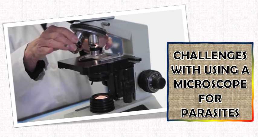
Microscopes can be a valuable tool for researchers, physicians, and others who work with parasites. However, they can also be challenging to use, as they can be expensive, require specialized training, and sometimes require special equipment. Additionally, they can be delicate, and if not handled properly, they can be damaged.
Another challenge with using a microscope is the difficulty of seeing small details, which is why it is essential to use a well-suited microscope for the task at hand. Additionally, it is necessary to take proper notes and make accurate measurements to diagnose and treat parasites correctly.
How much does a microscope for Parasites cost?
A microscope for parasites can cost anywhere from 50$ to 200$, depending on the type and quality of the microscope. The most popular models include the Nikon Coolpix S4200, Olympus BX51, and the Pentax WG-4. All of these microscopes offer high-quality images and are perfect for studying parasites.
When choosing a microscope, it is essential to consider the purpose for which it will be used. For instance, a cheaper model might be sufficient if you are looking for a microscope for school projects. If, however, you plan on using the microscope for professional purposes such as diagnosis or research, a higher-quality model may be more appropriate. It is also essential to consider the microscope’s size; a cheaper model might be sufficient if used in a small or limited space.
How to clean a microscope for Parasites?
Cleaning a microscope for parasites can be a daunting task, but with the help of a few simple steps, it can be done in a matter of minutes. The first step is to remove any dust or debris on the microscope using a vacuum cleaner. Then, use a mild soap and water solution to clean the eyepieces and lenses. Finally, dry the microscope using a soft cloth. Please store it in a clean and dry environment to keep it in good condition.
How to set up a microscope for Parasites?
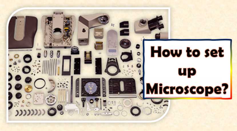
Setting up a microscope for parasites can be a fun and educational experience, and it can also be a valuable tool in your diagnostic arsenal. Microscopes are often used to view objects smaller than what the naked eye can see. This is great for examining parasites such as lice, bed bugs, and ticks.
To get started, you will need to gather the necessary materials. These include a slide, a microscope, and a light source. The slide should contain a section on the parasite you wish to examine. The microscope should have a high-power objective lens, and the light source should be a bulb that emits ultraviolet or infrared light.
Before viewing the parasites, it is essential to set up the microscope properly. This includes adjusting the focus and the magnification. Once everything is set, it is time to take a look! You will be able to see the parasites magnified up to 10,000 times.
How to store a microscope which is used for researching Parasites?
If you are a researcher who uses a microscope for studying parasites, it is essential to take proper care of it. The microscope should be stored in a dry and dark place to prevent damage. Additionally, it should be protected from moisture and dust. If you keep the microscope for an extended period, it is best to wrap it in a plastic bag.
The best way to store a microscope is to keep it in a dark and dry place. If you store it for an extended period, it is also essential to protect it from moisture and pests. Some suggested storage methods include using a corkboard or wooden frame or placing it in a glass or plastic container. It is also good to store the microscope in its original packaging to protect it from damage.
How do you fix a microscope for Parasites?
A few steps need to be taken to fix a microscope for parasites. The first step is to remove the lens if it is damaged or dirty. Next, clean the inside of the eyepiece and the objective lens with a soft cloth or a lens-cleaning solution.
Remove any dust, dirt, or other particles obstructing the view. Finally, you need to apply a solution of lens cleaning fluid to a clean cloth and wipe it across the lens and eyepiece. You can also use a blower to clean the eyepiece if it is difficult to access.
Final Words
I recommend selecting one of these microscopes above due to their outstanding features. The price range is also very affordable, and they have the flexibility that may answer what type of job you need your microscope for. Overall, I believe AmScope – M102C-PB10 40X-1000X Microscope is the best microscope for parasites because of its remarkable features and phenomenal price! It includes a powerful motor for focusing and easy handling. Besides all this, it comes with great accessories meaning that you do not need extra accessories like most microscopes.
To find the best microscope for your specific job needs, you can go to any laboratory supply store and make a comparison there. I hope this review has helped you find a suitable microscope to review school projects with!

I am an enthusiastic student of optics, so I may be biased when I say that optics is one of the most critical fields. It doesn’t matter what type of optics you are talking about – optics for astronomy, medicine, engineering, or pleasure – all types are essential.
Last update on 2025-07-15 / Affiliate links / Images from Amazon Product Advertising API
Table of Contents

Pingback: We Tested Wireless Digital Microscopes to See If They Work: Including a Comparison with Video Guide