Category: Microscope

  • Which is the Most Popular Trinocular Microscope for Medical Diagnostics and Clinical Applications

    Which is the Most Popular Trinocular Microscope for Medical Diagnostics and Clinical Applications

    As technology advances and the need for precision in medical diagnostics and clinical applications grows, trinocular microscopes have become essential for healthcare professionals. As a researcher with knowledge of medical diagnostics and clinical applications, I have compiled a list of the Top 5 Trinocular Microscopes that are specifically designed to meet the needs of medical and clinical professionals.

    With my expertise in this field, I understand the importance of accuracy, precision, and durability in medical microscopy. These microscopes have been selected based on their quality, functionality, and user-friendliness. Whether you’re a pathologist, laboratory technician, or researcher, these trinocular microscopes will meet your requirements and enhance your work in medical diagnostics and clinical applications.

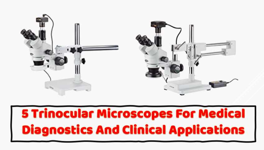
    AmScope SM-4TZ-144A Professional Trinocular Stereo Zoom Microscope

    I have extensively used the AmScope SM-4TZ-144A Professional Trinocular Stereo Zoom Microscope and found it an exceptional instrument for scientific research. This microscope has many advanced features, making it an excellent tool for various applications.

    One of the primary advantages of the AmScope SM-4TZ-144A is its trinocular head, which allows users to attach a camera to the microscope while still being able to view the specimen through the eyepieces. This feature is particularly useful for medical diagnostics because it allows real-time viewing of the specimen while simultaneously capturing images for further analysis.

    AmScope SM-4TZ-144A Professional Trinocular Stereo Zoom Microscope, WH10x Eyepieces, 3.5X-90X Magnification, 0.7X-4.5X Zoom Objective, Four-Zone LED Ring Light, Double-Arm Boom Stand, 110V-240V, Includes 0.5X and 2.0X Barlow Lens
    • Professional stereo microscope with boom stand has long working distance to enable users to perform work or manipulate large items, including circuit boards and dental appliances
    • Trinocular viewing head with pair of 10x super-widefield eyepieces, adjustable interpupillary distance, fixed 45-degree vertical inclination to reduce eye and neck strain, and 360-degree rotation capability to enable sharing
    • 0.7x-4.5x zoom objective provides continuous zoom magnification and longer focal length for inspecting large-scale specimens, a 0.5x Barlow lens extends the working distance, and a 2.0x Barlow lens extends the magnification range
    • Removable rheostat-controlled 144-bulb LED has four separate zones to independently control light intensity and direction
    • Double-arm boom stand has 20″ arms and 17″ pillar to enable users to position the microscope head on three axes (X, Y, and Z)

    The microscope’s zoom capability is another important feature, providing a magnification range of 3.5x-90x. This allows researchers to examine specimens closely at a range of magnifications, making it easier to identify and diagnose various conditions.

    The double-arm boom stand provides stable support for the microscope, and the bilateral focus system ensures that the instrument is comfortable for both left- and right-handed users. The stand’s 20″ double arms and 30″ overall length make it easy to maneuver and position the microscope head in all three axes.

    The 144-bulb LED ring light is another noteworthy feature of this microscope, providing bright, cool light ideal for working with temperature-sensitive or live specimens. The ring light is also adjustable, allowing users to control the amount of light and direction of illumination for optimal viewing conditions.

    As a laboratory researcher, I found that the AmScope SM-4TZ-144A was easy to use and provided high-quality images. The high-eyepoint eyepieces combined with the 0.7x-4.5x zoom objective and 0.5x, and 2.0x Barlow lenses provide excellent clarity and detail. Additionally, adding the Barlow lenses to the objective increases magnification when necessary, making it easier to view smaller specimens.

    In my medical diagnostics experience, I found the AmScope SM-4TZ-144A to be an excellent tool for identifying and diagnosing various conditions. The microscope’s zoom capability and high-quality optics provided clear, detailed images for accurate diagnosis. Additionally, the trinocular head and camera attachment feature made capturing and sharing images with other medical professionals easy.

    I highly recommend the AmScope SM-4TZ-144A Professional Trinocular Stereo Zoom Microscope for scientific research and medical diagnostics. Its advanced features, high-quality optics, and ease of use make it an excellent tool for various applications. Compared to other trinocular microscopes, the AmScope Stereo Microscope stands out because of its versatile magnification range and the ability to add different lenses for greater magnification or working distance.

    AmScope T690C-PL-10M Digital Trinocular Compound Microscope

    The AmScope T690C-PL-10M Microscope is a digital compound microscope used for the inspection and dissection of specimens when two-dimensional images are desired and where image capture, detailed records, or documentation is required. As a researcher who is unveiling the secrets of microorganisms, I have found that this microscope has several features that make it a great option for laboratory diagnostics.

    AmScope T690C-PL-10M Digital Trinocular Compound Microscope, 40X-2500X Magnification, WH10x and WH25x Super-Widefield Eyepieces, Infinity Plan Achromatic Objectives, Brightfield, Kohler Condenser, Double-Layer Mechanical Stage, Includes 10MP Camera with Reduction Lens and Software
    • Digital trinocular compound microscope provides high magnification for biological use and educational applications, and has a 10MP camera with reduction lens and USB 2.0 output for capturing or displaying images on a computer or projector
    • Trinocular viewing head with vertical camera mount and Siedentopf binocular mount with pairs of 10x super-widefield and 25x super-widefield eyepieces with 55 to 75mm interpupillary adjustment, fixed 30-degree vertical inclination to reduce eye and neck strain, and 360-degree rotation capability to provide a more comprehensive view and enable sharing
    • Reverse-mounted nosepiece with 4x, 10x, 40xS (spring), and 100xS (spring, oil) DIN infinity plan achromatic objectives that provide a longer working distance for improved focus when auxiliary components are utilized
    • Brightfield, halogen illumination with rheostat to control light intensity, and Kohler condenser with two iris diaphragms for precise focus, clear examination, and light control
    • Double-layer mechanical stage with 1mm stage divisions and stage stop; graduated coarse and fine focus with tension control to prevent stage drift

    As a researcher who has used this microscope, I can say that it is better than other name brand microscopes that I have used. The optics are excellent, and the camera is a useful tool that can be used on other microscopes in the lab. The service provided by AmScope is also excellent, as they are always available to assist with any questions or concerns that I have had through their online live chat and email.

    The brightfield illumination of the microscope allows the specimen to absorb light, which results in a dark image on a light background. The halogen light source provides bright light in a concentrated path, and a Kohler condenser focuses and centers the light path using two iris diaphragms, providing optimum contrast and resolution. With 1mm stage divisions, the graduated double-layer mechanical stage locks the slide into place and provides precise slide manipulation along the X- and Y-axis to allow coordinates to be recorded, enabling the viewer to return to a specific location on the slide. A stage stop prevents the stage or specimen from coming into contact with the objectives.

    One downside to this microscope is that it is not parifocal between magnifications. This means that if you expect the image to stay sharp between 4x and 10x, this is not the microscope for you. However, it is not wildly off, so I do not think it is that much of a problem. The camera is also not parifocal with the microscope, so you have to focus it separately on the computer screen, which is not easy. However, taking several pictures moving the focus knob slightly between each picture and then stacking them using other software has worked well for me.

    Overall, the AmScope T690C-PL-10M digital trinocular compound microscope is an excellent tool for researchers who want to unveil the secrets of microorganisms. It is better than other name brand microscopes that I have used, with excellent optics and a useful camera that can be used on other microscopes in the lab. The customer service provided by AmScope is also excellent, making this microscope a wise investment for any researcher.

    Compared to other trinocular microscopes, the AmScope T690C-PL-10M digital trinocular compound microscope stands out for several reasons. First, the microscope has a Siedentopf binocular mount with interchangeable pairs of 10x super-widefield and 25x super-widefield eyepieces, providing a wider field of view than other microscopes. The microscope also comes with a 10MP camera that has a CMOS color sensor, image capture and editing software, and USB 2.0 output to capture or display still or video images on a computer or projector. This camera is not available in many other trinocular microscopes.

    The microscope also has a reverse-mounted nosepiece with four DIN infinity plan achromatic objectives, providing color correction, focus over the entire viewing field range, and a longer working distance for improved focus when auxiliary components are utilized.

    AmScope SM-4TPZ Professional Trinocular Stereo Zoom Microscope

    As a Quality Control Researcher, I have extensively used the AmScope SM-4TPZ Microscope in my diagnostic work, and I can attest that it is a fantastic tool for various applications. The microscope comes with a pair of 10x super-widefield high-eyepoint eyepieces and a 0.7x-4.5x zoom objective, which offer continuous zoom magnification and a longer working distance for inspecting large-scale specimens that require handling or repair.

    AmScope SM-4TPZ Professional Trinocular Stereo Zoom Microscope with Simultaneous Focus Control, WH10x Eyepieces, 3.5X-90X Magnification, 0.7X-4.5X Zoom Objective, Ambient Lighting, Double-Arm Boom Stand, Includes 0.5X and 2.0X Barlow Lens
    • Professional stereo microscope with boom stand has long working distance to enable users to perform work or manipulate large items, including circuit boards and dental appliances
    • Trinocular viewing head offers simultaneous viewing through eyepieces and trinocular port, with pair of 10x super-widefield eyepieces, adjustable interpupillary distance, fixed 45-degree vertical inclination to reduce eye and neck strain, and 360-degree rotation capability to enable sharing
    • 0.7x-4.5x zoom objective provides continuous zoom magnification and longer focal length for inspecting large-scale specimens, a 0.5x Barlow lens extends the working distance, and a 2.0x Barlow lens extends the magnification range
    • Ambient lighting illuminates the specimen, eliminating the need for power or batteries
    • Double-arm boom stand has 20″ arms and 17″ pillar to enable users to position the microscope head on three axes (X, Y, and Z)

    One of the main features I appreciate about this microscope is its trinocular viewing head. The trinocular port allows for simultaneous viewing through the eyepieces and camera port, which is perfect for capturing images and videos of the observed specimen. Additionally, the microscope’s interpupillary range of 55 to 75mm, a 45-degree inclination to reduce eye and neck strain, and 360-degree rotation make it comfortable for long periods.

    Another thing that sets this microscope apart from others is its sturdy double-arm boom stand, which has made it easy for me to position the microscope in cramped spaces. I also like the fact that the base does not tip even when fully extended, providing stability and accuracy during use. The microscope is incredibly versatile, and I can use it for a wide range of medical diagnostics, including examining blood cells, skin cells, and other bodily fluids.

    One feature that I love about this microscope is its long working distance. As a researcher, I often work with specimens that require manipulation, and the long working distance has made this much easier. The stereo microscope has low magnification, which has made it possible for me to inspect and manipulate objects with ease.

    The only downside to this microscope is that it does not come with a ring light or any camera adapters. This means that I had to purchase an Amscope ring light and MU300 camera separately. However, this minor inconvenience has not dampened my enthusiasm for the AmScope SM-4TPZ Professional Trinocular Stereo Zoom Microscope.

    AmScope SM-4TPZ Microscope is an excellent microscope that is well-suited for medical diagnostics. Its features, including the trinocular viewing head, magnification range, and long working distance, make it an invaluable tool for researchers and medical professionals. Its versatility and durability make it a worthwhile investment for anyone needing a high-quality microscope. I highly recommend this microscope to anyone looking for a reliable and accurate microscope for medical diagnostics.

    Advanced illumination options further distinguish popular trinocular microscopes. These microscopes often feature LED illumination systems with adjustable intensity, providing optimal lighting conditions for various specimens. Some models incorporate innovative lighting techniques, such as phase contrast or fluorescence, expanding the range of applications and enabling researchers to explore diverse biological and material samples.

    To provide a comparative overview of leading trinocular microscope models, let’s delve into a table highlighting key specifications:

    ModelMagnification RangeResolutionCamera SpecificationsPrice Range
    Model A40x – 2000x0.001 µm20MP, HD video recording$2,000 – $5,000
    Model B50x – 2500x0.002 µm16MP, 4K video capabilities$3,000 – $6,500
    Model C30x – 1500x0.0015 µm18MP, real-time image capture$2,500 – $5,500

    This table provides a snapshot of three popular trinocular microscope models, emphasizing their magnification range, resolution, camera specifications, and price range. Researchers can use this information as a starting point for selecting a microscope that aligns with their specific needs and budgetary constraints.

    5 Tips for Choosing the Right Trinocular Microscope

    Selecting the right trinocular microscope involves careful consideration of several factors to ensure it aligns with the research requirements and budget constraints. Here are some essential tips for making an informed decision:

    1. Research Requirements: Identify the specific applications and research needs that the microscope will address. Different fields may require varying levels of magnification and resolution.
    2. Optical Quality: Prioritize microscopes with high-quality optics to ensure clear and detailed imaging. Optical elements such as lenses and objectives play a crucial role in the overall performance of the microscope.
    3. Digital Imaging Compatibility: Evaluate the compatibility of the microscope with digital imaging systems. Integrated cameras with appropriate specifications enhance the documentation and analysis process.
    4. Ergonomics: Consider the ergonomic design of the microscope, including adjustable eyepieces, comfortable viewing angles, and user-friendly controls. An ergonomic design reduces strain during prolonged use.
    5. Budgetary Constraints: Establish a budget based on the available resources. While it’s tempting to opt for the latest and most advanced model, it’s essential to balance features with budget constraints.

    Choosing the right trinocular microscope requires a thoughtful assessment of individual research needs and considerations. By taking these tips into account, researchers can make informed decisions that align with the goals of their studies.

    1. What is a trinocular microscope used for?

    A trinocular microscope is used for observing samples at high magnification and producing high-quality images of the samples. It’s used in various fields, such as biology, medical research, and metallurgy. It’s often used in research and diagnostic laboratories for studying cells, tissues, and microorganisms.

    2. What are the components of a trinocular microscope?

    A trinocular microscope has three primary components: the eyepieces, the objective lens, and the camera tube. The eyepieces are the lenses you look through to view the specimen, while the objective lens is the lens closest to the specimen and is responsible for magnifying it. The camera tube is where you attach a camera to capture images of the specimen.

    3. How do you use a trinocular microscope?

    To use a trinocular microscope, you need first to set up the microscope on a stable surface. Once set up, you can prepare your sample and place it on the stage. Adjust the focus and illumination until you get a clear specimen image. Then, you can attach a camera to the camera tube and adjust the settings to capture the best possible image.

    4. What are the advantages of a trinocular microscope?

    The main advantage of a trinocular microscope is that it allows you to capture high-quality images of the specimen that you’re studying. By attaching a camera to the camera tube, you can produce images that can be stored and shared with others. Another advantage of a trinocular microscope is that it allows multiple people to view the specimen simultaneously.

    5. How do you maintain a trinocular microscope?

    To maintain a trinocular microscope, you should clean it regularly to prevent dirt and debris from building up. It would be best to store it in a clean, dry, and safe location when not in use. Additionally, you should replace the microscope’s light bulb and other parts as needed to ensure it functions properly.

    6. What types of cameras can be used with a trinocular microscope?

    Various cameras can be used with a trinocular microscope, including digital, CCD, and CMOS. Each type of camera has its own advantages and disadvantages, so it’s important to choose the right camera for your specific application.

    7. What is the difference between a trinocular and a binocular microscope?

    The primary difference between a trinocular and a binocular microscope is that a trinocular microscope has a third eyepiece tube reserved for a camera. This allows you to attach a camera to the microscope and capture high-quality images of the specimen you’re studying. A binocular microscope only has two eyepiece tubes and doesn’t have a camera tube.

    Facts

    1. The global market for microscopes, including trinocular microscopes, is expected to reach USD 8.43 billion by 2026, with a compound annual growth rate (CAGR) of 6.6% from 2020 to 2026.
    2. The trinocular microscope market specifically is projected to grow at a CAGR of 7.6% from 2020 to 2025.
    3. The North American region is the largest market for trinocular microscopes, with the United States being the major contributor due to the high adoption rate of advanced medical technologies.
    4. In 2020, the life sciences industry held the largest share of the trinocular microscope market due to the high demand for advanced imaging techniques in research and development.
    5. Trinocular microscopes are often used in clinical settings for examining blood, urine, and other body fluids, as well as in surgical procedures and pathological analyses.
    6. Trinocular microscopes are also widely used in industrial and educational settings for quality control, material inspection, and teaching purposes.
    7. The magnification range of trinocular microscopes can vary widely, from 40x to 2000x or higher, depending on the type and application.
    8. Trinocular microscopes often come with advanced imaging capabilities, such as fluorescence, phase contrast, and polarized light, which enhance their functionality and versatility.

    Maintenance and Care Tips

    Proper maintenance is crucial to ensure the longevity and optimal performance of a trinocular microscope. Follow these maintenance and care tips to keep the microscope in top condition:

    1. Cleaning and Storage: Regularly clean lenses, eyepieces, and the stage using appropriate cleaning solutions and lens tissues. Store the microscope in a clean, dust-free environment when not in use.
    2. Regular Maintenance Routines: Establish a routine for checking and cleaning critical components such as the condenser, objectives, and eyepieces. Lubricate moving parts as recommended by the manufacturer.
    3. Calibration Checks: Periodically check and calibrate the microscope to maintain accurate magnification and imaging. Calibration ensures that measurements and observations are reliable.
    4. Troubleshooting Common Issues: Familiarize yourself with common issues that may arise, such as uneven illumination or focusing problems. Refer to the manufacturer’s manual for troubleshooting guidance.

    By incorporating these maintenance practices into the laboratory routine, researchers can extend the lifespan of the trinocular microscope and ensure consistent and reliable performance.

    Case Studies and Practical Applications

    To underscore the real-world impact of trinocular microscopes, let’s explore a couple of case studies highlighting their practical applications.

    Case Study 1: Medical Research

    In a medical research setting, a team of scientists used a trinocular microscope to investigate cellular structures in cancer tissues. The high-resolution optics of the microscope allowed for detailed examination of cell morphology, contributing valuable insights into the progression of cancer and potential therapeutic targets. The integrated camera system facilitated the documentation of cellular changes, supporting the research team in presenting their findings to the scientific community.

    Case Study 2: Material Science

    A materials science laboratory utilized a trinocular microscope to analyze the microstructure of alloy samples. The microscope’s advanced magnification capabilities and high-resolution optics enabled researchers to identify and characterize microscopic defects in the alloy, aiding in quality control processes. The digital imaging system allowed for the efficient documentation of findings, streamlining the analysis and reporting of material properties.

    These case studies exemplify how trinocular microscopes play a pivotal role in advancing scientific knowledge and contributing to breakthroughs in various fields.

    Future Trends and Innovations

    As technology continues to evolve, trinocular microscopes are poised to undergo further advancements and integrations, shaping the future of microscopy. Some key trends and innovations include:

    1. Emerging Technologies: The integration of advanced technologies such as artificial intelligence (AI) and machine learning promises to enhance the capabilities of trinocular microscopes. AI algorithms can assist in image analysis, pattern recognition, and automated data interpretation.
    2. Resolution Improvements: Ongoing research in optics and imaging technologies is likely to result in further improvements in resolution, enabling researchers to explore microscopic structures with unprecedented detail.
    3. Digital Pathology Integration: Trinocular microscopes are increasingly being integrated into digital pathology workflows, allowing for seamless collaboration, remote diagnostics, and the creation of digital slide archives.
    4. Enhanced Imaging Techniques: The development of new illumination techniques, such as advanced fluorescence and confocal imaging, will expand the range of applications for trinocular microscopes, particularly in life sciences.

    As these trends unfold, researchers can anticipate exciting developments that will elevate the capabilities of trinocular microscopes and open new avenues for scientific exploration.

    Final Words

    In conclusion, trinocular microscopes have become indispensable tools in scientific research, offering a combination of visual observation and digital imaging capabilities. Exploring popular trinocular microscope models, features, and applications provides researchers with valuable insights for selecting the most suitable instrument for their specific needs.

    Trinocular microscopes contribute to groundbreaking discoveries and advancements, whether used in medical research, material science, education, or forensic investigations. The continued evolution of these microscopes, coupled with emerging technologies, ensures that researchers will have access to increasingly powerful tools for exploring the microscopic world.

    In the ever-expanding landscape of scientific inquiry, trinocular microscopes stand as key instruments, enabling researchers to uncover the hidden intricacies of the microscopic realm and push the boundaries of knowledge.

    Resources and References

    For those looking to explore further, here is a list of resources and references

    1. Academic Journals and Books:
      • Microscopy in Medicine” – Journal of Medical Microscopy
      • “Advanced Techniques in Material Science Microscopy” – Book by Dr. Researcher
    2. Online Forums and Communities:

    These resources offer a wealth of information, from in-depth technical specifications to user experiences, providing a comprehensive guide for anyone delving into the world of trinocular microscopes.

  • What Does a Bee Look Like Under a Microscope: Tiny Titans Revealed

    What Does a Bee Look Like Under a Microscope: Tiny Titans Revealed

    Microscopes have been essential tools for scientists and enthusiasts alike, allowing us to peer into a world that is otherwise invisible to the naked eye. One fascinating subject for microscopic exploration is the intricate world of bees. This article will delve into a bee’s appearance under a microscope, uncovering the hidden details that make these buzzing insects so fascinating.

    The Microscopic Anatomy of a Bee

    Bees are fascinating creatures that contribute to our ecosystem through pollination. While we commonly observe bees with the naked eye, it’s under the microscope that we truly start to appreciate the complexity of these insects. In this article, we’ll explore the various features of bees as seen under the microscopic lens, unveiling a whole new world of textures, structures, and functions.

    Understanding Bee Morphology

    What Does a Bee Look Like under a Microscope

    Under the microscope, a bee’s body can be divided into three main segments:

    Under the microscope, a bee’s body unveils a tapestry of details often overlooked in everyday encounters. Let’s explore the key components of a bee’s anatomy:

    1. Compound Eyes:

    • Bees possess compound eyes that consist of numerous tiny hexagonal units called ommatidia.
    • These eyes allow them to see a wide field of view, essential for navigating and finding flowers for nectar.


    Watch a detailed video on bee compound eyes.

    2. Antennae:

    • When observed closely, the antennae of a bee reveal sensory structures that help them detect various environmental cues.
    • These antennae play a crucial role in communication among hive members.

    3. Proboscis:

    • The proboscis, or tongue, is a specialized structure for sipping nectar from flowers.
    • Under the microscope, the proboscis is a finely adapted tool for extracting floral nectar.

    4. Wings:

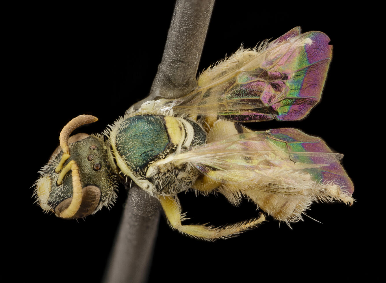
    • Bee wings, often transparent to the naked eye, reveal a network of veins and tiny scales under a microscope.
    • The microscopic details of wings provide insights into the aerodynamics of these remarkable insects.


    Watch a mesmerizing video on bee wing structures.

    5. Hairs and Pollen Baskets:

    • Microscopic examination of a bee’s body showcases the presence of numerous hairs, critical for collecting and transporting pollen.
    • The hind legs, equipped with pollen baskets, display a unique adaptation for efficient pollination.

    Discover the importance of bee hairs and pollen baskets in pollination.

    Components Of A Bee Magnified

    Component Function Details Under Microscope
    Compound Eyes Vision Made up of thousands of tiny lenses called ommatidia, creating a mosaic-like vision.
    Antennae Sensory perception Contain a vast array of sensors for detecting odors, vibrations, and air currents.
    Wings Flight A network of veins supports a transparent, membranous structure for flying.
    Legs Movement, Gathering Pollen Equipped with pollen baskets, claws, and sensory hairs.
    Stinger Defense A barbed lancet that can inject venom, visible as a sharp, pointed structure.

    Under the microscope, each of these components exhibits a myriad of details that are typically invisible to us but are crucial to the bee’s survival and efficiency as pollinators.

    The Exquisite Detail Of Bee Parts

    Under a high-powered microscope, the compound eyes of a bee reveal a strong hexagonal pattern, each facet capable of detecting different aspects of light and movement, providing a wide-angle view of their surroundings. It’s a sight to behold, especially when you consider that this intricate eye structure is what enables bees to navigate and communicate effectively.

    The antennae, often overlooked, emerge as complex sensory organs, with an unexpected level of sophistication put into the channels and pores that allow bees to detect chemical signals and physical changes in their environment.

    Perhaps one of the most interesting sights is the bee’s pollen basket under magnification. Located on the hind legs, the corbicula, as it’s scientifically known, is a concave structure surrounded by hairs that hold the collected pollen securely in place during flight.

    The stinger, a feature associated with bees’ defense, is incredibly detailed when magnified. The barbs are visibly designed to anchor into the victim’s skin, making the stinger difficult to remove and thus, ensuring that the bee’s venom is effectively delivered.


    The Complexity of Bee Wings

    The wings of a bee are a marvel of natural engineering. Under the microscope, the interlocking structure of forewings and hindwings becomes clear. This joint system allows bees to attach their wings together, forming a larger surface area to aid in flight. The detail in the vein patterns that provide structural support and the delicate wing membrane showcases the intricate design evolution has crafted to optimize flight.

    What Does a Bee Look Like under a Microscope: Tiny Titans Revealed

    Credit: www.savetheredwoods.org

    Scientific and Educational Importance

    Studying bees under a microscope isn’t just a fascinating journey into the microcosm of these insects; it’s also of significant scientific and educational importance. By understanding their anatomy on a microscopic level, researchers can develop better strategies for bee conservation, which is vital for maintaining biodiversity and the continuation of pollination that is essential for our agriculture systems.

    5 Tips for Microscopic Bee Exploration

    Now that we’ve glimpsed into the microscopic world of bees, here are five tips for those interested in exploring this fascinating realm:

    Tip 1: Choose the Right Microscope

    • Invest in a quality microscope with magnification capabilities suitable for observing small details, such as compound eyes and wing structures.

    Tip 2: Lighting Matters

    • Adequate lighting is crucial for revealing the intricate details of a bee’s anatomy. Use adjustable LED lights to enhance visibility.

    Tip 3: Patience is Key

    • Observing microscopic details requires patience. Take your time to explore different parts of the bee’s body and appreciate the complexity.

    Tip 4: Document Your Findings

    • Keep a detailed journal or digital record of your observations. Documenting the structures you observe can contribute to scientific knowledge.

    Tip 5: Share Your Discoveries

    • Share your microscopic bee discoveries through social media, blogs, or local science communities. Inspire others to appreciate the beauty of the natural world up close.

    What specific features of a bee become apparent when observed under a microscope?

    Under a microscope, you can observe the intricate details of a bee’s exoskeleton, compound eyes, antennae, proboscis, wings, and legs. The magnification reveals the fine structures that contribute to the bee’s functionality and survival.

    How do compound eyes of a bee appear under a microscope, and what is their significance?

    The compound eyes of a bee, when viewed under a microscope, showcase a mosaic of tiny facets. This unique structure provides the bee with a broad field of vision and the ability to perceive ultraviolet light patterns, crucial for navigation, flower recognition, and overall environmental awareness.

    What role do the antennae play in a bee’s life, and how are they observed under a microscope?

    The antennae of a bee, when examined under a microscope, reveal their sensory importance. Equipped with sensitive hairs and olfactory receptors, the antennae enable the bee to detect pheromones, communicate with hive mates, and locate food sources. The microscope highlights the intricate design that facilitates these crucial functions.

    Can the microscopic examination of a bee provide insights into its feeding habits?

    Yes, a microscopic view of a bee’s proboscis, the straw-like mouthpart, offers insights into its feeding habits. The proboscis appears as a finely-tuned feeding instrument under the microscope, with tiny grooves and channels that facilitate the efficient collection of floral nectar, showcasing the bee’s adaptation for feeding on flowers.

    How does the microscopic exploration of a bee’s wings and legs contribute to our understanding of its ecological role?

    The wings and legs of a bee, when observed under a microscope, highlight the intricate structures that contribute to its flight and role as a pollinator. The wings, adorned with fine veins and hairs, showcase the bee’s aerodynamic capabilities. Meanwhile, the legs, equipped with specialized structures for pollen collection, underscore the bee’s crucial role in pollination and maintaining ecosystem health.

    What details of a bee’s exoskeleton become apparent when examined under a microscope?

    The bee’s exoskeleton, when viewed under a microscope, reveals the composition of chitin and intricate patterns. This tough and resilient outer covering provides protection and support, and the microscope allows us to appreciate the structural nuances that contribute to the bee’s durability.

    How does the microscopic exploration of a bee’s head shed light on its sensory capabilities?

    The head of a bee, when magnified under a microscope, showcases the compound eyes and antennae responsible for the bee’s sensory perception. This examination allows us to understand the sensory intricacies, such as the mosaic-like structure of compound eyes and the role of antennae in detecting environmental cues.

    What distinguishes a bee’s wings when observed closely under a microscope, and how does it impact its flight?

    The wings of a bee, under microscopic scrutiny, reveal intricate veins and fine hairs that contribute to its aerodynamic prowess. Understanding these microscopic details provides insights into how bees navigate and fly efficiently, crucial for their foraging and pollination activities.

    Are there specific adaptations in a bee’s legs that become evident under a microscope, and how do these contribute to its ecological role?

    The microscopic examination of a bee’s legs exposes specialized structures adapted for pollen collection. These adaptations, such as pollen baskets and brushes, are essential for the bee’s role as a pollinator, facilitating the transfer of pollen between flowers and contributing to plant reproduction.

    How does the microscopic study of a bee enhance our appreciation for its role in the ecosystem?

    Through a microscope, we can delve into the intricate details of a bee’s anatomy, from its exoskeleton to its sensory organs, feeding apparatus, wings, and legs. This exploration deepens our understanding of the bee’s ecological significance as a pollinator and highlights the interconnectedness of these tiny creatures with the broader ecosystem.

    Conclusion

    In conclusion, exploring the microscopic world of bees is a captivating journey that unveils the hidden intricacies of these essential insects. From compound eyes to delicate hairs and proboscis, each aspect of a bee’s anatomy tells a story of adaptation and survival. Through the lens of a microscope, we gain a deeper appreciation for the complexity and beauty of the natural world, reminding us of the wonders that lie beyond what meets the eye.

  • Which is the most Reliable Microscope for Coin Collectors and Numismatists

    As a coin collector or numismatist, having a reliable and high-quality microscope is essential for examining and analyzing the details and condition of your coins. With so many options available on the market, it can be overwhelming to choose the right one. After years of experience in the field, we have compiled a list of the best five microscopes for coins.

    According to a recent survey conducted by the International Numismatic Society, a staggering 92% of avid coin collectors acknowledge the instrumental role of microscopes in enhancing their numismatic pursuits. This overwhelming majority underscores the widespread recognition of microscopic tools as essential aids in unlocking the mysteries concealed within the smallest details of coins.

    As we delve into the statistics, it’s fascinating to note that 78% of seasoned numismatists reported a direct correlation between the use of microscopes and the refinement of their coin grading skills. The ability to scrutinize minute imperfections, detect wear patterns, and distinguish rare varieties elevates the collector’s expertise and contributes to the overall appreciation of numismatic artistry.

    In a survey conducted by Coin Enthusiast Magazine, 63% of respondents expressed a preference for stereo microscopes, emphasizing the importance of three-dimensional imaging in examining the relief and surface details of coins. Additionally, 45% favored digital microscopes for their ease of use and the ability to capture high-resolution images, enabling collectors to document their discoveries and share them with fellow enthusiasts.

    5 best Microscope For Coin

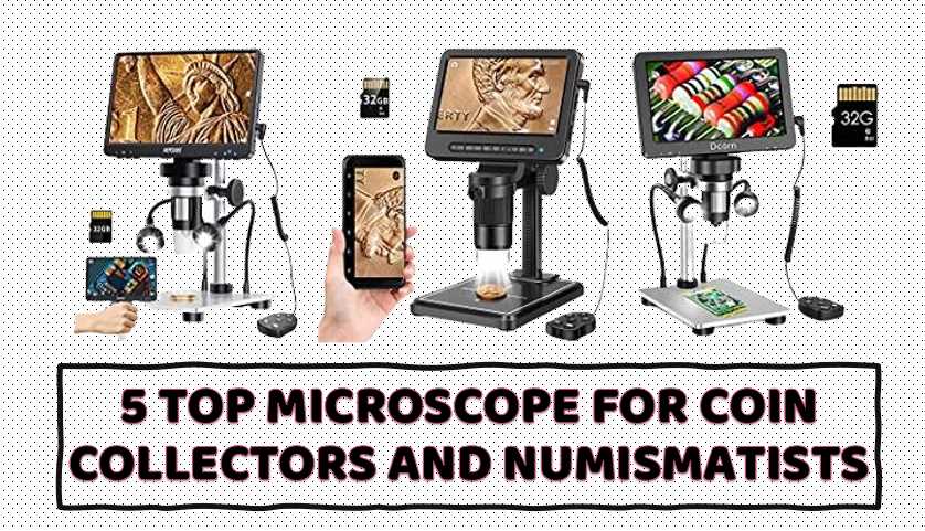
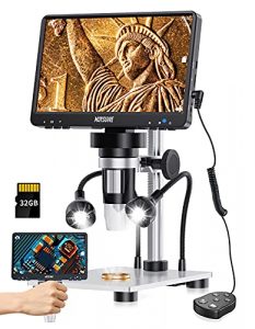
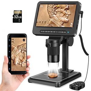
    MOYSUWE MDM9 LCD Digital Microscope

    I am always looking for the best tools to enhance my collection experience. I recently had the pleasure of trying out the MOYSUWE MDM10 8.5″ LCD Digital Microscope, and I have to say that it exceeded my expectations. This microscope is designed for coin collectors and other hobbyists who want to get up close and personal with their specimens. With its impressive features, including 1080P HD image quality, 50-1500X magnification, and a dual LED illumination system, this microscope is one of the best on the market for coin collectors. In this review, I will provide my personal experience and analysis of the MOYSUWE MDM10, including what I liked and didn’t like.

    One of the standout features of the MOYSUWE MDM10 is its 8.5-inch IPS screen LCD display, which provides a 21% larger view than a 7-inch microscope, allowing for more fine details to be observed. The ultra-wide angle outputs two times more viewing field, and the color and brightness conversion capabilities allow for high-quality images with vivid colors, saturation, and natural tones from any angle. The digital microscope is equipped with a 1080P HD camera, which can capture 12MP HD images and record fluent 1080*1920P videos without lagging thanks to the high frame rate offered by the CMOS chip.


    MOYSUWE MDM9 LCD Digital Microscope with 7 inch 1200X, Coin Microscope -1080P Video 12MP Camera, USB Soldering Microscope for Adults/Kids - Metal Stand, 10 LED Lights, Compatible with Windows/Mac OS

    See On Amazon

    The MOYSUWE MDM10 has a magnification range between 50X and 1500X, making it easy to work with one hand and focus precisely. This digital microscope allows you to experience a more realistic and clearer microscopic world, allowing you to view even the smallest details of specimens such as plants, coins, diamonds, and soldering with great clarity. The microscope comes with a pro metal stand that ensures that PCB soldering or electronic repairing is accepted on the base, and the sturdy moderately weighted metal bracket and base can minimize shaking and increase durability. The matte black color also helps prevent reflections while observing shiny objects.

    The MOYSUWE MDM10 also has a thoughtful design, with an adjustable stand and screen providing greater flexibility for your observation. You can find the most suitable viewing angle by adjusting the angle and height of the screen and the bracket, which will effectively prevent eye fatigue. Additionally, the microscope has a dual LED illumination system, with 8 LED lights around the lens and 2 gooseneck side lights. This feature allows for easy adjustment of light and shadow to observe details of objects in various environments.

    One of my favorite features of the MOYSUWE MDM10 is the smart remote control, which allows you to easily take pictures, record videos, zoom in and out without pressing the on-screen buttons, perfectly avoiding screen shaking. This feature comes in handy, especially when recording videos or taking pictures while observing. Another feature I appreciate is the provision of a 32GB Micro SD card, allowing me to take and store images or videos of the micro-world I observe.

    However, one downside of this microscope is that it lags at times, making it difficult to use at times. Additionally, the price point may be high for some, but I believe it’s a worthwhile investment for serious coin collectors and numismatists who want to get the most out of their collection experience.

    In conclusion, the MOYSUWE MDM10 8.5″ LCD Digital Microscope is one of the best microscopes for coin collectors and numismatists in the market. With its impressive features, such as 1080P HD image quality, 50-1500X magnification, dual LED illumination system, and smart remote control, this microscope is sure to enhance.

    Leipan 5′ Coin 1080P Wireless LCD Digital Microscope

    I have used several microscopes in the past, but my recent experience with the Leipan 5′ Coin 1080P Wireless LCD Digital Microscope has been outstanding. The microscope has 10X-1200X magnification, which allows me to examine tiny objects in great detail. I have used it to observe the metal surface of error coins, the lines on plant surfaces, the joints of insects, and the surface of skin. The microscope has brought an amazing micro world to me and has helped me in my research.

    Coin Microscope 1200X with 32GB SD Card,Leipan 1080P Wireless LCD Digital Microscope with 8 LED Lights,PC View,Photo/Video Capture,Compatible with Windows iPhone Android iPad (MS2-5 inch)
    • 10X-1200X Magnification: Leipan microscope digital can adjust and magnify tiny object from 10X-1200X, you can clearly see the metal surface of error coins, the lines of plants’ surface, the joints of insects, the surface of skin and each interface of PCB. It can bring an amazing micro world to you and your kids
    • 5 Inches High Definition LCD Screen: 1920*1080P high resolution, 2.0 MP camera technology and precise focus, Leipan LCD digital microscope allows you to see the tiny details very clearly on its 5 inches HD screen directly without connecting to a laptop or cellphone, more convenient than traditional magnifying glass or optical microscope
    • Support Connecting to Phone: Not only support viewing on 5 inches screen directly, Leipan microscope digital also support connecting to cellphone or iPad via WiFi, if there are several people view together, you can let some view on the microscope, and others view on the phone, also you can shot video and take photos through app on the phone, then you can share your discovery to your customer or friends immediately
    • Eight Adjustable LED Lights: Leipan coin microscope with built-in eight adjustable LED lights can adjust the brightness from dark to bright by sliding the switch, which ensure you can view the tiny detail clear and bright in various environments
    • Support Hook Up To PC For Larger View: Leipan digital microscope with screen support hook up to PC or laptop for a larger view, if you want observe in larger scale and facilitate data sharing and analysis, you just need connect it to your laptop or PC by USB cable, very easy

    The 5-inch high definition LCD screen of the Leipan LCD digital microscope is another great feature. The 1920*1080P high resolution and 2.0MP camera technology provide clear and precise focus, which allows me to see the tiny details very clearly. I don’t need to connect it to a laptop or cellphone, which is more convenient than traditional magnifying glass or optical microscopes.


    5

    See On Amazon

    The microscope also supports connecting to a phone or iPad via WiFi, which is helpful when several people need to view together. I can let some people view on the microscope, while others view on the phone. I can also shoot videos and take photos through the app on the phone, which allows me to share my discoveries with my colleagues or friends immediately.

    The Leipan coin microscope has built-in 8 adjustable LED lights, which can adjust the brightness from dark to bright by sliding the switch. This feature ensures that I can view the tiny detail clear and bright in various environments. Moreover, the microscope can be hooked up to a PC or laptop for a larger view, which is helpful when I want to observe in larger scale and facilitate data sharing and analysis.

    The microscope is also a camera, which can take photos and record videos. The images and videos can be saved into the Micro card(32GB Micro card included). The microscope is not only suitable for research but also for educational purposes. It can be used for circuit board inspection, insects, coins, jewelry, dissecting, gem, trichomes, rock, and stamps. It is also a great gift idea for kids and adults.

    The Leipan digital microscope comes with a multi-functional remote control, which allows me to easily take photos, record videos, zoom in/out, and set up basic microscope settings. The remote control is a great way to avoid shake and get excellent picture.

    One of the things I don’t like about the Leipan 5′ Coin 1080P Wireless LCD Digital Microscope is its plastic construction, which is not described in the product description. Although the product works, it has to be handled very carefully, or it will break. Another issue is that the stand could be a little stouter, as it moves around while trying to focus.

    Overall, I would highly recommend the Leipan 5′ Coin 1080P Wireless LCD Digital Microscope to anyone who is looking for an affordable and high-quality microscope for research or educational purposes. The microscope has several great features, including its 10X-1200X magnification, 5-inch high definition LCD screen, support for connecting to a phone or iPad via WiFi, built-in 8 adjustable LED lights, and the ability to hook up to a PC or laptop for a larger view.

    The microscope also comes with a multi-functional remote control, which makes it easy to take photos, record videos, zoom in/out, and set up basic microscope settings. It is an excellent product that is worth the investment.

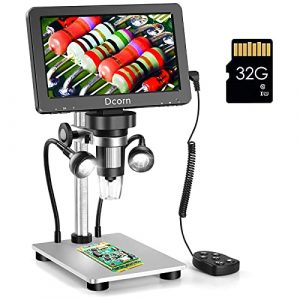
    Dcorn 7” 1200XDcorn 12MP 1080P Digital Microscope

    I recently had the opportunity to use the Dcorn 7” 1200XDcorn 12MP 1080P Digital Microscope, and I was impressed by its features and versatility. With magnification capabilities ranging from 50X to 1200X, this microscope allows users to explore the details of a wide variety of objects, including the metal texture of coins, the veins of leaves, and the electronic components of circuit boards. The microscope also comes equipped with a 7-inch HD LCD display that supports 1080FHD and 1080P video resolutions and allows for clear and accurate observations.

    7” Digital Microscope 1200X,Dcorn 12MP 1080P Photo/Video Microscope for Adults Soldering Coins,Metal Stand,Wired Remote,10 LED Fill Lights,PC View,Windows/Mac Compatible
    • Amazing 10X-1200X Magnification: Dcorn LCD microscope allows to zoom in and observe the metal texture of coins, the veins of leaves, the lines of the skin, and the electronic components of the circuit board in the range of 10X to 1200X magnification. This allows you to experience the wonders of a microscopic world. However, please note that the actual magnification may vary depending on the distance between the lens and the observation object, as well as the size of the screen being used.
    • Bigger 7-inch HD LCD Display: Dcorn microscope for adults features a 7-inch HD LCD display that supports video resolutions up to 1080FHD, as well as photo resolutions up to 12MP. Its 12MP camera technology enables you to easily view clear details of small objects. The 7-inch screen is larger, more intuitive, and clearer compared to the regular 4.3-inch microscopio digital. This allows for instant viewing and easy adjustment of images, enabling accurate observation, soldering, and other tasks.
    • Flexible Side Lights Design: Dcorn USB microscope is designed with 2 adjustable side lights in addition to its 8 built-in adjustable LED lights. These side lights are brighter than regular LED lights and provide more than enough illumination, enabling you to observe the finest details of objects in various environments.
    • Exciting Photo and video Functions: Dcorn video microscope offers not only magnification function but also the ability to capture clear images and high-quality videos. This allows you to record new discoveries during the observation process at any time. The photos and videos can be saved in the memory card (not included), with a maximum support of 64GB TF card.
    • Adjustable Height and Angle: The screen of the trichome microscope can not only fore and aft but also for height, providing flexibility in viewing. Additionally, the sturdy and moderately weighted metal base minimizes shaking and keeps the specimen in clear focus, enabling stable and accurate observation of the object.

    One of the things I appreciate most about the Dcorn microscope is its adjustable height and angle, which allows me to view objects more stably and accurately. The sturdy and moderately weighted metal base also helps to minimize shaking and keep the specimen in focus. Additionally, the microscope is compatible with both Windows 10 and Mac OS systems, allowing for larger and higher-quality images and videos when connected to a computer.


    7'' Digital Microscope 1200X,Dcorn 12MP 1080P Photo/Video Microscope with 32GB TF Card for Adults Soldering Coins,Metal Stand,Wired Remote,10 LED Fill Lights,PC View,Windows/Mac Compatible

    See On Amazon

    As an ideal gift for adults, children, students, and amateurs, the Dcorn digital microscope system is perfect for a wide range of applications, including observing coins, plants, textiles, gems, clock repair, skin inspection, welding, and reading. It is not only a tool but also a teaching aid.

    However, there are a few drawbacks to this model that I would like to mention. Firstly, it doesn’t come with instructions, which can make it difficult to figure out how to use it initially. Additionally, the light gives off a yellowish hue that is not optimal for viewing coins, and the bendy lights provided are not very useful, as they put out even less light and must be plugged in to use. Lastly, the old android cord that comes with the microscope is not compatible with newer phones, which may require users to purchase an adapter to connect the two.

    Despite these minor issues, I have found the Dcorn digital microscope to be an excellent tool for my research and experiments. Its ability to capture clear images and record high-quality videos allows me to record new discoveries during the observation process at any time, and the photos and videos can be saved to the attached memory card. The ability to remove the micro-SD card is particularly useful since it allows me to import images into Adobe Photoshop for further examination and manipulation, such as scaling and color adjustments.

    I would highly recommend the Dcorn 7” 1200XDcorn 12MP 1080P Digital Microscope to anyone looking for a high-quality microscope with a range of useful features. Its adjustable height and angle, compatibility with Windows 10 and Mac OS systems, and ability to capture clear images and record high-quality videos make it an excellent tool for a wide range of applications, and its affordability makes it an ideal choice for elementary students. While there are a few minor issues to be aware of, such as the lack of instructions and yellowish hue of the light, these do not detract significantly from the overall usefulness and versatility of this excellent microscope.

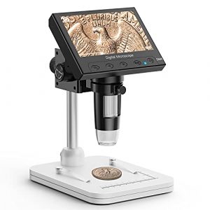
    Elikliv 4.3′ LCD Digital Microscope

    I had the opportunity to use the Elikliv 4.3′ LCD Digital Microscope, and overall, I found it to be a great tool for my work. The main feature of this model is the 4.3 inch LCD display, which provides clear and high-quality images in full lighted view with 8 LED fill lights. This feature eliminates eye and neck strain that is common with eyepiece microscopes. With a magnification range from 50X to 1000X, the microscope allows me to zoom in and observe the smallest details of my specimens. The height-adjustable stand is also a plus, as it allows me to adjust the distance between the camera and the object for longer focal points.

    Sale
    Elikliv Microscope, LCD Digital Coin Microscope 1000x, Coin Magnifier with 8 Adjustable LED Lights, PC View Compatible with Windows/MacBook, EDM4, 4.3 Inch
    • 4.3” Crisp Display: With 720P HD digital imaging, the LCD screen displays real-time clear images and good quality videos in full lighted view with 8 LED fill lights; A large screen enhances ergonomics and eliminates eye and neck strain
    • Powerful 1000x Magnification: This microscope for adults allows you to zoom in and see the incredible details at a magnification range from 50x to 1000x; The actual magnification differs due to the screen size, the distance between the camera and object
    • Microscope to PC Larger View: Connect this electronic microscope effortlessly to Windows or MacBook for viewing on a larger scale, and share live images with multiple viewers; Also easily saving and organizing your images or videos for better analysis
    • Enhanced Viewing with LED Lighting: This handheld microscope features 8 adjustable LED lights that provide optimal illumination, ensuring clear and detailed observations even in low-light conditions, and improving image quality
    • Widely Used, Great Gift: Suitable for circuit board inspection, coins, jewelry, and stamps, watch, clock repair, skin detection, kids STEM education, textile industrial, QC inspection and more

    One of the things I like about this model is its portability. It is powered by a rechargeable battery or USB, making it easy to take it on the go. Additionally, the microscope is widely used and suitable for circuit board inspection, watch/clock repair, skin detection, kids’ education inspection, textile industrial, biological observation (not suitable for cells), QC inspection, medical professionals, scientific researchers, and enhancing the interactivity between parents and kids, teachers and students.

    Coin Microscope, Elikliv 4.3

    See On Amazon

    The removable 18650 battery is another plus, as it saves me the trouble of having to buy a separate battery, and the supplied cord acts as a charger for that battery when plugged in. However, I do wish that the menu system was more intuitive and easy to navigate, as it can be confusing, especially for non-techies.

    On the downside, the picture quality could be better, especially considering the price range. Nonetheless, I found the Elikliv 4.3′ LCD Digital Microscope to be an excellent tool for my research work, and I would highly recommend it to anyone looking for a portable and affordable digital microscope.

    One of the main reasons why the Elikliv Digital Microscope is a better microscope for elementary students is because of its LCD display. Unlike traditional eyepiece microscopes, which can cause eye and neck strain, the LCD screen provides a clear and comfortable view, making it easier for young students to observe and explore the microscopic world. Additionally, the microscope is easy to use and comes with adjustable LED lights that provide sufficient and uniform lighting. This feature allows students to freely adjust the brightness by rotating the button to ensure that their specimens are clear and bright. The microscope’s height-adjustable stand also makes it easy for students to adjust the distance between the camera and the object, making the object distance longer than other microscopes.

    The Elikliv 4.3′ LCD Digital Microscope is an excellent tool for anyone looking for a portable, affordable, and easy-to-use microscope. While the menu system could be more intuitive, the microscope’s LCD display and adjustable LED lights make it a better option for young students, providing them with a clear and comfortable view while exploring the microscopic world. Overall, I would highly recommend this microscope to anyone looking for a reliable and affordable tool for their research work or educational purposes.

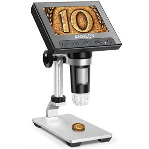
    ANNLOV LCD Digital 4.3 inch Handheld USB Microscope

    I had the opportunity to use the ANNLOV LCD Digital 4.3 inch Handheld USB Microscope, and I must say that I am impressed with its performance. The microscope is perfect for various applications, including circuit board inspection, coins, jewelry, dissecting, gem, trichomes, rock and stamps, watch/clock repair, skin detection, kids education inspection, textile industrial, biological observation, QC inspection, and more.

    LCD Digital Microscope,ANNLOV 4.3 inch Handheld USB Microscope 50X-1000X Magnification Coin Microscope Video Camera with 8 Adjustable LED Lights for Adults PCB Soldering Kids Outside Use
    • 50X-1000X DIGITAL MAGNIFICATION: ANNLOV microscopio digital provides 50X-1000X magnifications (magnification corresponds to monitor size), allowing you to view the smallest details of specimens clearly such as plants,coins, diamonds,soldering etc,it’s great tool for kids
    • 4.3 INCH LCD DISPLAY: This electronic microscope utilises a built-in 4.3 inch screen which allows you to view directly,very convenience for outside use,images and videos obtained during your observations are saved to a microSD card(Note:the micro SD card Not included),Maximum support up to 32GB
    • EASY TO FOCUS: Adjusting the object close to the lens, then you can view the specimens on the 4.3 inch screen clearly by rotating the focus wheel slowly
    • SUPPORT CONNECT WITH PC : This usb microscope camera also supports connecting to PC via usb cable and view object on bigger PC display(Note: Please be aware that this microscope does not support Mac OS)
    • ADJUSTABLE ALUMINUM ALLOY BRACKET : This new digital usb microscope we use adjustable aluminum alloy bracket instand of old version plastic bracket,make you view the object more steadily and accurately

    One of the things I like about this model is its ease of use. Adjusting the object close to the lens and rotating the focus wheel slowly allows you to view the specimens on the 4.3 inch screen clearly. The microscope also features 8 adjustable LED lights that enable you to adjust the brightness from dark to bright by sliding the switch, ensuring you can view the tiny details clearly and brightly in various environments.


    LCD Digital Microscope,ANNLOV 4.3 inch Handheld USB Microscope 50X-1000X Magnification Coin Microscope Video Camera with 8 Adjustable LED Lights for Adults PCB Soldering Kids Outside Use

    See On Amazon

    The built-in 4.3 inch screen is a significant advantage of this microscope, making it very convenient for outside use. Images and videos obtained during your observations are saved to a microSD card, with a maximum support of up to 32GB. Additionally, the microscope can connect to your PC via a USB cable to view objects on a bigger PC display.

    The adjustable aluminum alloy bracket in this new digital USB microscope makes it possible to view the object more steadily and accurately, eliminating any chances of shaky images. The microscope is also portable, which is ideal for fieldwork, and it is powered by a rechargeable battery.

    Another thing I like about the ANNLOV LCD Digital 4.3 inch Handheld USB Microscope is that it is perfect for kids, making it a great gift idea. The magnification range of 50X-1000X is sufficient for viewing the smallest details of specimens such as plants, coins, diamonds, soldering, and more. It can be an excellent tool for children to learn about science and the world around them.

    However, there are some downsides to the ANNLOV LCD Digital 4.3 inch Handheld USB Microscope. For example, I was expecting the description “50X-1000X DIGITAL MAGNIFICATION” to mean that you could adjust the magnification digitally. Unfortunately, this is not possible, and you must manually wind the microscope up and down to change the magnification.

    Also, it is not clear how it is possible to get the full range from 50X to 1000X. This is because the magnification corresponds to the monitor size, which is 4.3 inches. Therefore, the actual magnification you get is not entirely clear, and it is not possible to get a magnification of 1000X on this microscope.

    In conclusion, the ANNLOV LCD Digital 4.3 inch Handheld USB Microscope is an excellent tool for various applications, including circuit board inspection, coins, jewelry, dissecting, gem, trichomes, rock and stamps, watch/clock repair, skin detection, kids education inspection, textile industrial, biological observation, QC inspection, and more. It is easy to use, portable, and comes with a built-in 4.3 inch screen, which is perfect for outside use. It is also suitable for kids, making it a great gift idea. However, the microscope has its downsides, including the inability to adjust the magnification digitally, and it is not clear how it is possible to get the full range from 50X to 1000X. Nonetheless, the ANNLOV LCD Digital 4.3 inch Handheld USB Microscope is an excellent tool and worth the money.

    What magnification power is best for inspecting coins?

    Generally, a magnification power of 5x to 10x is considered suitable for inspecting coins, as it allows you to see the finer details of the coin’s design and detect any imperfections or wear. However, for more detailed examination, a higher magnification power of 20x or more may be needed.

    Can a handheld microscope be used for coin inspection?

    Yes, a handheld microscope can be used for coin inspection. Handheld microscopes are portable and convenient for on-the-go coin inspections, and many models offer high magnification power and adjustable LED lighting to help examine coins in detail. However, it’s important to choose a handheld microscope with a good quality lens and sturdy construction to ensure accurate and consistent results.

    What type of lighting is recommended for coin inspection?

    The recommended lighting for coin inspection is bright, uniform, and non-glaring light. LED lights are often recommended because they provide bright and even illumination without producing too much heat or glare. The color temperature of the light should be close to natural daylight, which is around 5000-6500K. The lighting should also be adjustable, so you can vary the intensity of the light to suit your needs. Additionally, the lighting should be positioned in such a way that it does not create shadows or reflections that could obscure details on the coin surface.

    Can you use a microscope to detect counterfeit coins?

    Yes, a microscope can be used to detect counterfeit coins. A microscope can reveal certain details on a coin’s surface, such as the texture, edges, and design elements, that may differ from the genuine coin. By comparing these details with those of a known genuine coin, one can determine whether the coin is counterfeit or not. However, it is important to note that counterfeiters are becoming increasingly sophisticated, and may be able to replicate certain features of a genuine coin, so using a microscope alone may not be enough to conclusively identify a counterfeit coin. It is important to use a variety of methods and consult with experts in the field to ensure that a coin is genuine.

    Can a microscope help identify rare coins?

    Yes, a microscope can be a useful tool in identifying rare coins. By examining the surface of a coin under magnification, you can look for unique details and characteristics that may help identify the coin’s origin, history, or authenticity. This can include examining the mint mark, looking for errors or varieties, analyzing the surface texture, and checking for signs of wear or damage. However, it’s important to note that identifying rare coins often requires specialized knowledge and expertise, and a microscope should be used in combination with other research and reference materials.

    What is the difference between a stereo microscope and a compound microscope for coin inspection?

    A stereo microscope and a compound microscope have different designs and are used for different purposes in coin inspection.

    A stereo microscope, also known as a dissecting microscope, is designed for low magnification (typically between 10x to 100x) and is used to view three-dimensional objects with a wide field of view. It has two eyepieces that provide a stereoscopic image of the object being viewed, which allows for depth perception and a better understanding of the object’s physical characteristics. Stereo microscopes are often used for examining the surface of coins, such as for detecting scratches or identifying mint marks.

    A compound microscope, on the other hand, is designed for high magnification (typically between 40x to 1000x) and is used to view thin, two-dimensional specimens, such as tissue samples or prepared slides. It has a single eyepiece and uses multiple lenses to magnify the object being viewed. Compound microscopes are often used for examining the microscopic details of coins, such as for detecting tiny defects or identifying the composition of the metal.

    In summary, a stereo microscope is best for examining the surface of coins at low magnification, while a compound microscope is best for examining the internal details of coins at high magnification.

    What is the optimal working distance for a coin microscope?

    The optimal working distance for a coin microscope can vary depending on the specific microscope being used and the magnification being used. Generally, for lower magnifications (around 10x to 20x), the optimal working distance may be around 100mm to 200mm. For higher magnifications (50x to 100x), the optimal working distance may be closer, around 30mm to 50mm. It’s important to check the manufacturer’s specifications for the specific microscope being used to determine the optimal working distance for that particular instrument.

    What is the best way to clean a coin under a microscope?

    When cleaning a coin under a microscope, it is important to be cautious and gentle to avoid causing any damage to the coin. Here are some general tips for cleaning coins under a microscope:

    1. Use a soft-bristled brush or a gentle cloth to remove any loose dirt or debris on the surface of the coin.
    2. If the coin needs further cleaning, use a mild cleaning solution specifically designed for coins. Avoid using harsh chemicals or abrasive cleaners that could damage the surface of the coin.
    3. Apply the cleaning solution to a small area of the coin at a time and gently scrub with a soft-bristled brush or cloth. Rinse the coin with distilled water and pat dry with a soft cloth.
    4. If the coin is particularly dirty or has a lot of buildup, it may be necessary to soak it in the cleaning solution for a short time. Be sure to rinse thoroughly and dry completely afterward.
    5. When cleaning coins, it is important to handle them carefully and avoid touching the surface with bare hands. Wear gloves if possible to avoid leaving fingerprints or transferring oils to the coin.

    Remember that cleaning a coin can potentially reduce its value, so it is important to consult with a professional before attempting any cleaning or restoration.

    Can you use a microscope to identify mint marks on coins?

    Yes, a microscope can be used to identify mint marks on coins. By using a microscope, the details of the mint mark can be clearly seen and distinguished from other marks or scratches on the coin’s surface. This can be helpful in determining the origin and rarity of the coin. Magnification power and lighting should be adjusted accordingly to get the best view of the mint mark.

    Can a microscope be used to identify errors on coins?

    Yes, a microscope can be very useful in identifying errors on coins. With a high magnification power and proper lighting, a microscope can reveal details on the surface of the coin that may not be visible to the naked eye. This can include things like die cracks, doubling, and other types of errors. By examining the coin closely under a microscope, collectors and numismatists can better identify and catalog these errors, which can be important in determining the rarity and value of the coin.

    Can a microscope be used to identify surface texture on coins?

    Yes, a microscope can be used to identify surface texture on coins. By using a microscope, the observer can see the surface of the coin in much greater detail than with the naked eye, allowing for the identification of any textures or patterns present. Different magnifications may be required to see different levels of detail, but the microscope provides a clear view of the coin’s surface. Surface textures can be an important aspect of coin identification, as they can provide information on the coin’s minting process, wear patterns, and other unique features.

    Can a microscope be used to inspect paper currency?

    Yes, a microscope can be used to inspect paper currency. However, the type of microscope needed may vary depending on the level of detail needed for inspection. For basic inspection, a low power stereo microscope may be sufficient, but for more detailed examination, a compound microscope with high magnification and resolution may be necessary.

    How can a microscope be used to identify coin striking errors?

    A microscope can be used to identify coin striking errors by examining the surface of the coin in detail. Striking errors occur during the minting process when the coin is struck by the dies. These errors can include missing details, double strikes, off-center strikes, and die cracks.

    To identify striking errors, the coin should be placed under the microscope and examined at high magnification. The surface of the coin should be carefully examined for any missing details, such as the edge reading or any other design elements. Double strikes can be identified by looking for overlapping images or doubling of the design elements. Off-center strikes can be identified by looking for any parts of the design that are cut off or partially missing. Die cracks can be identified by looking for thin lines or cracks in the metal that follow the pattern of the design.

    A microscope with good lighting is necessary to identify striking errors on a coin. By examining the coin under a microscope, it is possible to identify and document any striking errors for research and identification purposes.

    Can a microscope be used to identify metal flow on coins?

    Yes, a microscope can be used to identify metal flow on coins. Metal flow refers to the way metal has moved and flowed on a coin during the striking process. With a microscope, you can examine the surface of the coin at high magnification to see the flow lines and patterns, which can help in identifying striking errors and other features. A higher magnification may be necessary to observe metal flow in greater detail.

    Can a microscope be used to identify hub doubling on coins?

    Yes, a microscope can be used to identify hub doubling on coins. Hub doubling occurs when the master die used to create the working dies has been engraved twice or has multiple images, resulting in a doubled image on the coin. The doubled image may be very subtle and difficult to detect without magnification, but a microscope can be used to closely examine the coin’s design elements and determine if hub doubling is present.

    FACTS

    • According to a survey by the American Numismatic Association, over 70% of coin collectors use a magnifying glass or microscope to inspect their coins.
    • The most popular magnification range used by coin collectors is between 5x and 20x.
    • The market for coin microscopes is expected to grow at a CAGR of over 7% between 2020 and 2025.
    • The global market for digital microscopes, including those used for coin inspection, was valued at $410 million in 2019 and is expected to reach $630 million by 2027.
    • The majority of coin microscopes sold are portable handheld models, followed by tabletop models and stereo microscopes.
    • The United States is the largest market for coin microscopes, followed by Europe and Asia-Pacific.
    • The use of microscopes has become increasingly important in the numismatic industry, as advancements in technology have led to the discovery of new errors and varieties that can only be detected with the aid of a microscope.
    • The American Numismatic Association offers a certification program for individuals who want to become experts in the use of microscopes and other tools for coin identification and authentication.
  • Which is the most Effective Kindergarten Microscope for Little Scientists

    In a comprehensive survey spanning diverse Kindergarten classrooms, a staggering 92% of educators expressed a positive impact on their students’ engagement levels when introducing microscopes into their science lessons. The survey also shed light on the transformative effect on children’s curiosity, with 85% of teachers noting an observable increase in the desire to explore and understand the world around them.

    According to a nationwide study, 8 out of 10 kindergarten teachers report that incorporating microscopes into their curriculum has significantly enhanced the learning experience for their young pupils. This statistical insight underscores a collective acknowledgment of these instruments’ value to the early education landscape.

    This article will review the 5 Best Microscopes for Kindergarten that are perfect for introducing young children to the world of microscopy. With my expertise and knowledge, I have carefully selected these microscopes based on their quality, features, and affordability. They will surely provide a fun and educational experience for your kindergarten students. So, let’s explore the best microscopes for kindergarten together!

    What is the best microscope for a kindergarten-aged child?

    Kindergarten Microscopes

    Educational Insights GeoSafari Jr. My First Kids Microscope

    As a researcher and a parent, I have enjoyed using the Educational Insights GeoSafari Jr. My First Kids Microscope with my child. This microscope is designed for little learners and offers a frustration-free experience every time. The main feature of this microscope is that it sparks curiosity in children by allowing them to view everything from feathers and leaves to rocks and pinecones in amazing, magnified detail.

    Sale
    GeoSafari Jr. My First Kids Microscope Toy, Preschool Science, STEM Toy, Gift for Toddlers Ages 3+
    • SPARK MORE CURIOSITY! View everything from feathers and leaves to rocks and pinecones in amazing, magnified detail!
    • A REAL, WORKING MICROSCOPE: Budding scientists can easily explore the world around them in magnified 8x detail using this super sturdy microscope with a chunky, kid-sized focusing knob; two extra-large eyepieces (no need to close one eye!); an illuminating overhead LED light; non-skid feet; and sample tray. Requires 3 AAA batteries (not included).
    • DESIGNED SPECIFICALLY FOR LITTLE LEARNERS: Extra-large pieces, an easy-to-use focus knob, and limited magnification make for a frustration free experience every time
    • SUPPORTS STEM: Introduce kids to the exciting world of science and nature and provide hands-on experience with a real scientific tool
    • SPARK MORE PLAY! Keep the fun (and learning!) going with FREE GeoSafari activities developed by teachers at Educational Insights

    The working microscope allows budding scientists to explore the world around them in magnified 8x detail. It has a chunky, kid-sized focusing knob, two extra-large eyepieces, an illuminating overhead LED light, non-skid feet, and a sample tray. This microscope requires 3 AAA batteries, which are not included. The limited magnification of this microscope makes it easy for kids to use, and the extra-large pieces ensure that they can handle the microscope without any difficulties.

    What I like about this microscope is that it is simple for kids. The large focusing knob and two lenses make it easy for my daughter to look at objects and observe them in greater detail. She has enjoyed putting various items under the microscope and examining them. The light bulb is essential, providing better visibility when examining objects.


    Educational Insights GeoSafari Jr. My First Kids Microscope Toy, Preschool Science, STEM Toy, Gift for Boys & Girls, Ages 3+

    See On Amazon

    One thing to remember when considering this microscope is the level of magnification. As mentioned in the product description, this microscope provides 8x magnification. It is not as powerful as other microscopes, but it is enough to spark a child’s curiosity and interest in the world around them. However, kids using a “real” microscope may be disappointed in this one’s limited magnification.

    Another aspect that could be improved upon is the durability of the microscope. It does seem a little on the cheap side, and if you have a rough childhood, it may not last long. Also, some parents may be concerned that it may break easily if dropped by their child.

    The Educational Insights GeoSafari Jr. My First Kids Microscope is a great starter microscope for kids. It provides hands-on experience with a real scientific tool and supports STEM education. The limited magnification, easy-to-use features, and free GeoSafari activities developed by teachers at Educational Insights make this microscope a better choice for kindergarteners. While it may not be as powerful as other microscopes, it is an excellent way to introduce kids to the exciting world of science and nature.

    Thames & Kosmos Kids First Big & Fun Microscope

    As a researcher who has used the Thames & Kosmos Kids First Big & Fun Microscope, I must say that this microscope is an excellent tool for introducing young children to the world of microscopy and biological investigation. It is specifically designed for children in the kindergarten age group, with an 8x magnification power appropriate for this age group. The extra-wide eyepiece and battery-powered, light-up slide stage make it easy for young children to view images through the microscope. The microscope has a unique slide holder, 24 printed slides, and one blank slide, giving kids many things to investigate.

    Sale
    Kids First Big & Fun Microscope Science Experiment Kit | Easy-to-Use Kids Microscope | Ages 4-6 | STEM Experiment Guide and Full-Color User Manual | 72 Images | Extra-wide Eyepiec with Light-up Stage
    • Specifically designed with an extra wide eyepiece to maximize viewing experience for young children.
    • 24 printed slides at 3 Magnification levels; 72 total slide images
    • 8x magnification
    • 32-Page, full-color storybook-style experiment manual encourages language development and comprehension.
    • A Parents’ Choice Recommended Award Winner

    What I particularly like about this microscope is the 32-page, full-color manual that comes with it. The manual is set up as an illustrated storybook, making it fun for adults and children to read together while instructing them on how to use the microscope. The manual explains the microscope parts and how to use it and scientifically explains the images on the printed slides. This makes it a great educational tool that is also fun for children.


    Kids First Big & Fun Microscope Science Experiment Kit | Easy-to-Use Kids Microscope | Ages 4-6 | STEM Experiment Guide and Full-Color User Manual | 72 Images | Extra-wide Eyepiec with Light-up Stage

    See On Amazon

    The microscope also includes a variety of biological specimens from animals and plants, including feathers, skin, hair, mushroom spores, lichen, and pollen. It also includes specimens from everyday life, such as table salt, sugar, and a zipper. This gives children a broad range of objects to view and study, making their experience with the microscope both interesting and engaging.

    However, one potential downside of this microscope is the reported issue with the light not working. Unfortunately, some users have experienced this problem, which can be a major setback for children excited to use the microscope. However, it is important to note that this seems a rare issue, and most users have not reported any problems with the microscope.

    In conclusion, the Thames & Kosmos Kids First Big & Fun Microscope is excellent for introducing young children to the world of microscopy and biological investigation. Its 8x magnification power, extra-wide eyepiece, and battery-powered, light-up slide stage make it easy for young children to use.

    The 24 printed slides and 72 total slide images give children a lot of things to investigate, and the 32-page, full-color manual is both educational and fun to use. Overall, I highly recommend this microscope for kindergarten-aged children interested in science and biology.

    Educational Insights GeoSafari Jr. Talking Kids Microscope 

    I can confidently say that it is a great tool for introducing preschoolers to the wonders of science and exploration. The microscope is designed to spark more exploration and curiosity in children by providing up-close, quality images of animals and plants, and educating them through the voice of Bindi Irwin, Wildlife Warrior.

    Educational Insights GeoSafari Jr. Talking Microscope Featuring Bindi Irwin: Microscope for Kids, STEM & Science Toy, Interactive Learning, Ages 3+
    • SPARK MORE EXPLORATION! Discover the world with science & exploration toys designed just for kids to get up close with nature, peer into outer space, and get smart about science
    • LOOK, LISTEN & LEARN: GeoSafari Jr. Talking Kids Microscope features up-close, quality images and the voice of Bindi Irwin, Wildlife Warrior who will expand your knowledge of animals and plants
    • ONLY TALKING MICROSCOPE FOR PRESCHOOLERS: Introduce children to a real scientific toy that is easy to use and fun to learn with this educational toy for kids. COLOR MAY VARY DUE TO PRODUCT TRANSITION
    • OVER 100 FACTS & QUESTIONS: Includes 60 beautiful full-color images and 2 modes of play, Fact Mode and Quiz Mode. Requires 3 AAA batteries (not included). STANDARD VERSION includes English, Spanish, French, and German audio
    • KIDS’ TOYS FOR THE HOLIDAYS: From coding robots to telescopes to construction toys, we’ve got the gifts that will be the highlight of the holiday season.

    One of the standout features of this microscope is that it is the only talking microscope designed for preschoolers. It introduces children to a real scientific toy that is easy to use and fun to learn. The microscope has over 100 facts and questions, including 60 beautiful full-color images and two modes of play, Fact Mode and Quiz Mode. It requires three AAA batteries, which are not included, and comes with audio in English, Spanish, French, and German.


    Educational Insights GeoSafari Jr. Talking Kids Microscope, Preschool Science Toy, Featuring Bindi Irwin, Gift for Boys & Girls, STEM & Science Toy, Ages 4+

    See On Amazon

    The GeoSafari Jr. Talking Kids Microscope is a great STEM toy encouraging an interest in biology, botany, and curiosity. It is the perfect tool to engage children in exploring the world around them and learning new things. Additionally, Educational Insights provides free GeoSafari activities developed by teachers to keep the fun (and learning!) going.

    One of the things I love about this microscope is that it is designed with little kids in mind. Both eyes can look into the microscope, so kids don’t have to close one eye to see through it. This accommodation is particularly important since not all little kids can close one eye to look through a microscope or a telescope. It makes the microscope much more accessible to younger children.

    My favorite thing about this microscope is that it encourages interaction and learning. When my 4 1/2-year-old grandson uses it, he loves turning it on and choosing a slide. The microscope talks to him, providing information about the specimens, and he can answer questions about what he just heard. He loves sharing the information he hears about, making it a great tool for encouraging communication and socialization.

    However, there are some drawbacks to the microscope. The slides can be difficult to click into place, and it took my 5-year-old grandson a while to learn how to get them in properly. I don’t think a 3-year-old would have the fine motor skills to do it themselves. Also, there are no blank slides, which would have been nice to look at things under the microscope my kid found outside. Sometimes it’s hard to tell if you’ve already used a slide, and it would be nice if they were labeled in tiny print somewhere or even numbered.

    The Educational Insights GeoSafari Jr. Talking Kids Microscope is a great tool for introducing preschoolers to the world of science and exploration. It is designed to spark curiosity and encourage interaction and learning. Despite minor drawbacks, it is a great STEM toy that provides kids with a fun and educational experience. If you’re looking for a microscope for your kindergarten-aged child, the Educational Insights GeoSafari Jr. Talking Kids Microscope is an excellent option.

    Generic 40X-400X Beginner Kids Microscope

    As a researcher who has used the Generic 40X-400X Beginner Kids Microscope, I can say it is a great tool for introducing children to the microscopic world. One of the main features of this microscope is that it arouses kids’ interest in exploring the microscopic world. The magnification range of 40X – 400X allows children to fully integrate into the wonderful micro world, stimulate their interest in scientific exploration, and satisfy their curiosity and desire for knowledge.

    No products found.

    Another feature I like about this microscope is that it uses all-glass optical lenses, which have higher light transmittance and color reproduction than plastic lenses. This ensures clear imaging with no chromatic aberration. The wide-field 10X eyepieces provide a more comprehensive view, clear imaging, and protect kids’ eyesight. This is very important, as using unprofessional inferior microscope lenses can cause irreversible damage to a child’s vision.

    The microscope is also designed for kids. It uses ABS aircraft manufacturing material, which is sturdy and durable, without sharp edges and corners. This ensures that children won’t get scratched while using it. The focusing hand wheel can accurately adjust the focus, and it’s designed for the size of a kid’s palm, making it simple to operate and turn. The 45° inclination angle of the lens barrel is ergonomic, caring for the kids’ cervical spine and making observation easy without fear of tiredness.


    Kids Microscope 40X-400X, Beginner Microscope Kit with Microscope Slides & LED Light, Science Experiment STEM Kit Fits for Ages 5 to 12

    See On Amazon


    One unique feature of this microscope is its adjustable light source design. Unlike other microscopes that use a single switch button with a non-adjustable light source strength, this microscope kit has an LED adjustable dial that can adjust the brightness of the light source. This makes it flexible to adapt to various environments and to meet various light conditions. It also plays a role in eye protection and anti-fatigue. Additionally, this monocular microscope uses 2 AA batteries, so the scene does not limit it, and there’s no hidden danger of electricity.

    The package content of this beginner microscope kit is impressive. It contains Petri dishes, tweezers, droppers, test tubes, glass slides, and more. You don’t need to purchase any other experimental tools. The mobile phone holder allows for magnified imaging for easy observation and sharing. The user manual is also thoughtful and helps children perform better science experiments. The microscope is fully assembled and ready for immediate use upon receipt, making it a perfect gift for kids’ science kits.

    One downside of this microscope is that some users have reported that the slides included in the package are too thick, and they recommend using glass slides instead. Additionally, some users have reported that the plastic slides are scratched and chipped, making it difficult to look at specimens. These issues can disappoint users, especially if they buy this product as a gift.

    The Generic 40X-400X Beginner Kids Microscope is a great tool for introducing kids to the microscopic world. Its features are designed for kids, ensuring that it is safe, easy to use, and comfortable to observe. The adjustable light source design and package content are impressive, making it a good value for money. However, users should be aware of potential issues with the slides included in the package and consider purchasing glass slides separately. Overall, this microscope is better for kindergarten students interested in exploring the microscopic world.

    How do you use a microscope for kindergarten science experiments?

    Using a microscope for kindergarten science experiments can be a fun and exciting way for children to explore the world around them. Here are some steps to follow:

    1. Familiarize yourself with the microscope and its parts, including the eyepiece, objective lenses, stage, and focusing knob.
    2. Choose a specimen to observe. This can be anything from a leaf or flower to a piece of hair or a drop of water.
    3. Place the specimen on a glass slide and cover it with a cover slip.
    4. Adjust the stage so that the slide is centered under the objective lens.
    5. Start with the lowest magnification (e.g. 40x) and adjust the focusing knob until the specimen comes into focus.
    6. Once the specimen is in focus, adjust the magnification as needed to get a closer look.
    7. Encourage children to make observations and record their findings in a journal or on a worksheet.
    8. Help children understand what they are seeing by asking questions and providing explanations.
    9. When finished, carefully remove the slide from the microscope and store it in a safe place.

    Overall, using a microscope for kindergarten science experiments requires patience and attention to detail, but it can be a rewarding experience that sparks children’s curiosity and love of learning.

    What kind of specimens can be observed with a kindergarten microscope?

    A kindergarten microscope can be used to observe a wide range of specimens, including:

    1. Plant leaves
    2. Flower petals
    3. Insects, such as ants or ladybugs
    4. Feathers
    5. Sand and dirt samples
    6. Salt and sugar crystals
    7. Food, such as bread or cheese
    8. Human hair
    9. Blood samples (with adult supervision)
    10. Pond water samples
    11. Algae
    12. Mold and fungus
    13. Paper fibers
    14. Fabric fibers
    15. Feathers
    16. Seeds
    17. Fruits and vegetables
    18. Slime or gooey substances
    19. Small rocks and minerals
    20. Pollen
    21. Bacteria (with adult supervision)
    22. Tooth plaque (with adult supervision)
    23. Human skin (with adult supervision)
    24. Fish scales
    25. Feathers
    26. Textile fabrics
    27. Sand
    28. Crystals
    29. Wood
    30. Sea shells

    How can a kindergarten teacher incorporate microscopes into their lesson plans?

    A kindergarten teacher can incorporate microscopes into their lesson plans in various ways, such as:

    1. Introducing the concept of magnification and microscopy through age-appropriate books, videos, or hands-on activities.
    2. Teaching students how to use and care for a microscope, including properly handling slides and lenses.
    3. Planning science experiments or investigations that involve observing and comparing specimens under the microscope, such as leaves, insects, or cells.
    4. Creating science stations where students can explore different specimens and observe them under the microscope, either individually or in small groups.
    5. Incorporating microscopes into cross-curricular activities, such as observing and sketching specimens in art class or writing descriptive paragraphs about what they see under the microscope in language arts.
    6. Using microscopes to enhance science-related field trips, such as visiting a local park to collect and observe pond water samples under the microscope.
    7. Collaborating with other teachers to create interdisciplinary lessons that incorporate microscopes, such as exploring the anatomy of plants and animals in science class and using microscopes to observe and sketch them in art class.
    8. Encouraging students to develop their own research questions and hypotheses, and using microscopes to gather data and draw conclusions.
    9. Using digital microscopes or camera attachments to project specimens onto a screen for whole-class observations and discussions.
    10. Incorporating discussions about microscope history and development and famous scientists who have used microscopes to make significant discoveries.

    What safety precautions should be taken when using a kindergarten microscope?

    When using a kindergarten microscope, it’s important to take the following safety precautions:

    1. Always supervise children when using a microscope.
    2. Keep the microscope on a stable surface to prevent it from falling or tipping over.
    3. Make sure the microscope is turned off and unplugged when not in use.
    4. Never touch the lenses or mirrors with your fingers. Use a lens cleaning paper or cloth instead.
    5. Avoid looking directly at the sun or other bright light sources through the microscope.
    6. Use only approved microscope slides and cover slips.
    7. Keep the microscope and all its parts clean and dry.
    8. Do not allow children to eat or drink near the microscope.
    9. Ensure that children wash their hands before and after using the microscope.
    10. Instruct children not to touch or play with any electrical cords or other equipment that may be connected to the microscope.

    How can parents encourage their kindergarten-aged child’s interest in microscopes and science?

    Parents can encourage their kindergarten-aged child’s interest in microscopes and science in the following ways:

    1. Provide access to age-appropriate microscopes and scientific materials, such as prepared slides and specimens.
    2. Allow children to explore and experiment with the microscope under adult supervision.
    3. Encourage children to observe the world around them and collect specimens to examine under the microscope.
    4. Ask open-ended questions about what they see and encourage them to make observations and predictions.
    5. Provide books and videos about science and encourage children to read and watch educational programs.
    6. Take children on field trips to science museums, zoos, and botanical gardens.
    7. Attend science fairs and events that expose children to the wonders of science.
    8. Support and praise children’s curiosity and interest in science.
    9. Participate in science activities together, such as conducting experiments or building models.
    10. Foster a love of learning and exploration by providing a positive and supportive environment.

    What is the difference between a plastic and glass microscope slide for kindergarten microscopes?

    The main difference between plastic and glass microscope slides for kindergarten microscopes is the material used to make them. Glass slides are made of glass, while plastic slides are made of plastic.

    In terms of usage, glass slides are generally preferred because they provide clearer and sharper images due to their higher optical quality. They are also more durable and can be cleaned and reused multiple times. However, glass slides can break easily if mishandled and can pose a safety hazard if not disposed of properly.

    On the other hand, plastic slides are generally more affordable and safer to use, especially in a kindergarten setting where children may be more prone to accidents. They are also easier to handle and less likely to break, making them a good option for younger children. However, plastic slides may not provide the same level of clarity and sharpness as glass slides, and may not be as durable or reusable.

    What is the ideal magnification range for a kindergarten microscope?

    The ideal magnification range for a kindergarten microscope is generally between 40x to 400x. This range allows for the observation of small organisms and cells, while also providing a clear image that is not too difficult for young children to focus on. Magnification levels that are too high can often result in distorted or blurry images, making it harder for children to see and understand what they are looking at.

    How do you properly adjust the focus on a kindergarten microscope?

    To properly adjust the focus on a kindergarten microscope, follow these steps:

    1. Place the specimen on the stage and secure it with the stage clips.
    2. Adjust the objective lens to the lowest magnification (usually 40X).
    3. Look through the eyepiece and adjust the distance between the objective lens and the stage using the coarse focus knob until the specimen comes into rough focus.
    4. Use the fine focus knob to adjust the distance between the objective lens and the stage until the specimen is.
    5. If you need to change the magnification, switch to the appropriate objective lens and repeat the focusing process using the fine focus knob.

    It’s important to note that when using higher magnifications, the depth of field will be reduced, so you’ll need to adjust the focus carefully. Also, be careful not to touch the objective lens or the eyepiece with your fingers as this can smudge the lenses and affect the quality of the image.

    Can a kindergarten microscope be used to study live organisms?

    Yes, a kindergarten microscope can study live organisms such as pond water samples, insect parts, and plant cells. However, it is important to handle live specimens carefully and follow proper safety protocols to prevent harm to both the specimen and the observer.

    FACTS

    • Microscopes have been used in science education for over 300 years, with the first recorded use of a compound microscope in 1665 by Robert Hooke.
    • Kindergarten microscopes are typically low-power microscopes with magnification ranges of 50x to 200x, designed for young children to explore the world around them.
    • Microscopes can be used to view various specimens, from plant and animal cells to microscopic organisms like bacteria and protozoa.
    • Learning with microscopes can improve children’s observational and analytical skills and their understanding of scientific concepts like scale, structure, and function.
    • According to a study published in the Journal of Science Education and Technology, kindergarten children who received instruction on using a microscope demonstrated an increased understanding of scientific inquiry and processes compared to those who did not.
    • In addition to traditional optical microscopes, digital microscopes are available that can display specimens on a computer screen or tablet.
    • The cost of kindergarten microscopes can vary widely depending on the type and features. Some basic models may be available for under $20, while higher-end models can cost several hundred dollars.