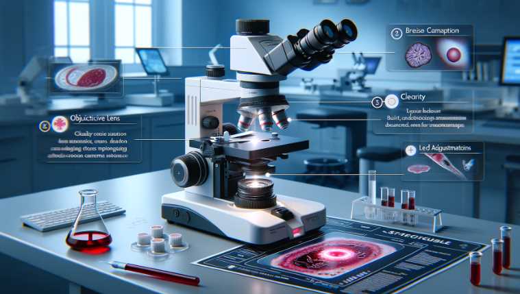
A compound microscope is commonly used for blood analysis.
Compound microscopes are versatile instruments that allow for the observation of microscopic structures in biological samples, including blood. They provide high magnification and resolution, enabling detailed examination of blood cells, such as red blood cells, white blood cells, and platelets. Additionally, specialized microscopes like the dark-field microscope or phase-contrast microscope may be employed for specific blood analysis applications.
I have worked with various microscopes as a laboratory technician with several years of blood analysis experience. Through my experiences, I have comprehensively understood the pros and cons of microscopes used for blood analysis.
| Image | Product | Detail | Price |
|---|---|---|---|
 | Carson MicroBrite Plus 60x-120x LED Lighted Pocket Microscope |
| See on Amazon |
 | Elikliv LCD Digital Coin Microscope |
| See on Amazon |
 | AmScope M150 Series Portable Compound Microscope |
| See on Amazon |
 | PalliPartners Compound Microscope for Adults & Kids |
| See on Amazon |
 | Skybasic 50X-1000X Magnification WiFi Portable Handheld Microscopes |
| See on Amazon |
| Microscope Type | Application in Blood Analysis |
|---|---|
| Compound Microscope | General observation of blood components. |
| Dark-Field Microscope | Enhanced visualization of unstained blood cells. |
| Phase-Contrast Microscope | Improved contrast for transparent blood components. |
| Fluorescence Microscope | Detection of fluorescently labeled blood elements. |
| Electron Microscope | Ultrastructural analysis of blood cell components. |
In this article, I aim to share my knowledge and provide an in-depth overview of the different types of microscopes available for blood analysis, their features, and the factors to consider when choosing the right one for your laboratory needs. Whether you are a student, a research scientist, or a medical professional, this article will help you understand the intricacies of blood analysis microscopy and assist you in making an informed decision when choosing the best microscope for your needs.
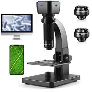
Digital Microscope 2000X Biological Microscope
As a seasoned user of digital microscopes, I was intrigued by the WSWXFWJ Digital Microscope and its features. After using it for a few weeks, I must say that I am impressed with its performance.
The dual-lens system, which includes a digital and microbial lens, provides a clear and sharp image with magnifications ranging from 50X to 2000X. The 5.0MP camera technology combined with precise focus makes it easier to view even the smallest details of the specimen.
The 11 adjustable LED lights are another standout feature of this microscope. The brightness can be easily adjusted, providing excellent details and definition, resulting in clearer images and videos.
One of the best aspects of this microscope is its portability. The rechargeable lithium-ion battery (1800mAh) provides continuous use for up to 5 hours and makes it a useful tool for outdoor nature hikes or for taking on the go.

- 🔬【Digital/microbial dual lens】Digital microscope has 5.0MP camera technology and precise focus. The microscope magnification is 50X to 2000X, allowing you to clearly view the smallest details of the specimen, such as plants, coins, diamonds, Welding, etc., can help you easily see the clear details of tiny objects.
- 🔬【11 Adjustable LED Lights】 Microscope has built-in 11 adjustable LED lights. The brightness can be adjusted from dark to bright by sliding the switch. Excellent details and best definition, the user’s image and Video can improve the quality of clarity.
- 🔬【Easy to Carry】It is very easy to charge and the charge lasts for a long time. It makes for a very useful and fun tool to always have with you in the outdoors. You can enjoy the portable mini pocket microscope on your nature hikes.
- 🔬【1800 mA rechargeable battery】 Built-in rechargeable lithium-ion battery (1800mAh) can work continuously for 5 hours. It is very suitable for hobbyists, quality control inspectors and scientific researchers.
- 🔬【A Funny Tool】 Microscopes can be widely used in circuit board inspection, insects, coins, jewelry, gems, trichomes, rocks and stamps, clocks/clock repairs, skin inspections, children's education inspections, textiles Industrial, QC inspection (not suitable for cell and medical purposes), and can also be used for children's learning exploration, observing flowers, insects, coins, etc., to improve children's curiosity Desire opens the door to the micro world.
The WSWXFWJ Digital Microscope can be used for a wide range of applications, including circuit board inspection, jewelry inspection, and even educational purposes for children. It comes with a comprehensive set of accessories, including slides, data lines, and a cleaning cloth.
In conclusion, I highly recommend the WSWXFWJ Digital Microscope for its excellent performance and versatility. The dual lens system, adjustable LED lights, and portability make it an excellent tool for hobbyists, quality control inspectors, and scientific researchers. Additionally, the company’s 24-hour professional after-sales service provides peace of mind for any issues or concerns.
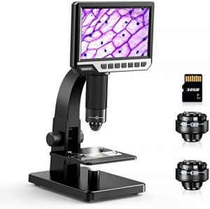
TOMLOV DM11 LCD Biological Microscope
I have been impressed by its versatility and convenience. The microscope comes with a digital and microbial lens, allowing for the observation of macroscopic objects and microscopic specimens with a magnification range of 50-2000X. The 7″ IPS display and 12MP camera provide clear, color-rich images and videos that can be saved onto the included SD card.

One of the key features I appreciate is the compatibility with Windows and Mac OS, which allows for easy connection to a computer and viewing on a larger scale without the need for additional software downloads. The ability to switch between taking photos and recording videos with just one button is also a convenient feature.
However, there has been one issue that I have encountered. The included battery did not seem to be working, even after being fully charged, which resulted in the unit not turning on. I reached out to the company for support but have yet to receive a response. This has been a frustrating experience and has detracted from my overall satisfaction with the product.
The microscope can be connected to a PC compatible with Windows and Mac OS (10.5 and above). This allows for larger-scale observation and the option to save images and videos on an SD card (included). The microscope has two slides, but you can make your own glass slides.

However, I was disappointed that my unit would not turn on even after being fully charged. I reached out to the company but have yet to receive a response. Additionally, it was only 3.5 volts after checking the battery, indicating a potential issue with the battery charging system.
Overall, while the TOMLOV DM11 LCD Digital Microscope offers great features, my experience with the battery and lack of response from the company is a concern.
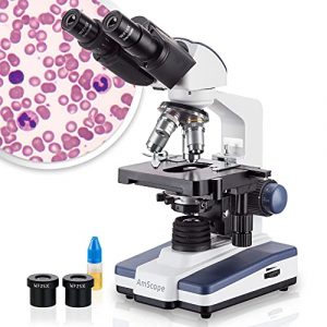
AmScope B120C Siedentopf Compound Microscope
I can say that this is a high-quality, versatile microscope designed for a range of biological and educational applications. The Siedentopf binocular head features widefield eyepieces and 53 to 77mm inter-pupillary adjustment, providing a more comprehensive view and enabling easy user sharing.
The forward-facing nosepiece houses a range of DIN achromatic objectives, which correct for color distortion and provide a range of magnifications from 40X to 2500X. The LED illumination, Abbe condenser with iris diaphragm, and double-layer mechanical stage with 1mm stage divisions all contribute to clear examination and precise slide manipulation.
However, I did have a few issues with my AmScope B120C. The first unit I received was missing parts and appeared to be used, so I had to request a replacement from Amazon. The new unit arrived quickly, but unfortunately it had oil all over the stage, and the two eyepieces were so badly aligned that I saw double. I tried to find information in the manual or the AmScope site to see if the eyepieces could be aligned, but I did not find anything easily. This was a bit disappointing, as it would have been helpful if the company had provided information on how to resolve the issue.

- Magnification Excellence: The AmScope B120 compound microscope offers a broad magnification range from 40X to 2500X, enabling detailed examination of microscopic specimens
- Advanced LED Illumination: Equipped with energy-efficient LED light, this binocular microscope provides bright, daylight-balanced illumination with a specialized fly-eye lens
- Ergonomic Design: The microscope features a 360-degree rotatable Siedentopf head with adjustable interpupillary distance for comfortable and flexible viewing
- Precision Stage Control: A 3D mechanical stage and coaxial coarse and fine focus knobs allow for smooth and precise adjustments
- About AmScope: We have the industry's leading collection of microscopes, microscopes cameras, accessories and other related products
In conclusion, the AmScope B120C Siedentopf Binocular Compound Microscope is a well-designed and high-performing microscope, but there were some problems with the quality of the units I received. Despite these issues, I would recommend this microscope to others looking for a versatile, high-magnification microscope for biological and educational applications.
TOMLOV DM9 7′ LCD Digital Microscope
The TOMLOV DM9 7″ LCD Digital Microscope is a high-quality microscope that offers a lot of features and capabilities to its users. With its rotatable 7-inch FHD screen and 12-megapixel ultra-precise focusing camera, this microscope creates high-quality images and videos and offers an incredible experience for observing the micro world. The magnification range of 50X to 1200X and the 10 LED fill lights make it easy to see the tiniest details in a full lighted view.
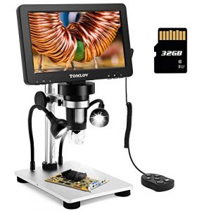
One of the great things about this microscope is that it can be connected to a PC, which allows for larger views and easier data sharing and analysis. The microscope is compatible with both Windows and Mac OS and does not require any extra software downloads. A 32GB SD card is included, which is an excellent bonus for saving photos and videos. The solid metal frame construction offers durability and long-term use, making it great for micro-soldering or repairing printed circuit boards.
One issue that some users have reported is that the port where you plug in the charger can break off inside. This can cause the microscope to no longer be able to be charged or turned on. It is recommended that users consider purchasing the longest warranty available or a cheaper model, as some have reported that their microscope did not last long.

- Tons of Fun and Applications: With easy-to-use operation and a wide range of applications, from micro soldering and rock inspection to coin and stamp observation, this coin microscope with screen is perfect for adults, students, and young learners, enhancing interaction between parents and kids, as well as teachers and students
- 7-Inch Rotatable FHD Screen: Equipped with a 12MP ultra-precise focusing camera and 1080P high-definition imaging, this LCD digital microscope captures high-quality images and videos, delivering an incredible microscopic viewing experience. The rotatable (90-degree) screen design improves ergonomics and reduces eye and neck strain
- 5X-1200X Magnification: Allows you to zoom in and observe the tiniest details with a magnification range from 5X to 1200X. The 8 LED fill lights, plus 2 additional gooseneck lights, ensure a well-lit view. The actual magnification varies depending on the screen size and the distance between the camera and objects
- Hook Up to PC for a Larger View: Compatible with Windows and macOS, the PC view allows you to observe on a larger screen and facilitates data sharing and analysis. No additional software required—simply use the default apps: "Windows Camera" for Windows and "Photo Booth" for iMac/MacBook
- 32GB SD Card Included: This coin magnifier with light includes a 32GB Micro SD card, allowing you to store numerous photos and videos. Press and hold the menu button for 3 seconds to switch between “Photographing,” “Video Recording,” and “Playback” modes
Despite this issue, many users have found this microscope a great investment. Its solid construction quality and wide range of applications, from micro soldering to coin or stamp observing, make it a versatile tool for both adults and young learners. Its compatibility with PC view and ability to save photos and videos are also great bonuses.
In conclusion, the TOMLOV DM9 7″ LCD Digital Microscope is an excellent option for those looking for a high-quality microscope with a range of features and capabilities. However, users should be aware of the potential for the charger port to break and consider purchasing a warranty or a cheaper model.
Plugable USB 2.0 Digital Microscope
The Plugable USB Digital Microscope is a handy and user-friendly tool for hobbyists and professionals interested in exploring the microscopic world. The 2.0-megapixel microscope camera offers up to 250x magnification and includes an LED halo light with brightness control, making it suitable for inspecting objects and specimens in fine detail.
The microscope also features a flexible arm stand with an observation pad that can be used as a handheld device or with graduated marks for easy measurement. The microscope is compatible with Windows, Mac, and Linux operating systems, making it a versatile and accessible tool for many users.
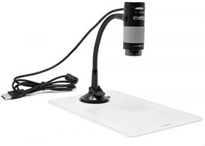
However, the Plugable microscope is not a plug-and-play device. The software must be downloaded from the manufacturer’s website, and in some cases, additional setup may be required for users with an Oculus Rift. This may be a hassle for some users looking for a more straightforward setup process.
In addition, the suction cup and board included with the microscope are not heavy enough to securely hold the microscope in place, even when adjusting the camera. This can lead to frustration when trying to get the microscope in the right position.
On the positive side, the driver package is easy to install and contains nothing malicious. Once installed, the microscope works well, although it takes a little bit of time to get used to adjusting the focal range. The flexible arm, while not super stable, is a useful feature, though users may opt to 3D print an adjustable arm for increased stability in the future.
The picture quality is not as sharp as some users may like, but for a hobbyist-level microscope, it’s acceptable. The LED lighting is adjustable and provides enough illumination for most applications. The 2-year warranty from Plugable is an added bonus, and the Seattle-based email support provides users peace of mind.
In conclusion, the Plugable USB Digital Microscope is an excellent option for casual and hobbyist users looking for an affordable and accessible tool for macroscopic inspection. Its broad compatibility, LED lighting, and flexible arm make it a useful and fun tool for students, collectors, testers, and anyone interested in exploring the microscopic world. However, users who require a more professional-level microscope may want to consider a more advanced model.
Is a microscope necessary for blood analysis?
A microscope is a useful tool for blood analysis and can provide a significant amount of information about a sample, but it is not always necessary. In many cases, other types of diagnostic tests, such as a complete blood count (CBC) or a blood chemistry panel, can provide the information needed without the use of a microscope.
The decision to use a microscope for blood analysis depends on the specific needs and goals of the analysis and should be made in consultation with a healthcare provider or laboratory professional.
How do I choose the best microscope for blood analysis?
When choosing the best microscope for blood analysis, consider the following factors:
Magnification: A good microscope for blood analysis should have a magnification power between 100x and 400x.
Quality of Optics: Ensure the microscope has high-quality lenses, which are essential for getting a clear and detailed image.
Bright Field Illumination: This is the most commonly used type of illumination for blood analysis. Choose a microscope that has adjustable brightness control to reduce glare and increase clarity.
Size and Weight: Blood analysis often involves moving the microscope around, so it’s essential to choose a model that is compact and lightweight, making it easier to move and handle.
Quality of Construction: Blood analysis can be a messy process, so it’s important to choose a microscope that is built with quality materials that can withstand regular cleaning and decontamination.
Compatibility: Choose a microscope compatible with your laboratory equipment and software, ensuring that you can integrate it seamlessly into your workflow.
Price: Blood analysis microscopes can range from basic to high-end models. Consider your budget and choose a microscope that meets your needs and fits within your budget.
Warranty and Support: A good warranty and technical support can save you time and money in the long run. Look for a microscope that comes with a good warranty and customer support.
Benefits of using
There are several benefits of using a microscope for blood analysis:
Accuracy: Microscopes provide a highly accurate and detailed view of blood cells and their characteristics, making it easier to diagnose various diseases and conditions.
Early Detection: By examining blood cells, healthcare professionals can detect diseases and conditions at an early stage, which often leads to better outcomes.
Cost-effective: In many cases, blood analysis through a microscope is a cost-effective method of diagnosis compared to more expensive imaging tests or biopsies.
Non-invasive: Microscopic examination of the blood is a non-invasive procedure, meaning it does not involve any surgical procedures or incisions.
Versatile: Microscopes can be used to examine a wide variety of blood cells, including red blood cells, white blood cells, and platelets, allowing for comprehensive blood analysis.
Rapid Results: Microscopic examination of the blood is a relatively rapid procedure, with results available in a matter of minutes or hours.
Portable: Many modern microscopes are portable and lightweight, making them ideal for use in a clinical setting or in remote areas where medical facilities are limited.
Overall, microscopes play a critical role in the diagnosis and treatment of various diseases and conditions by providing an accurate and cost-effective method of blood analysis.
How does a microscope work for blood analysis?
A microscope is a crucial tool for blood analysis, as it allows for a detailed examination of blood cells and other components. In blood analysis, a blood sample is typically taken from a patient and stained with special dyes. The stained blood sample is then placed on a microscope slide and viewed under high magnification. The microscope’s light source illuminates the sample, and the user can adjust the focus to view the different components of the blood, such as red and white blood cells, platelets, and other particles.
Red blood cells, which carry oxygen to tissues, appear as biconcave disks under the microscope. White blood cells, which are involved in fighting infections, appear in a variety of shapes, including spherical, rod-like, and amorphous. Platelets, which are involved in blood clotting, appear as small, dark circles.
In blood analysis, a hematologist or a medical laboratory technologist will use the microscope to count the number of red and white blood cells and to determine the size and shape of the cells. They may also look for any abnormal cells or cellular structures, such as clumps or fragments, which could indicate disease or injury. This information can then diagnose and treat various medical conditions, including anemia, infection, and cancer.
Overall, the microscope plays a critical role in blood analysis, allowing for the detailed examination of blood cells and components to aid in the diagnosis and treatment of medical conditions.
What are the limitations of a microscope for blood analysis?
There are several limitations of a microscope for blood analysis:
Magnification: While a microscope provides high magnification, there are limits to the level of detail that can be seen and the accuracy of the results.
Sample preparation: Proper sample preparation is crucial for accurate results, and errors during this stage can lead to false results.
Experience of the operator: The interpretation of the results depends on the experience and expertise of the person using the microscope.
Quality of the microscope: The quality of the microscope, including the optics and illumination, affects the accuracy of the results.
Background interference: Interference from the background can make it difficult to see the cells clearly and accurately.
Dynamic range: The dynamic range of the microscope may not be adequate to detect all the changes in the blood cells, especially in cases where the sample contains a range of cell sizes and densities.
Limited view: The view through the microscope is limited to a single focal plane, which may not provide enough information to accurately diagnose conditions based on changes in cell shape or size.
False results: The potential for false results is always present when analyzing blood samples, especially when dealing with the limitations of the microscope.
It is important to consider these limitations and choose a microscope with the right specifications and capabilities for the intended use. Additionally, proper training and sample preparation techniques can help minimize these limitations.
What should I do if I find a problem with my microscope while doing blood analysis?
If you encounter any issues or problems with your microscope while conducting a blood analysis, it’s essential to take the following steps:
- Check the manual: Review the manual or documentation that came with your microscope to see if there is any information that can help you resolve the problem.
- Contact the manufacturer: If you cannot resolve the issue, contact the manufacturer’s customer support team. They should be able to provide you with further assistance.
- Check for maintenance: Ensure your microscope is regularly maintained and cleaned to ensure optimal performance. If necessary, consult a professional to perform maintenance or repairs.
- Consider upgrades: If your microscope is outdated or not capable of performing the analysis you need, consider upgrading to a newer model that is better suited to your needs.
- Double-check results: If the problem affects the accuracy of the results, it may be necessary to double-check your results or conduct the analysis again using a different instrument.
It is important to address any problems with your microscope as soon as possible to ensure accurate and reliable results in your blood analysis.
How do I use a microscope for blood analysis?
To use a microscope for blood analysis, follow these steps:
- Prepare a blood sample: Obtain a fresh blood sample, either by a finger prick or through a venipuncture. Transfer the blood to a glass slide, either by using a dropper or by smearing the blood onto the slide.
- Staining the slide: To make the blood cells visible under the microscope, the slide needs to be stained with a particular stain. The most commonly used stain for blood analysis is a Romanowski stain, such as Wright’s or Giemsa stain.
- Mounting the slide: After staining, allow the slide to air dry. After drying, mount the slide onto the microscope stage using a cover slip.
- Focus the microscope: Turn on the microscope light and adjust the focus knobs to bring the blood cells into focus. Start with low-power objectives and then switch to higher-power objectives for better magnification.
- Observing the blood cells: Observe the blood cells under the microscope, counting the different types of cells present and evaluating their shape, size, and color. Record your observations and make a note of any abnormal cells.
- Clean up: After completing the blood analysis, turn off the microscope light and clean the slide with a damp cloth. Store the slide and your notes for future reference.
It is important to keep in mind that blood analysis through microscopy is a complex process and requires a trained professional to interpret the results accurately.
Final Words
Microscope plays an important role in blood analysis and is necessary for proper and accurate diagnoses. The Plugable USB Digital Microscope with Flexible Arm Observation Stand is an excellent option for hobbyists and students looking to explore the microscopic world. It offers a high-definition image with up to 250x magnification and is compatible with Windows, Mac, and Linux operating systems. The flexible arm stand and integrated LED lighting make it convenient to use.
However, it is essential to note that it is not a plug-and-play device and requires software installation. Additionally, the suction cup and board are not well-suited for each other, and the picture quality may not be as crisp as desired. Nevertheless, it is a great choice for casual, hobbyist-level macroscopic inspection. When using a microscope for blood analysis, it is important to maintain the device and troubleshoot any issues properly. With proper care and use, a microscope can provide valuable insights into blood analysis and contribute to accurate diagnoses.

Fahim Foysal is a well-known expert in the field of binoculars, with a passion for exploring the great outdoors and observing nature up close. With years of experience in the field, Fahim has honed his skills as a binocular user and has become a go-to resource for those seeking advice on choosing the right binoculars for their needs.
Fahim’s love for the natural world began during his time at The Millennium Stars School and College and BIAM Laboratory School, where he spent much of his free time exploring the outdoors and observing the wildlife around him. This passion for nature led him to pursue a degree in Fine Arts from the University of Dhaka, where he gained a deep understanding of the importance of observation and attention to detail.
Throughout his career, Fahim has used his expertise in binoculars to help others discover the beauty of the natural world. His extensive knowledge of binocular technology and optics has made him a trusted advisor for amateur and professional wildlife observers alike. Whether you’re looking to spot rare birds or observe animals in their natural habitats, Fahim can help you choose the perfect binoculars for your needs. With his guidance, you’ll be able to explore the outdoors with a newfound appreciation for the beauty of the natural world.
Last update on 2025-07-03 / Affiliate links / Images from Amazon Product Advertising API
Table of Contents
