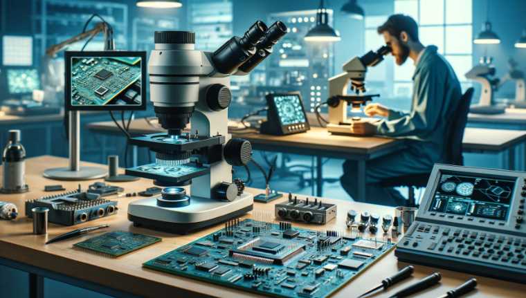
Under a microscope, nail fungus typically appears as hyphae invading and spreading within the nail tissue.

What Does Nail Fungus Look Like under Microscope: Revealing Close-Ups!

Under a microscope, nail fungus (onychomycosis) typically appears as hyphae (thread-like structures) invading and spreading within the nail tissue. Nail fungus is primarily caused by dermatophyte fungi, commonly Trichophyton rubrum, Trichophyton mentagrophytes, or Candida species. When observed under a microscope, these fungi exhibit distinctive characteristics. The hyphae, which are branching filaments, penetrate and invade the […]

What Does Nail Fungus Look Like under Microscope: Revealing Close-Ups! Read More »

What Does Chlamydia Look Like under Microscope

What Does Chlamydia Look Like under Microscope? Unseen Details Revealed

Chlamydia is a bacteria that cannot be seen with the naked eye. Under a microscope, Chlamydia appears as small, round, or oval-shaped cells. Chlamydia trachomatis is an obligate intracellular bacterium, meaning it can only survive and replicate inside host cells. When observed under a microscope, Chlamydia cells typically appear as small, gram-negative, cocci or rod-shaped

What Does Chlamydia Look Like under Microscope? Unseen Details Revealed Read More »

Are Atoms Visible under Electron Microscope

Are Atoms Visible under Electron Microscope? Find Out How!

No, individual atoms are not directly visible under a standard electron microscope. The wavelength of electrons used in electron microscopes is much shorter than that of visible light, allowing for much higher resolution. However, even with this high resolution, the individual atoms are generally not directly visible. Instead, electron microscopes can provide detailed images of

Are Atoms Visible under Electron Microscope? Find Out How! Read More »

What Does Trichomoniasis Look Like under the Microscope

What Does Trichomoniasis Look Like under the Microscope: Unveiling the Microbial World

Under the microscope, Trichomoniasis appears as pear-shaped protozoa with a jerky, irregular motion. Trichomoniasis is caused by the protozoan parasite Trichomonas vaginalis. When observed under a microscope, the organism typically has a pear-shaped or oval form, measuring about 10 to 20 micrometers in length. It has a single flagellum (whip-like appendage) that propels its movement,

What Does Trichomoniasis Look Like under the Microscope: Unveiling the Microbial World Read More »