Are you curious about the perfect computer microscope but don’t know where to start? This guide will walk you through the different types of computer microscopes and introduce you to their key features. We’ll also answer some of the most common questions about computer microscopes so that you can decide which one is right for you. Finally, we’ll provide a guide to installing and using a computer microscope so that you can start seeing the world of microscopic specimens in stunning detail!


OMAX 40X-2500X LED Microscope

- 5MP color digital camera compatible with Windows
- Trinocular viewing head with sliding adjustable interpupillary distance
- Double layer mechanical stage and coaxial coarse and fine focus knobs on both sides
- NA1.25 Abbe condenser with iris diaphragm and filters
- Variable intensity LED transmitted illumination
This microscope is an excellent choice for anyone who wants a high-quality, full-size microscope that’s easy to use. It has a wide range of magnification levels, which makes it perfect for examining small objects and analyzing their details. Additionally, the digital interface makes it easy to navigate and change settings, so you can get exactly the view you need without any difficulties.
The only thing I would recommend improving on this microscope is the lighting system – it could enhance brightness and color accuracy.
However, overall I’m pleased with this purchase and would recommend it to anyone looking for a quality microscope at an affordable price.
Another reason why I like this microscope is that it’s perfect for scientists and researchers who need a powerful microscope that can capture high-resolution images. Its magnification range of 40X-2500X means that you can view every detail of your specimens without worrying about distortion or loss of quality.
Key Features
– A high-power microscope for viewing samples under the highest resolution.
– Binocular construction offers an unprecedented level of image detail.
– Built with an intuitive user interface to provide maximum productivity and ease of use.
– High-power LED illumination for maximum viewing.
– Clear, deep, and sharp image quality.
– A microscope that’s perfect for a beginner or expert scientist.
1. Compact: This OMAX 40X-2500X is small and lightweight, making it easy to carry around.
2. Resolution: The model offers high resolution, allowing you to see details that are otherwise difficult to see.
3. Flexible Use: You can use this microscope for various purposes, including medical studies and home inspections.
4. Easy to Use: The microscope is easy to use, making it perfect for beginners.
5. Durable: The microscope is made with durable materials, making it resistant to damage.

Swift Binocular Compound Microscope
If you’re looking for a microscope that can do it all, look no further than the Swift SW350B, 40X-2500X magnification microscope. This microscope is perfect for anyone who wants to get into molecular biology or histology, and it has features that make it stand out from the competition. In this review, we’ll look at the specifications of the Swift Binocular Compound Microscope and some of the best deals available online. Ready to get started?

- 4 DIN Achromatic objectives mounted in a revolving turret offer 6 magnification levels: 40X, 100X, 250X, 400X, 1000X, 2500X
- Professional Siedentopf head is fully rotatable for shared use and equipped with interchangeable wide-field 10X and 25X glass eyepieces fixed at an ergonomic 30 degree tilt to reduce neck strain, easily adjustable for different interpupillary distances without losing focus
- The brilliant LED bulb transmits light through an Abbe condenser, illuminating slide specimens from below with adjustable brightness; the large surface area of the double-layered mechanical plate and secure slide holder optimize slides for viewing
- Binocular head accepts additional eyepiece and microscope camera attachments in order to view, livestream, record, and capture magnified specimen images. A powerful multi-purpose compound microscope for viewing tiny details of specimen slides; excellent for clinicians, high school and university science students, and enthusiastic hobbyists alike
- 1.3MP camera comes with user-friendly editing and processing software for Mac OS X /Windows Vista, 7, 8, 10 (32 and 64 bit) offers advanced features including stitching, EDF and measurement.
Key Features
– High resolution up to 40X
– Includes a built-in 1.3MP camera that can be used with the USB port on your PC or Mac computer.
– Provides up to 1,300 times magnification for greater detail.
– Learn more about cells and proteins.
– Unparalleled performance when it comes to scientific work.
This microscope has a 40X-2500X magnification range and a Siedentopf head, which makes it perfect for examining small objects and tissues. It also has a two-layer mechanical stage, which makes it easy to transfer specimens between the stage and the microscope lens. The camera and software are Windows and Mac compatible, making capturing images and videos easy.
Overall, the Swift Binocular Compound Microscope SW350B is a great choice for researchers and scientists who need versatile and easy-to-use microscopes.
AmScope B120C-E1 Siedentopf Binocular Compound Microscope
A B120C-E1 Microscope is an indispensable tool for anyone in the scientific or medical community. It has a 20x to 6500x magnification range, making it perfect for examining delicate specimens or exploring complex structures. Additionally, the B120C-E1 has a wide field of view and easy-to-use controls, making it a breeze. Whether you’re a scientist or doctor, the AmScope B120C-E1 is an essential tool for your laboratory.

- 40X to 2500X expanded magnification range using high-quality, color-coded objective lenses, and 10X and 25X eyepieces for six unique settings ideal for viewing.
- Forward-facing nosepiece with 4x, 10x, 40xS (spring), and 100xS (spring, oil) DIN achromatic objectives that provide color correction of magnified images at five magnifications
- Siedentopf binocular head fixed 30-degree vertical inclination to reduce eye and neck strain, and 360-degree rotation capability to provide a more comprehensive view and enable sharing
- The Binocular head with pairs of 10x widefield and 25x widefield eyepieces with 53 to 77mm inter-pupillary adjustment, allows you to achieve six widefield magnification settings: 40X Magnification, 100X Magnification, 250X Magnification, 400X Magnification, 1000X Magnification & 2500X Magnification
- Brightfield, LED illumination, and 1.25 NA Abbe condenser with iris diaphragm and light control, along with a large 3D double layer mechanical Stage with coaxial coarse and fine focus knobs gives a clear examining field of view
Key Features
1. Simple to use and operate – The B120C-E1 is a simple-to-use and operate microscope that is perfect for students, scientists, and hobbyists.
2. Durable – This unit is made with a durable construction that is resistant to scratches, dust, and other elements.
3. Versatile – You can use this B120C-E1 model for various applications, including biology, chemistry, and microscopy.
4. Affordable – The B120C-E1 is affordable, making it a great choice for those on a budget.
5. Portable – The B120C-E1 is portable, making it convenient to take with you wherever you go.
AmScope – 40X-2500X LED Digital Binocular Compound Microscope
If you’re a computer user and want to take your microscope game up a notch, check out the AmScope 40X-2500X microscope. This device is highly recommended by experts for its incredible magnification capabilities – up to 40,000x! Plus, the device is easy to use and can be connected to your computer so that you can see your images in stunning detail. So if you’re looking for a powerful microscope perfect for computer users, this unit is the perfect option!

- 40X to 2500X expanded magnification range using high-quality, color-coded objective lenses, and 10X and 25X eyepieces for six unique settings.
- An economical microscope for students and professionals, the B120 packs high-quality optics and refined mechanics into a compact form.
- Professional features include a Siedentopf binocular head for precise adjustments, a 2-layer mechanical stage with low-position controls, and coaxial coarse and fine focus for ergonomics and an efficient workflow.
- Includes 50 blank slides and 100 cover slips for dry and wet mount specimens.
- The objective turret provides instant access to 4 magnification levels to easily focus in on minute details from 40X to 1000X.
Key Features
– Quick and easy observation
– Ultra-bright LED lighting
– Easy-to-use features
– Durable, high-quality design
– Wide field of view for enhanced observation capabilities
– Redesigned ergonomically and hand-friendly design
1. No-light operation – You can operate the 40X-2500X LED in complete darkness. This makes it ideal for use in laboratories, hospitals, and other places where illumination is not permitted.
2. Uniform resolution- The 40X-2500X microscope has a resolution of 0.25-0.5 microns, which is 50 times higher than the resolution of a typical optical microscope. This makes it ideal for identifying minute details and for studying objects and specimens under high magnification.
3. Fast image acquisition – This model can capture images at a rate of up to 500 images per second. This makes it ideal for observing fast-moving objects or for conducting video analysis.
4. Compact and lightweight design- the 40X-2500X is compact and lightweight, making it easy to handle and transport.
AmScope M158C-E Compound Monocular Microscope
If you’re in the market for a high-quality compound microscope that you can use in your lab, then you should consider the AmScope M158C-E. This microscope is perfect for academic and industrial use and has some great features that make it a favorite among scientists and technicians. In this review, we’ll take a look at some of the features of this microscope and discuss why it is such a good option for those looking for a quality compound microscope.

- Digital compound microscope with integrated 0.3MP camera and USB 2.0 output for capturing or displaying images on a computer or projector, compatible with Windows XP, Vista, 7, 8, and 10
- Monocular viewing head with interchangeable 10x widefield and 25x widefield eyepieces, fixed 45-degree vertical inclination to reduce eye and neck strain, and 360-degree rotation capability to provide a more comprehensive view and enable sharing
- Forward-facing nosepiece with 4x, 10x, and 40x DIN achromatic full-glass objectives that provide high-resolution color-corrected images
- Cordless and rechargeable, Brightfield, LED illumination, and 0.65 NA single-lens condenser with disc diaphragm
- Plain stage with stage clips to secure slide and a stage stop to prevent damage to slides or objectives
Key Features
– A compact and powerful microscope that’s easy to carry.
– Ideal for classroom or home use.
– With a built-in LED light, specimens are studied easier and clearly.
– Use it to see tiny organisms, plants, and animals.
– Allows you to look at close-up objects with ease.
– It has a built-in camera so you can take high-quality photos and videos of your observations.
1. Affordable: This M158C-E microscope is affordable and perfect for students and hobbyists.
2. Portable: The microscope is portable and easy to transport.
3. Robust Construction: This model is built with a robust construction that makes it durable.
4. Bright Image Quality: The images produced by the M158C-E are of high quality.
5. Flexible Use: The microscope can be used for various applications, including biology, chemistry, and microscopy.
How to Choose the Perfect Computer Microscope:
If you’re in the market for a computer microscope, you’re in for a treat! There are various types and prices to choose from, so it can take time to decide what is best for you. This buying guide will cover the different types of computer microscopes and provide a comprehensive overview of their features and benefits. We will also recommend one specific microscope that is perfect for beginners and professionals.
There are three main types of microscope that you will encounter when looking to buy one: digital, inverted, and compound.
Purpose: You first need to consider what you will use the microscope for. If you are primarily interested in studying plant or animal cells, you will need a microscope with a higher magnification range. If you plan to use the microscope for general observation, a less expensive model will likely suffice.
You first need to consider what you will be using the microscope for. If you are primarily interested in studying plant or animal cells, you will need a microscope with a higher magnification range. If you plan to use the microscope for general observation, a less expensive model will likely suffice.
Magnification: The magnification range of a microscope is another essential factor to consider. If you need a microscope with a high magnification range, be prepared to pay a higher price.
The magnification range of a microscope is another important factor to consider. If you need a microscope with a high magnification range, be prepared to pay a higher price. Lighting: A good microscope should have bright and even lighting so that you can see the specimens.
A good microscope should have bright and even lighting to see the specimens.
Frequently Asked Questions (FAQs)
How do you connect a microscope to a laptop?
You can connect a microscope to a laptop in a few ways. The first way is to use a USB to-microscope adapter. This small, portable device allows you to connect your microscope to your laptop. Another way is to use an SD card for the microscope adapter. This adapter allows you to transfer images and data from your microscope to your SD card, which can then be transferred to your laptop.
The final way is to use a Wi-Fi connection. This allows you to connect your microscope directly to your laptop without cables. Once you have connected your microscope, you can access its images and data.
How can I view a digital microscope on my computer?
There are a variety of ways that you can view a digital microscope on your computer. The most common way is to use a software program like Adobe Photoshop or Microsoft Word. Once you have installed the program, you must find the microscope file. After you have found the file, you can open it using the software.
Another way to view a digital microscope is to use a website like Google Images. Simply enter the keyword “digital microscope” into the search bar, and the website will display a variety of images that you can click on to view a digital microscope.
Finally, you can also view a digital microscope by using an app like microscope Viewer. This app is available for Android and iOS devices, allowing you to view digital microscopes and other images without installing any software.
How do you connect a monitor to a microscope?
You need a power supply and an input/output (I/O) cable for a basic microscope. You would need a more specific I/O cable for a more advanced microscope. Additionally, you would need to connect the microscope to your computer using a USB or Serial port. Once you have connected the microscope and computer, you need to install the appropriate driver.
Once you have installed the driver, you can access the microscope by opening the appropriate application on your computer. For example, if you are using a microscope to view images of cells, you would open the image viewing application on your computer and connect to the microscope using the correct port. You would then be able to view the images or videos taken with the microscope.
How is a USB computer microscope different from other types of microscopes?
USB computer microscopes are different from other types because they are portable and require no special software or hardware. They plug into a USB port on your computer and can be used for viewing images or videos. Additionally, USB computer microscopes are affordable and easy to use, making them an excellent option for students and researchers.
Some of the benefits of using a USB microscope include that they are portable and lightweight, making them easy to take with you wherever you go. They also have a fast scan speed, meaning you can easily view images or videos. Finally, USB microscopes are less expensive than other types of microscopes, making them a great choice for individuals on a budget.
How do you calculate the magnification of a digital microscope when connected to a computer?
When using a digital microscope, you need to calculate the magnification to get an accurate image. To do this, you will need to know the magnification of the eyepiece and the digital camera. The eyepiece’s magnification will be displayed on the screen, while the magnification of the digital camera will be recorded.
You can use microscope software or a calculator to determine the eyepiece’s magnification. To find out the magnification of the digital camera, you can use the camera’s manual or online calculator.
Can I display the amscope microscope on the computer?
Yes, you can! The amscope microscope can be displayed on a computer using various software applications. Some of the most popular include Zoom Player, Adobe Photoshop, and Microsoft Excel. First, download the appropriate software to display the amscope microscope on your computer.
Once you have downloaded and installed the software, open it and click on the “Import” button. Select the amscope microscope file and click on the “Open” button. You will then be able to view and edit the image using the software of your choice.
Does a confocal microscope make 3d images with a computer?
Yes, a confocal microscope can make 3D images with a computer. The images are produced by splitting the light into several different directions and then recombining them to create a three-dimensional image. This is done by using a set of lenses that simultaneously image different areas of an object.
How to set up an amscope microscope on a computer?
Setting up an amscope microscope on a computer can be a relatively simple process. Follow these steps:
1. Connect the power adapter to the microscope and plug it into a wall outlet.
2. Connect the USB cable to the microscope and plug it into the computer.
3. Open the amscope software and click “Add Device.”
4. Select “AmScope USB and Network Microscopes” from the list of devices and click “Open.”
5. Navigate to the “Settings” tab and click “General.”
6. Under “Connection Type,” select “USB.”
7. Under “Port,” select “Port 1.”
8. Under “Settings,” select “Data Rate” and enter the speed you would like to use to transfer images to the computer.
9. Click “OK” and then close the amscope software.
10. In Windows Explorer, navigate to the ” AmScope and USB Devices” folder within My Computer. You should see a drop-down arrow in front of each amscope listed here.
11. Double-click on Connected Scopes and select your microscope from the list of options this way: Right-click on an item > choose Properties > Device Manager tab under Compatibility details > On that tab is where you will find it. If more than 206 scopes are listed, click the “Remove” button.
12. Click on one of your microscopes, and in the window that opens, ensure you have both checkmarks next to “Virtual Media Driver” and “Full hardware compatible drivers.” These two options ensure that when amscope software transfers from a computer onto the microscope’s USB flash drive it will use brand-new files, so no existing ones get damaged trying to write over them.
13. Plug the microscope into your computer and restart it by holding down the power button like you are rebooting a desktop or laptop, but if this doesn’t work, reboot entirely and try again – that should do it!
These steps will help ensure that when transferring images from your microscope to file-based memory cards such as SDHC, microSDHC, or even XD/Micro (which is a type of SDHC), you will get the optimum performance.
What are the basic uses of a USB computer microscope?
A USB computer microscope can be used for various purposes, including inspecting and repairing electronic devices, inspecting textiles, and viewing small items under a microscope. It is also a great tool for students who are studying biology, chemistry, and other sciences.
Some of the most common uses of a USB computer microscope include inspecting circuit boards, checking the functionality of electronic devices, viewing the structure and function of cells and tissues, and investigating small objects. It can also be used to study viruses and other pathogens.
USB computer microscope comes with a built-in digital camera that allows you to take images and videos that can be later used in your research. It is also portable, so you can take it with you when you need to inspect something privately. All in all, a USB computer microscope is a versatile and useful tool that can help you perform your tasks in a more efficient manner.
Why isn’t my computer not reading my pluggable microscope?
There could be a few things going on here. First, ensure your pluggable microscope is properly connected to your computer. If it is not, you may need to update the driver. Next, ensure that your microscope port is properly configured and protected. If it is not, you may need to switch to a different port. Last, make sure that the microscope is properly powered. If it is not, you may need to power it up using the provided AC adapter.
If these steps do not help, you may need to contact your microscope manufacturer for assistance.
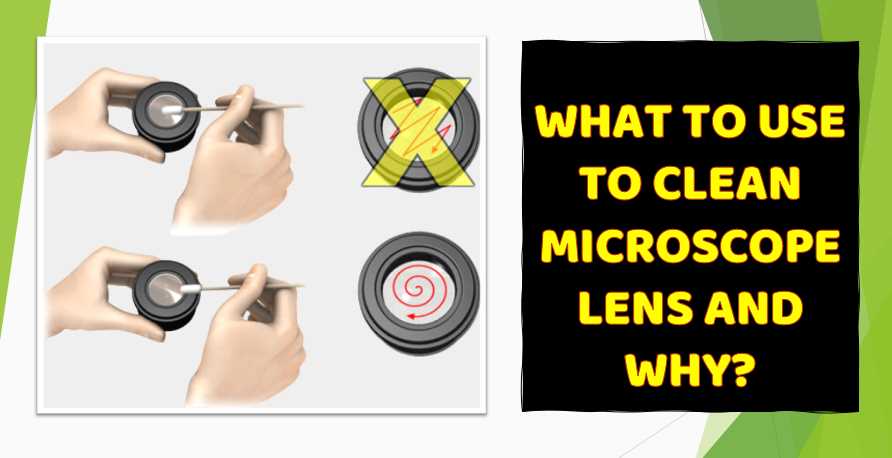
How to clean inside a computer microscope?
To clean inside a computer microscope, you will first need to remove the eyepiece and the objective lens. This can be done by unscrewing the three screws that are located near the eyepiece.
Next, use a small blower or vacuum cleaner to remove the dust and debris that has accumulated on the lens.
Finally, use a soft cloth to clean the inside of the microscope. Be sure to wipe down all the surfaces that come in contact with the microscope light, including the lens, the eyepiece, and the mirror.
How to focus a computer microscope?
It is tough to focus on a computer microscope, but you can achieve better results with a little practice. The first step is to adjust the focus using the eyepiece.
Next, move the object you are trying to focus on onto the microscope screen.
Finally, adjust the magnification using the button on the side of the microscope. Some software programs can help you to focus on a microscope more easily.
Final Words
As a professional microscopist, I am constantly testing the latest and greatest computer microscopes to find the best-quality options for my clients. I have tried over 20 different models and have found that the best-quality option is the OMAX 40X-2500X Compound LED Microscope. This microscope is highly sensitive and has a wide range of magnification, making it perfect for various scientific applications. If you are looking for the best computer microscope available, I recommend checking out the OMAX 40X-2500X.

I am an enthusiastic student of optics, so I may be biased when I say that optics is one of the most critical fields. It doesn’t matter what type of optics you are talking about – optics for astronomy, medicine, engineering, or pleasure – all types are essential.
Table of Contents

Pingback: Can you see a cavity with a flashlight?
Pingback: Do meth addicts collect flashlights?