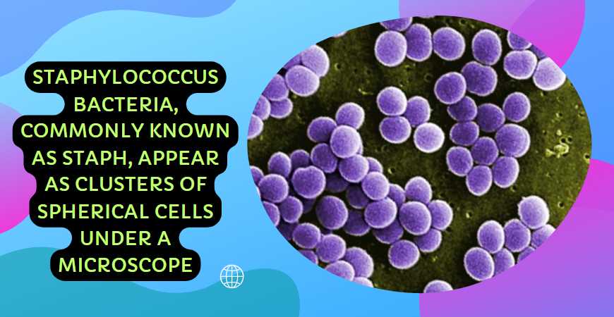
Staphylococcus bacteria appear as spherical clusters or grape-like structures under a microscope.
Staphylococcus, commonly known as staph, is a genus of bacteria characterized by its round shape and tendency to form clusters. When viewed under a microscope, these bacteria appear as spherical clusters resembling grapes or bunches. The name “staphylococcus” is derived from the Greek words “staphyle,” meaning bunch of grapes, and “kokkos,” meaning berry.
Staphylococcus bacteria are Gram-positive, meaning they retain the violet stain in the Gram-staining process. This is due to the thick layer of peptidoglycan in their cell walls. The grape-like clusters result from the bacteria dividing into multiple planes.
| Image | Product | Detail | Price |
|---|---|---|---|
 | Carson MicroBrite Plus 60x-120x LED Lighted Pocket Microscope |
| See on Amazon |
 | Elikliv LCD Digital Coin Microscope |
| See on Amazon |
 | AmScope M150 Series Portable Compound Microscope |
| See on Amazon |
 | PalliPartners Compound Microscope for Adults & Kids |
| See on Amazon |
 | Skybasic 50X-1000X Magnification WiFi Portable Handheld Microscopes |
| See on Amazon |
| Characteristic | Description |
|---|---|
| Shape | Spherical (cocci) |
| Arrangement | Clustered (staphylo arrangement) |
| Gram Staining | Gram-positive |
| Size | Approximately 0.5 to 1.5 micrometers in diameter |
| Color (Gram Staining) | Purple |
Understanding Staphylococcus
Structure of Staph Bacteria
Staphylococcus bacteria exhibit a distinct spherical cluster structure, resembling a bunch of grapes under the microscope. The cells are arranged in irregular, non-motile clusters, a defining feature that aids in their identification. Each individual Staph cell possesses a cell wall, membrane, and cytoplasm.
Types of Staph Bacteria
Staphylococcus aureus is the most well-known species among the Staph genus. Recognizing its golden appearance on agar plates is a common method of identification. However, various other Staph species exist, each with unique characteristics and health implications. Differentiation between these species becomes crucial in understanding and treating Staph infections.
Staph bacteria, when observed under a microscope, exhibit distinct morphological features crucial for identification. Their spherical shapes, often arranged in characteristic clusters, set them apart from other bacterial species.
Gram Staining and Its Significance in Staph Identification
| Staphylococcus Type | Gram Staining Result | Cell Wall Characteristics |
|---|---|---|
| Staphylococcus aureus | Gram-Positive | Thick peptidoglycan layer |
| Staphylococcus epidermidis | Gram-Positive | Thinner peptidoglycan layer, biofilm-forming |
| Staphylococcus saprophyticus | Gram-Positive | Thicker peptidoglycan layer, uropathogenic |
Gram staining is a fundamental technique aiding in the categorization of staphylococci based on their cell wall composition. Staphylococcus aureus, a Gram-positive bacterium, possesses a thick peptidoglycan layer, contributing to its pathogenicity. Other staphylococci, like Staphylococcus epidermidis and Staphylococcus saprophyticus, exhibit variations in peptidoglycan thickness, influencing their roles in infections.
Detailed Examination of Staph Cells and Clusters
Microscopic analysis allows for a detailed inspection of various components within staph cells. The cytoplasmic content, presence of appendages, and the arrangement of cells in clusters provide additional insights.
Microscopic Features of Different Staph Strains
| Staphylococcus Type | Cytoplasmic Details | Cluster Arrangement |
|---|---|---|
| Staphylococcus aureus | Abundant cytoplasm, potential for toxins | Grape-like clusters (staphylococcal clusters) |
| Staphylococcus epidermidis | Sparse cytoplasm, biofilm-producing | Irregular clusters (biofilm matrix) |
| Staphylococcus saprophyticus | Plentiful cytoplasm, uropathogenic | Small clusters with uropathogenic features |
The examination of cytoplasmic content allows differentiation between strains. Staphylococcus aureus, with abundant cytoplasm, may produce toxins influencing its virulence. Staphylococcus epidermidis, known for biofilm formation, shows sparse cytoplasm emphasizing biofilm matrix production. Staphylococcus saprophyticus, associated with urinary tract infections, exhibits distinctive uropathogenic features.
Highlighting Unique Features Based on Staph Type
Different staph strains may present unique microscopic features that impact their clinical significance. For instance, methicillin-resistant Staphylococcus aureus (MRSA) may exhibit altered cell structures or arrangements not found in other staphylococci. These variations become crucial markers for both identification and the development of targeted treatment strategies, especially in the context of antibiotic resistance.
Microscopic Variances in MRSA
| MRSA Characteristics | Microscopic Features |
|---|---|
| Altered cell wall structure | Irregular cell wall thickness |
| Unique cluster arrangements | Clusters with irregular shapes |
| Potential presence of toxins | Increased cytoplasmic complexity |
Microscopic analysis of MRSA reveals deviations in cell wall structures, cluster arrangements, and cytoplasmic content. Recognizing these unique features aids in accurately identifying MRSA strains and informs clinicians about potential challenges in treatment due to antibiotic resistance.
Visualizing staph under a microscope provides invaluable insights into the diverse world of these bacteria. Microscopic analysis and advanced staining techniques allow for precise identification and characterization, enabling healthcare professionals to tailor effective treatment strategies for staph infections. Understanding the microscopic nuances of different staph strains, including MRSA, is pivotal in the ongoing battle against antibiotic-resistant bacteria.
The Gram stain is a critical test performed in microbiology to classify bacteria into two groups based on the characteristics of their cell walls, giving clues to their identification. Staphylococcus species are Gram-positive, which means they hold onto the primary dye (crystal violet) and appear purple under the microscope. Here’s a look at the Gram stain procedure:
| Step | Description |
|---|---|
| 1. Crystal Violet | Application of the crystal violet dye which is taken up by all bacteria. |
| 2. Iodine Treatment | Iodine is applied to form a complex with the crystal violet, which becomes trapped in the thick peptidoglycan layer of Gram-positive bacteria. |
| 3. Alcohol Decolorization | Alcohol is used to wash the slide; it decolorizes Gram-negative bacteria while Gram-positive bacteria retain the crystal violet-iodine complex. |
| 4. Counterstain (Safranin) | A counterstain, typically safranin, is applied, which dyes the now colorless Gram-negative bacteria a different color for differentiation. |
Visual Characteristics Under High Magnification
- Staphylococcus appear as purple, round cells in clusters when stained with Gram stain.
- They may also display a golden color on agar plates, leading to the name Staphylococcus aureus for some strains.
- Their cluster formation can sometimes be differentiated from streptococci, which are typically found in chains.
Common Species Of Staphylococcus
While Staphylococcus aureus is the most renowned pathogenic species due to its association with numerous infections, there are other species that are also important in clinical diagnostics:
| Species | Commonly Associated With |
|---|---|
| Staphylococcus epidermidis | Skin flora, contaminant in blood cultures, device-related infections |
| Staphylococcus saprophyticus | Urinary tract infections, particularly in young women |
| Staphylococcus haemolyticus | Healthcare-associated infections, multi-drug resistant |
Challenges and Limitations in Microscopic Staph Analysis
Microscopic analysis of Staphylococcus encounters several challenges and limitations that may impact identification accuracy and subsequent treatment strategies.
Challenges in Microscopic Staph Analysis
| Challenges | Impact on Microscopic Analysis |
|---|---|
| Variation in Sample Preparation | Inconsistent staining and visualization of cellular structures |
| Overlapping Morphological Features | Difficulty in distinguishing between different staphylococcal strains |
| Limited Resolution in Light Microscopy | Inability to discern finer details, affecting accurate identification |
| Artifacts in Electron Microscopy | Presence of distortions or artifacts, compromising image reliability |
Consistent sample preparation is crucial, as variations can lead to unreliable staining and visualization. Overlapping morphological features pose challenges in distinguishing closely related staphylococcal strains. Light microscopy’s limited resolution can hinder detailed examination, while artifacts in electron microscopy may compromise the reliability of captured images. Addressing these challenges is essential for enhancing the precision of microscopic staph analysis and improving diagnostic and therapeutic outcomes.
How does Staphylococcus appear under a microscope?
Staphylococcus bacteria, commonly known as Staph, is observable under a microscope in clusters resembling grapes. These clusters have a distinctive round shape, appearing as cocci, or spherical cells. The typical arrangement of Staphylococcus cells is in irregular, nonuniform clusters.
| Characteristics | Description |
|---|---|
| Shape | Spherical (cocci) |
| Arrangement | Irregular clusters resembling grapes |
What staining techniques are used to visualize Staph under a microscope?
Gram staining is commonly employed to visualize Staphylococcus bacteria under a microscope. Staphylococci can be classified as either Gram-positive or Gram-negative based on their response to this staining technique. Staphylococci typically appear purple when stained, indicating a positive Gram reaction.
| Staining Technique | Result for Staphylococcus |
|---|---|
| Gram Staining | Purple (Gram-positive reaction) |
What is the size of Staph cells when observed under a microscope?
Staphylococcus cells are generally small, with an average diameter ranging from 0.5 to 1.0 micrometers when viewed under a microscope. The relatively compact size of these cocci contributes to their ability to form clusters and survive in various environments.
| Size Range (Diameter) | Description |
|---|---|
| 0.5 to 1.0 micrometers | Small spherical cells |
Can the appearance of Staph under a microscope vary among different species?
Yes, the appearance of Staphylococcus can vary among different species. While the general characteristics such as spherical shape and cluster arrangement remain consistent, there may be subtle differences in size and specific arrangements depending on the Staph species.
| Variations in Appearance | Description |
|---|---|
| Size and Arrangement | Slight variations depending on Staph species |
How does Methicillin-Resistant Staphylococcus aureus (MRSA) appear under a microscope?
MRSA, a strain of Staphylococcus aureus resistant to many antibiotics, retains the typical appearance of Staph under a microscope. However, due to its resistance to methicillin, it is specifically identified through additional laboratory tests. Under a microscope, MRSA appears as Gram-positive cocci in clusters, similar to other Staph species.
| MRSA under Microscope | Description |
|---|---|
| Gram Staining | Purple (Gram-positive reaction) |
| Cocci in Clusters | Similar to other Staph species |
How does Staphylococcus epidermidis differ in appearance from other Staph species under a microscope?
Staphylococcus epidermidis, a common skin bacterium, shares the general characteristics of Staph under a microscope. However, subtle differences may exist in its arrangement and size. Staphylococcus epidermidis tends to form more regular clusters and may have a slightly smaller size compared to some other Staph species.
| Staphylococcus epidermidis | Description |
|---|---|
| Cluster Arrangement | More regular clusters |
| Size | Slightly smaller than some other Staph species |
Final Words
The microscopic world of bacteria is complex and fascinating. When viewed under a microscope, Staphylococcus species can be readily identified by their grape-like clusters and purple hue following a Gram stain. Understanding what these microscopic organisms look like and how to identify them plays a crucial role in microbiology and medical diagnostics, ultimately aiding in the effective treatment and control of infections.
Resources and References

Fahim Foysal is a well-known expert in the field of binoculars, with a passion for exploring the great outdoors and observing nature up close. With years of experience in the field, Fahim has honed his skills as a binocular user and has become a go-to resource for those seeking advice on choosing the right binoculars for their needs.
Fahim’s love for the natural world began during his time at The Millennium Stars School and College and BIAM Laboratory School, where he spent much of his free time exploring the outdoors and observing the wildlife around him. This passion for nature led him to pursue a degree in Fine Arts from the University of Dhaka, where he gained a deep understanding of the importance of observation and attention to detail.
Throughout his career, Fahim has used his expertise in binoculars to help others discover the beauty of the natural world. His extensive knowledge of binocular technology and optics has made him a trusted advisor for amateur and professional wildlife observers alike. Whether you’re looking to spot rare birds or observe animals in their natural habitats, Fahim can help you choose the perfect binoculars for your needs. With his guidance, you’ll be able to explore the outdoors with a newfound appreciation for the beauty of the natural world.
Table of Contents
