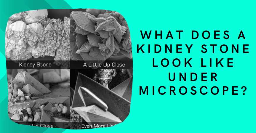
Under a microscope, a kidney stone typically appears as a crystalline structure with various shapes and sizes, such as jagged edges or smooth surfaces, depending on the composition of the stone.
Kidney stones are formed from accumulating substances in the urine, such as calcium, oxalate, and phosphate. These substances can crystallize and aggregate, leading to the formation of kidney stones. The appearance of a kidney stone under a microscope depends on its composition. Common types of kidney stones include calcium oxalate stones, calcium phosphate stones, uric acid stones, and struvite stones.
| Image | Product | Detail | Price |
|---|---|---|---|
 | Carson MicroBrite Plus 60x-120x LED Lighted Pocket Microscope |
| See on Amazon |
 | Elikliv LCD Digital Coin Microscope |
| See on Amazon |
 | AmScope M150 Series Portable Compound Microscope |
| See on Amazon |
 | PalliPartners Compound Microscope for Adults & Kids |
| See on Amazon |
 | Skybasic 50X-1000X Magnification WiFi Portable Handheld Microscopes |
| See on Amazon |
- Calcium Oxalate Stones: These stones often appear as small, sharp crystals with pointed edges. They can be colorless or have a yellowish-brown tint.
- Calcium Phosphate Stones: These stones may have a more irregular shape with a softer, grainy texture. They can range in color from light brown to dark brown.
- Uric Acid Stones: Uric acid stones are usually smooth and may have a yellow or brown color. They can be translucent and look somewhat like a rhombus or a diamond.
- Struvite Stones: These stones are often large and can have a staghorn shape. They may appear as a combination of crystals and organic material.
Here’s a general overview of the prevalence of different types of kidney stones:
| Type of Kidney Stone | Prevalence (%) |
|---|---|
| Calcium Oxalate | 70-80 |
| Calcium Phosphate | 5-10 |
| Uric Acid | 5-10 |
| Struvite | 10-15 |
| Cystine | <1 |
These percentages can vary based on factors such as diet, genetics, and geographical location. It’s essential to note that an accurate diagnosis of the stone type is crucial for effective treatment and prevention strategies.
The Microscopic Structure of Kidney Stones
When observed under high magnification, kidney stones reveal a fascinating array of shapes, sizes, and compositions. The most common types of kidney stones include calcium oxalate, uric acid, struvite, and cystine stones. Here is a detailed look at the microscopic appearance of these stones:
| Type of Stone | Microscopic Characteristics |
|---|---|
| Calcium Oxalate Stones |
|
| Uric Acid Stones |
|
| Struvite Stones (Infection Stones) |
|
| Cystine Stones |
|
Kidney stones, though small in size, can cause significant discomfort and health issues. Understanding the microscopic structure of these stones is essential for devising effective prevention and treatment strategies. In my exploration of this microscopic world, I uncovered the intricate details that contribute to the formation and composition of kidney stones.
Formation of Kidney Stones
Kidney stones, medically known as renal calculi, originate from the crystallization of minerals and salts in the urine. The process begins when certain substances in the urine, such as calcium, oxalate, and phosphate, exceed their solubility limits. As these elements precipitate, they form microscopic crystals that gradually aggregate into larger structures.
Types of Kidney Stones
The microscopic composition of kidney stones varies, leading to different types based on their mineral content. The four main types are calcium stones, struvite stones, uric acid stones, and cystine stones.
Calcium Stones
Approximately 80% of kidney stones are calcium stones, which primarily consist of calcium oxalate or calcium phosphate crystals. The microscopic examination of these stones reveals intricate arrangements of crystals, often resembling spiky structures.
Struvite Stones
Struvite stones, also known as infection stones, form in the presence of urinary tract infections. Microscopically, these stones exhibit a combination of magnesium, ammonium, and phosphate crystals, resembling branching or staghorn structures.
Uric Acid Stones
Uric acid stones result from elevated levels of uric acid in the urine. Microscopic analysis reveals these stones as smooth, yellow-brown crystals, often forming in acidic environments.
Cystine Stones
Cystine stones are rare and arise from a genetic disorder leading to the accumulation of cystine in the urine. Microscopically, these stones display hexagonal crystals, emphasizing their unique composition.
Microscopic Examination Techniques
Unraveling the microscopic structure of kidney stones requires advanced examination techniques. Two primary methods employed in this exploration are light microscopy and scanning electron microscopy.
Light Microscopy
Light microscopy involves the examination of thin sections of kidney stones under a microscope. This technique provides detailed information about the internal structure and mineral composition of the stones at the microscopic level. It allows scientists and medical professionals to identify the type of crystals present and understand their arrangements.
Scanning Electron Microscopy (SEM)
Scanning Electron Microscopy takes the exploration a step further by providing three-dimensional images of the kidney stones’ surfaces. This technique allows for a more detailed examination of the external morphology of the stones, revealing the intricate patterns and textures of the crystals.
Clinical Significance
Understanding the microscopic structure of kidney stones is crucial for tailoring effective treatment plans and preventive measures. Different types of stones may require distinct approaches for dissolution or removal, and identifying the composition guides dietary and lifestyle recommendations to prevent recurrence.
Additionally, microscopic analysis aids in determining the underlying causes of stone formation. Whether it’s related to dietary factors, genetic predispositions, or metabolic disorders, a precise understanding of the stone’s composition informs a holistic approach to patient care.
Microscopic Examination Techniques
Sample Collection
- Patient Sample Retrieval
- Collecting tiny kidney stone fragments from patients during medical procedures.
- Ensuring representation of stone composition for accurate microscopic analysis.
- Non-Invasive Techniques
- Utilizing non-invasive methods like ultrasound to locate and extract stones.
- Minimizing patient discomfort and promoting ease of sample collection.
| Techniques | Advantages | Challenges |
|---|---|---|
| Invasive | Accurate representation | Patient discomfort |
| Non-invasive | Minimized patient discomfort | Limited stone availability |
Sample Preparation for Microscopy
- Slide Preparation
- Fixing stone fragments on glass slides for microscopic observation.
- Enhancing visibility and facilitating detailed analysis.
- Staining Techniques
- Applying stains to highlight specific features of kidney stones.
- Improving contrast for better identification under the microscope.
| Techniques | Advantages | Challenges |
|---|---|---|
| Slide fixation | Enhanced visibility | Potential artifacts |
| Staining | Improved contrast | Standardization required |
Microscopic Techniques
- Light Microscopy
- Using visible light to observe kidney stone structures.
- Identifying general morphology and crystal patterns.
- Electron Microscopy
- Employing electron beams for higher magnification.
- Examining ultrastructural details of kidney stones.
| Techniques | Advantages | Challenges |
|---|---|---|
| Light Microscopy | Quick examination | Limited resolution |
| Electron Microscopy | High magnification for detailed analysis | Complex sample preparation |
- Scanning Electron Microscopy (SEM)
- Producing detailed three-dimensional images of kidney stone surfaces.
- Offering insights into surface characteristics.
- Transmission Electron Microscopy (TEM)
- Passing electrons through thin sections for internal structure analysis.
- Providing detailed information on crystal composition.
| Techniques | Advantages | Challenges |
|---|---|---|
| SEM | 3D imaging of surfaces | Limited depth details |
| TEM | Internal structure analysis | Thin section preparation |
Note: Combining these techniques enhances the comprehensive understanding of kidney stone composition and structure.
Microscopic Appearance of Kidney Stones
Kidney stones, when viewed under a microscope, unveil intricate details that aid in understanding their composition and origin. Here, we explore the microscopic appearances of three common types of kidney stones: Calcium Oxalate, Uric Acid, and Struvite stones.
Calcium Oxalate Stones Under Microscope
Calcium Oxalate stones, the most prevalent type, exhibit distinct characteristics when examined microscopically. The table below outlines their crystalline structure, coloration, and texture.
| Microscopic Feature | Description |
|---|---|
| Crystalline Structure | Elongated crystals with pointed edges |
| Coloration and Texture | Varies from brown to yellow; rough surface |
These microscopic traits contribute to the identification of Calcium Oxalate stones, aiding in targeted diagnostic and treatment approaches.
Uric Acid Stones Under Microscope
Uric Acid stones, formed in acidic urine conditions, possess unique microscopic characteristics. The following table details their distinctive features when observed under a microscope.
| Microscopic Feature | Description |
|---|---|
| Unique Characteristics | Smooth, flat crystals with a diamond-like shape |
| Comparison with Other Types | Clear appearance; differs from Calcium Oxalate |
Understanding these microscopic details helps differentiate Uric Acid stones from other types, ensuring accurate diagnosis and tailored treatment.
Struvite Stones Under Microscope
Struvite stones, commonly associated with bacterial infections, present distinctive features when examined microscopically. The table below delineates their microscopic characteristics.
| Microscopic Feature | Description |
|---|---|
| Presence of Bacteria | Clumps of crystals encasing bacterial debris |
| Distinctive Morphology | Large, coffin-shaped crystals |
Recognizing these microscopic attributes aids in identifying Struvite stones, highlighting the connection between microbial activity and stone formation.
How do kidney stones appear under a microscope?
Kidney stones exhibit diverse appearances under a microscope, depending on their composition. Here’s a breakdown:
| Type of Kidney Stone | Microscopic Appearance |
|---|---|
| Calcium Oxalate | Crystalline structures resembling envelopes or dumbbells. These can vary in size and shape, often appearing sharp-edged. |
| Struvite | Prismatic crystals with a rectangular or coffin-like shape. These stones may also feature a “coffin lid” appearance under certain conditions. |
| Uric Acid | Rhomboid or diamond-shaped crystals. These crystals are usually yellow or brown and may be transparent or colored. |
| Cystine | Hexagonal plates or crystals, often arranged in a stacked or honeycomb pattern. These stones may be smaller and more fragile compared to other types. |
Can kidney stones be identified based on color under a microscope?
Yes, the color of kidney stones under a microscope can provide valuable information about their composition:
| Stone Color | Possible Composition |
|---|---|
| Yellow/Brown | Uric acid or calcium oxalate stones. |
| White | Calcium phosphate or cystine stones. |
| Green | Struvite or certain types of calcium stones. |
Analyzing the color, along with other microscopic features, aids in identifying the specific type of kidney stone.
Do kidney stones show any signs of damage or inflammation under a microscope?
Examination under a microscope may reveal signs of tissue damage and inflammation associated with kidney stones:
| Microscopic Signs | Indications |
|---|---|
| Crystal Aggregation | Accumulation of crystals in renal tissues. |
| Inflammatory Infiltration | Presence of immune cells, indicating an inflammatory response. |
| Tissue Erosion | Evidence of damage to the renal epithelium. |
| Fibrosis | Formation of fibrous tissue in response to chronic irritation. |
These signs help clinicians understand the impact of kidney stones on the kidneys and guide treatment decisions.
Are there specific patterns in the arrangement of crystals within kidney stones?
Yes, the microscopic arrangement of crystals can vary, providing additional diagnostic insights:
| Crystal Arrangement | Possible Kidney Stone Type |
|---|---|
| Radial or Spherical | Calcium oxalate stones or cystine stones. |
| Stacked or Honeycomb | Cystine stones. |
| Prismatic or Coffin-Like | Struvite stones. |
| Randomly Oriented | Uric acid stones. |
Identifying these patterns aids in determining the type of kidney stone and guides treatment strategies.
How do doctors use microscopic analysis to customize treatment plans?
Microscopic analysis plays a crucial role in tailoring treatment for kidney stones:
| Aspect of Analysis | Treatment Considerations |
|---|---|
| Stone Composition | Prescribes dietary changes to prevent recurrence. |
| Inflammatory Response | Guides anti-inflammatory or immune-modulating interventions. |
| Tissue Damage and Fibrosis | Indicates the need for interventions to minimize scarring. |
| Crystal Arrangement and Size | Influences the choice of medications or surgical approaches. |
Healthcare providers can develop personalized plans to manage kidney stones effectively by understanding these microscopic details.

Fahim Foysal is a well-known expert in the field of binoculars, with a passion for exploring the great outdoors and observing nature up close. With years of experience in the field, Fahim has honed his skills as a binocular user and has become a go-to resource for those seeking advice on choosing the right binoculars for their needs.
Fahim’s love for the natural world began during his time at The Millennium Stars School and College and BIAM Laboratory School, where he spent much of his free time exploring the outdoors and observing the wildlife around him. This passion for nature led him to pursue a degree in Fine Arts from the University of Dhaka, where he gained a deep understanding of the importance of observation and attention to detail.
Throughout his career, Fahim has used his expertise in binoculars to help others discover the beauty of the natural world. His extensive knowledge of binocular technology and optics has made him a trusted advisor for amateur and professional wildlife observers alike. Whether you’re looking to spot rare birds or observe animals in their natural habitats, Fahim can help you choose the perfect binoculars for your needs. With his guidance, you’ll be able to explore the outdoors with a newfound appreciation for the beauty of the natural world.
Table of Contents

Pingback: What Does Staph Look Like Under Microscope: A Close-Up View