| Image | Product | Detail | Price |
|---|---|---|---|
 | Carson MicroBrite Plus 60x-120x LED Lighted Pocket Microscope |
| See on Amazon |
 | Elikliv LCD Digital Coin Microscope |
| See on Amazon |
 | AmScope M150 Series Portable Compound Microscope |
| See on Amazon |
 | PalliPartners Compound Microscope for Adults & Kids |
| See on Amazon |
 | Skybasic 50X-1000X Magnification WiFi Portable Handheld Microscopes |
| See on Amazon |
| Category | Data |
|---|---|
| Global Prevalence | Estimated 156 million cases annually |
| Affected Population | Both men and women, but higher in women |
| Transmission | Primarily through sexual contact |
| Symptoms (Women) | Vaginal discharge, itching, discomfort |
| Symptoms (Men) | Urethral discharge, discomfort |
| Diagnostic Methods | Wet mount microscopy, PCR, culture |
| Treatment | Metronidazole or tinidazole |
| Complications | Increased risk of HIV, preterm birth |
| Prevention | Safe sex practices, regular screenings |
Understanding Trichomoniasis
Before we dive into the microscopic realm, it’s important to get a grasp of what Trichomoniasis is. Trichomoniasis is a sexually transmitted infection caused by the flagellated protozoan parasite, Trichomonas vaginalis. It primarily infects the urogenital tract and can cause symptoms such as itching, burning, and unusual discharge, though many carriers can be asymptomatic.
The Role of Microscopy in Trichomoniasis Diagnosis
Diagnosing Trichomoniasis typically involves microscopic examination of urine, discharge, or swab samples. Microscopy is used to directly observe the characteristic movements and morphology of the Trichomonas vaginalis parasite within these samples.
| Feature | Description |
|---|---|
| Size | Around 10-20µm in length |
| Shape | Pear-shaped with an undulating membrane |
| Flagella | Four anterior flagella and a fifth forming the undulating membrane |
| Movement | Jerky, motile movement |
| Nucleus | Central, singular nucleus |
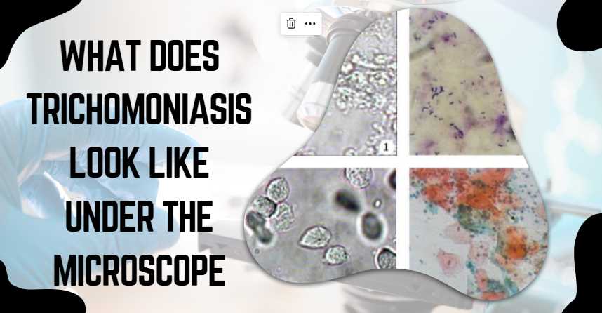
Visualizing Trichomonas vaginalis under the Microscope
When preparing a sample for microscopic evaluation, a wet mount is commonly used. This requires placing a drop of the fluid sample on a microscope slide and covering it with a cover slip. Under the microscope, several defining features can be observed:
- Pear-shaped Structure: Trichomonas vaginalis is identified by its unique pear shape. This shape is distinctive among the various protozoa that could potentially be present in a sample.
- Motility: One of the key diagnostic characteristics is the jerky motion of Trichomonas vaginalis, due to its flagella.
- Flagella: The organism possesses multiple flagella that can sometimes be seen moving in live samples.
- Undulating Membrane: Alongside the flagella, an undulating membrane along one side of the parasite adds to its distinctive swimming pattern.
- Nucleus: The central nucleus can be visualized under higher magnifications and is important for conclusive identification.
What Staining Techniques are Used for Trichomoniasis Diagnosis?
To enhance the visibility of Trichomonas vaginalis under the microscope, specific staining techniques are employed. The most common method is the use of a wet mount preparation, but additional stains like Giemsa and Papanicolaou can also be utilized.
| Staining Technique | Description |
|---|---|
| Wet Mount | Direct observation of live, unstained organisms |
| Giemsa Stain | Stains cellular elements, enhancing visibility |
| Papanicolaou Stain | Utilized for cytological examination of smears |
How Effective is Microscopy in Detecting Trichomoniasis?
Microscopic examination remains a valuable tool for diagnosing trichomoniasis, offering a quick and direct visualization of the parasite. However, it is worth noting that the sensitivity of microscopy can vary, and false-negative results may occur. To enhance accuracy, complementary diagnostic methods such as nucleic acid amplification tests (NAATs) are often employed.
| Diagnostic Method | Sensitivity |
|---|---|
| Microscopy | Variable, may yield false-negative results |
| Nucleic Acid Amplification Tests | Increased sensitivity and specificity |
Can Trichomoniasis Be Diagnosed Without Microscopy?
Yes, besides microscopy, several alternative methods can be employed for diagnosing trichomoniasis. Nucleic acid amplification tests (NAATs) are highly sensitive and specific, detecting the genetic material of the parasite. Additionally, culture methods and rapid antigen tests are available, offering alternative approaches to diagnosis.
| Diagnostic Method | Description |
|---|---|
| Nucleic Acid Amplification Tests | Detect genetic material of Trichomonas vaginalis |
| Culture Methods | Growing the parasite in a suitable culture medium |
| Rapid Antigen Tests | Detecting specific antigens of Trichomonas |
What Are the Symptoms of Trichomoniasis?
While some individuals with trichomoniasis may remain asymptomatic, common symptoms include vaginal discharge, itching, and discomfort during urination or sexual intercourse. It’s important to note that symptoms can vary, and some people may not experience any noticeable signs.
| Symptoms | Description |
|---|---|
| Vaginal Discharge | Unusual, often with a strong odor |
| Itching | Irritation in the genital area |
| Discomfort During Urination | Pain or burning sensation |
| Discomfort During Intercourse | Pain or irritation during sexual activity |
How Is Trichomoniasis Treated?
Trichomoniasis is typically treated with antibiotics, most commonly metronidazole or tinidazole. It is crucial to complete the full course of medication as prescribed by a healthcare provider to ensure complete eradication of the parasite.
| Treatment | Medications |
|---|---|
| Antibiotics | Metronidazole or Tinidazole |
| Treatment Duration | Full course as prescribed by a healthcare provider |
Can Trichomoniasis Recur After Treatment?
Yes, trichomoniasis can recur, and reinfection is possible if exposure to the parasite occurs again. To prevent recurrence, individuals treated for trichomoniasis should abstain from sexual activity until both partners have completed treatment. Condom use can also help reduce the risk of reinfection.
| Recurrence | Possibility of recurrence and reinfection |
|---|---|
| Abstaining from Sex | Recommended during and after treatment |
| Condom Use | Reduces the risk of reinfection |
Are There Limitations to Microscopic Detection of Trichomoniasis?
Microscopic detection of Trichomonas vaginalis, while valuable, does have limitations. The sensitivity of the method can be affected by factors such as the skill of the technician, the quality of the sample, and the presence of coexisting infections. To mitigate these limitations, healthcare providers may use additional diagnostic methods for confirmation.
| Limitations | Sensitivity affected by various factors |
|---|---|
| Technician Skill | Skill of the individual conducting the test |
| Sample Quality | Quality of the collected sample |
| Coexisting Infections | Presence of other infections may impact accuracy |
Recap
The accurate identification of Trichomonas vaginalis is crucial for appropriate treatment. Traditional microscopic examination may sometimes be supplemented with molecular tests for improved accuracy, especially in asymptomatic carriers who may still spread the infection. Antiprotozoal medication, such as metronidazole or tinidazole, is generally prescribed to treat Trichomoniasis once diagnosed.

Fahim Foysal is a well-known expert in the field of binoculars, with a passion for exploring the great outdoors and observing nature up close. With years of experience in the field, Fahim has honed his skills as a binocular user and has become a go-to resource for those seeking advice on choosing the right binoculars for their needs.
Fahim’s love for the natural world began during his time at The Millennium Stars School and College and BIAM Laboratory School, where he spent much of his free time exploring the outdoors and observing the wildlife around him. This passion for nature led him to pursue a degree in Fine Arts from the University of Dhaka, where he gained a deep understanding of the importance of observation and attention to detail.
Throughout his career, Fahim has used his expertise in binoculars to help others discover the beauty of the natural world. His extensive knowledge of binocular technology and optics has made him a trusted advisor for amateur and professional wildlife observers alike. Whether you’re looking to spot rare birds or observe animals in their natural habitats, Fahim can help you choose the perfect binoculars for your needs. With his guidance, you’ll be able to explore the outdoors with a newfound appreciation for the beauty of the natural world.
Table of Contents

Pingback: What Does a Kidney Stone Look Like Under Microscope: A Microscopic Marvel