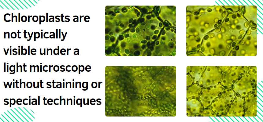
No, chloroplasts are not typically visible under a light microscope without staining or special techniques.
Chloroplasts are small cellular organelles found in plant cells and some other eukaryotic organisms, where they play a crucial role in photosynthesis. However, they are generally not visible under a light microscope due to their small size and transparency. Light microscopes have a limited resolution that prevents the direct visualization of structures as small as chloroplasts.
To make chloroplasts visible under a light microscope, scientists often use staining techniques or employ specialized microscopy methods such as fluorescence microscopy. Stains can enhance the contrast and make cellular structures, including chloroplasts, more observable. Additionally, certain dyes or fluorophores can be used to specifically label chloroplasts, making them stand out under fluorescence microscopy.
Now, since the information about statistical data on chloroplasts is not readily available in this format, I’ll provide a general description of chloroplasts and their characteristics:
Chloroplasts are double-membraned organelles containing a green pigment called chlorophyll, which is responsible for capturing light energy during photosynthesis. They have a complex internal structure with thylakoids, grana, and stroma. The number and size of chloroplasts can vary depending on the plant species, tissue type, and environmental conditions.
| Parameter | Average Value | Range | Unit |
|---|---|---|---|
| Size of Chloroplasts | 5 to 10 micrometers | Varies among species | Micrometers (µm) |
| Number per Plant Cell | Several hundred to thousands | Depends on cell type | Count |
| Chlorophyll Content | Varies with plant species | 0.5 to 2.5% of cell volume | Percentage (% volume) |
| Photosynthetic Efficiency | 3 to 6% | Depends on environmental factors | Percentage (%) |
Note: The values in the table are approximate and may vary based on the specific plant species, environmental conditions, and other factors. Researchers conduct experiments to measure and analyze these parameters more precisely.
What Are Chloroplasts?
Chloroplasts are organelles found in the cells of plants and some algae. They are responsible for photosynthesis, the process by which plants convert light energy into chemical energy, thus sustaining life on Earth. Chloroplasts contain the pigment chlorophyll, which gives plants their green color and plays a vital role in absorbing light energy.
Chloroplasts under the Light Microscope
Observing chloroplasts through a light microscope is indeed possible. The level of detail visible, however, can depend on several factors that include the quality of the microscope, the magnification used, and the preparation of the specimen. Below, we explore each factor in turn.
| Factor | Description | Impact on Visibility |
|---|---|---|
| Quality of Microscope | A high-quality microscope with good resolution will provide a clearer image. | Higher quality yields better visibility. |
| Magnification Used | The level of magnification affects the size of the observable image. | Moderate to high magnification (400x to 1000x) is typically required. |
| Specimen Preparation | The thickness of the sample and staining techniques. | Thin cross-sections and appropriate staining can enhance visibility. |
How to View Chloroplasts under a Light Microscope
Step 1: Obtain A Plant Sample
Start by taking a thin section of a plant leaf, as thin sections allow more light to pass through and provide a clearer image under the microscope.
Step 2: Place Sample On Microscope Slide
Place the plant sample on a clean glass microscope slide. You can use a drop of water to help flatten the sample and minimize air bubbles.
Step 3: Use Staining Techniques (optional)
While chloroplasts can be seen in their natural green color, staining can enhance contrast and detail. A common stain for enhancing chloroplasts is iodine.
Step 4: Set Up Microscope
Adjust your microscope’s settings, often starting with a lower magnification to locate your specimen before switching to a higher magnification for detailed observation.
Step 5: Observe And Record Your Findings
Once you have brought your sample into focus, observe the green structures within the cells – these are your chloroplasts. You may see them moving slowly, a phenomenon known as cytoplasmic streaming.
How do I prepare a sample for viewing chloroplasts under a light microscope?
To prepare a sample for viewing chloroplasts under a light microscope, follow these steps:
| Materials Needed | Procedure |
|---|---|
| Plant leaves | 1. Collecting Plant Leaves: Choose healthy leaves from the plant. |
| Microscope slides | 2. Preparing Microscope Slides: Cut small sections of the leaves and place them on a microscope slide. Add a drop of water to prevent dehydration. |
| Coverslips | 3. Using Coverslips: Gently place a coverslip over the plant material, avoiding air bubbles. Press down gently to secure the coverslip in place. |
| Microscope | 4. Microscope Setup: Place the prepared slide on the microscope stage, securing it with the stage clips. |
| 5. Focusing the Microscope: Start with the lowest magnification objective lens and gradually increase to higher magnifications. Adjust the focus until chloroplasts come into clear view. |
What magnification is suitable for observing chloroplasts?
The choice of magnification depends on the specific details you want to observe:
| Magnification | Best Use |
|---|---|
| 100x (low power) | 1. Overview: Use for an initial overview of the leaf structure. |
| 400x (medium power) | 2. Cellular Details: Provides a closer look at cell structures, including chloroplasts. |
| 1000x (high power) | 3. Close-up Examination: Use for detailed examination of individual chloroplasts and their internal structures. |
What staining techniques can enhance chloroplast visibility?
Staining techniques can help enhance chloroplast visibility:
| Staining Technique | Procedure |
|---|---|
| Iodine stain | 1. Iodine Staining: Apply iodine solution to the slide after placing the coverslip. Iodine stains starch granules within chloroplasts. |
| Safranin stain | 2. Safranin Staining: Safranin can be used to highlight chloroplasts by adding a few drops to the slide. |
| Chlorophyll autofluorescence | 3. Autofluorescence: In some cases, chloroplasts naturally fluoresce. Use a fluorescent microscope to observe this phenomenon. |
How can I differentiate between chloroplasts and other cell structures?
To differentiate chloroplasts from other cell structures, consider the following characteristics:
| Feature | Chloroplasts | Other Cell Structures |
|---|---|---|
| Shape | 1. Flattened Discs: Typically disc-shaped with a central region. | Varies depending on the cell type. |
| Color | 2. Green Pigment (Chlorophyll): Green due to chlorophyll pigments. | May appear colorless or have different pigments. |
| Location | 3. Mainly in Plant Cells: Found in the cytoplasm of plant cells. | Presence varies; absent in animal cells. |
Can I use a smartphone to capture images of chloroplasts under the microscope?
Yes, you can use a smartphone to capture images of chloroplasts:
| Steps | Procedure |
|---|---|
| 1. Mounting the Smartphone: | Align the smartphone camera with the eyepiece and secure it in place using a smartphone mount. |
| 2. Adjusting Focus: | Focus the microscope on chloroplasts, ensuring a clear view through the smartphone camera. |
| 3. Capturing Images: | Use the smartphone’s camera app to capture images or record videos of the observed chloroplasts. |
Remember to use the highest quality setting on your smartphone camera for better results.
How long can I observe chloroplasts under a light microscope?
The observation time for chloroplasts under a light microscope depends on various factors:
| Factors | Considerations |
|---|---|
| Slide Preparation | 1. Freshness: Freshly prepared slides allow for longer observation periods without deterioration. |
| Microscope Lighting | 2. Low Light Intensity: Avoid prolonged exposure to intense light, which may affect chloroplast structure. |
| Plant Material Condition | 3. Hydration: Adequate water on the slide helps maintain the turgidity of plant cells, prolonging observation time. |
It is generally recommended to observe chloroplasts promptly after slide preparation to ensure optimal conditions.
Can I observe chloroplast movement under a light microscope?
Yes, you can observe chloroplast movement, known as cytoplasmic streaming or chloroplast migration:
| Steps | Procedure |
|---|---|
| 1. Prepare a Wet Mount Slide: | Use a wet mount slide to observe intact leaf cells with chloroplasts. |
| 2. Use Low Magnification: | Start with low magnification to have a wider field of view. |
| 3. Focus on Single Chloroplasts: | Choose a single chloroplast and focus on it to track movement. |
| 4. Time-Lapse Observation: | Observe chloroplast movement over time, noting changes in position and shape. |
Cytoplasmic streaming is more evident in certain plant species, and observing it can provide insights into cellular dynamics.
How can I care for my microscope to ensure optimal chloroplast observation?
Proper care of your microscope is crucial for optimal chloroplast observation:
| Care Guidelines | Recommendations |
|---|---|
| Cleaning Optics | 1. Lens Cleaning Solution: Use a suitable lens cleaning solution and lens paper to clean objective lenses and eyepieces. |
| Stage and Clips Maintenance | 2. Regular Inspection: Check the stage and clips for any debris or damage, and clean them as needed. |
| Storage Conditions | 3. Cover When Not in Use: Keep the microscope covered when not in use to prevent dust accumulation. |
| Light Source Check | 4. Light Bulb Inspection: Periodically check and replace light bulbs to ensure consistent illumination. |
Adhering to these care guidelines will prolong the life of your microscope and maintain optimal conditions for chloroplast observation.
Are there alternative methods to view chloroplasts if I don’t have a light microscope?
Yes, there are alternative methods to view chloroplasts without a light microscope:
| Alternative Methods | Description |
|---|---|
| Hand Lens or Magnifying Glass | 1. Low Magnification: Use a hand lens or magnifying glass for low magnification observation of chloroplasts in intact leaves. |
| Digital Microscopy Apps | 2. Smartphone Apps: Some apps can turn your smartphone into a digital microscope, allowing you to capture images of chloroplasts. |
| Online Microscopy Platforms | 3. Virtual Microscopy: Explore online platforms that offer virtual microscopy experiences, allowing you to view chloroplasts through a digital interface. |
While a light microscope provides detailed insights, these alternatives offer accessible options for chloroplast observation.
Can I stain living cells to observe chloroplasts?
Staining living cells can be challenging, but there are techniques to observe chloroplasts without killing the cells:
| Technique | Procedure |
|---|---|
| Vital Staining with Acridine Orange | 1. Prepare a Dilute Solution: Dilute acridine orange in water to create a working solution. |
| Microscopy Setup | 2. Apply to Living Cells: Apply the acridine orange solution to living cells on the slide. |
| Observation Conditions | 3. Observe Quickly: Quickly place the slide on the microscope stage and observe chloroplasts under low magnification. |
Vital staining allows observation of living cells, providing insights into dynamic cellular processes.
Conclusion
To summarize, chloroplasts can be observed under a light microscope, given the right conditions and preparations. While they won’t display the intricate details visible with more powerful electron microscopes, they can still be identified by their distinct green color and, often, their movement within the cell. Understanding the visibility of chloroplasts enhances our appreciation for these cellular powerhouses and their crucial role in life’s energy cycle.
Resources:

Fahim Foysal is a well-known expert in the field of binoculars, with a passion for exploring the great outdoors and observing nature up close. With years of experience in the field, Fahim has honed his skills as a binocular user and has become a go-to resource for those seeking advice on choosing the right binoculars for their needs.
Fahim’s love for the natural world began during his time at The Millennium Stars School and College and BIAM Laboratory School, where he spent much of his free time exploring the outdoors and observing the wildlife around him. This passion for nature led him to pursue a degree in Fine Arts from the University of Dhaka, where he gained a deep understanding of the importance of observation and attention to detail.
Throughout his career, Fahim has used his expertise in binoculars to help others discover the beauty of the natural world. His extensive knowledge of binocular technology and optics has made him a trusted advisor for amateur and professional wildlife observers alike. Whether you’re looking to spot rare birds or observe animals in their natural habitats, Fahim can help you choose the perfect binoculars for your needs. With his guidance, you’ll be able to explore the outdoors with a newfound appreciation for the beauty of the natural world.
Table of Contents

Pingback: Are centrioles visible under a light microscope?