Author: Fahim Foysal

  • What Does Telophase Look Like Under Microscope: A Micro View

    What Does Telophase Look Like Under Microscope: A Micro View

    Under a microscope, telophase appears as a stage of mitosis where separated chromatids or chromosomes reach the opposite poles of the cell. Two distinct nuclei start forming, and the cell begins to undergo cytokinesis, forming two separate daughter cells.

    Telophase is the final stage of mitosis or meiosis, during which the separated chromatids or chromosomes reach the opposite poles of the dividing cell. The key microscopic features of telophase include:

    1. Chromosomes at Opposite Poles: Chromosomes, which were duplicated during earlier stages of cell division, are now clearly visible as distinct structures. They align at opposite poles of the cell.
    2. Nuclear Envelope Formation: As the chromosomes reach their respective poles, a new nuclear envelope or membrane begins to form around each set of chromosomes. This process marks the reformation of the nuclei in the daughter cells.
    3. Cell Division Completion: Telophase is followed by cytokinesis, where the cell undergoes physical division to form two separate daughter cells, each with its nucleus enclosed within a nuclear envelope.

    To provide a clearer understanding, here is a table summarizing the microscopic features of telophase:

    Microscopic FeatureDescription
    Chromosome ArrangementChromosomes align at opposite poles of the cell.
    Nuclear Envelope FormationNew nuclear envelopes begin to form around each set of chromosomes.
    NucleiTwo distinct nuclei become visible as the cell prepares to divide.

    Cell Division Process: Mitosis and Meiosis

    Cell division is a fundamental process that ensures the growth, development, and reproduction of organisms. There are two primary types of cell division: mitosis, responsible for the formation of somatic cells, and meiosis, which is specific to the generation of gametes.

    Key Stages Leading Up to Telophase:

    Both mitosis and meiosis share common stages leading up to Telophase, where the nucleus divides, and two daughter cells are formed.

    Key Stages of Mitosis:

    Mitosis is the process by which a single eukaryotic cell divides into two identical daughter cells. The key stages leading up to Telophase in mitosis include:

    1. Prophase: Chromosomes condense, the nuclear envelope disintegrates, and spindle fibers begin to form.
    2. Metaphase: Chromosomes align along the cell’s equator, known as the metaphase plate, ensuring an equal distribution of genetic material.
    3. Anaphase: Sister chromatids separate and move toward opposite poles of the cell, pulled by spindle fibers.
    4. Telophase: Chromosomes de-condense, a nuclear envelope reforms around each set of chromosomes, and the cell undergoes cytokinesis, resulting in two daughter cells.
    Key Stages of Meiosis:

    Meiosis is the process of cell division that produces haploid gametes (sperm and egg cells). The stages leading up to Telophase in meiosis are:

    1. Prophase I: Homologous chromosomes pair up in a process called synapsis, and genetic recombination occurs through crossing-over.
    2. Metaphase I: Homologous chromosome pairs align at the metaphase plate.
    3. Anaphase I: Homologous chromosomes separate and move to opposite poles, reducing the chromosome number by half.
    4. Telophase I: Chromosomes may partially de-condense, and the cell undergoes cytokinesis, resulting in two haploid cells.
    5. Prophase II, Metaphase II, Anaphase II, and Telophase II: These stages mirror those of mitosis, resulting in the formation of four haploid daughter cells, each with half the chromosome number of the original cell.

    Outcome of Telophase:

    The culmination of Telophase in both mitosis and meiosis is the formation of two daughter cells, each genetically identical in mitosis and genetically diverse in meiosis. This process is fundamental for organismal development, growth, and the generation of reproductive cells.

    Sample Preparation for Microscopic Observation

    Telophase microscopic observation requires meticulous sample preparation to ensure clear and detailed imaging. The process involves several key steps that preserve cellular structures and enhance visibility under the microscope.

    6 Steps in Sample Preparation:

    1. Cell Harvesting: Begin by carefully harvesting the cells of interest. This may involve cell culture techniques, tissue sectioning, or other methods depending on the sample type.
    2. Fixation: The crucial step of fixation involves preserving the cellular structure by arresting cellular activities and preventing decay. Common fixatives include formaldehyde or glutaraldehyde.
    3. Permeabilization: To allow staining agents to penetrate the cells, permeabilization is employed. This step is particularly important for fluorescent microscopy, enabling dyes to access cellular components.
    4. Staining: Staining enhances the contrast and visibility of cellular structures. Various dyes target specific components, such as DAPI for DNA, and Phalloidin for actin filaments.
    5. Mounting: After staining, the sample is carefully mounted on a slide using a mounting medium. This not only secures the sample but also helps maintain its three-dimensional structure.
    6. Cover Slipping: A cover slip is placed over the sample, minimizing dehydration and protecting it during microscopic observation.

    Importance of Proper Fixation:

    Proper fixation is a cornerstone in sample preparation as it preserves cellular morphology and prevents degradation. It serves the following crucial purposes:

    1. Structural Integrity: Fixation immobilizes cellular structures, preventing distortions or alterations during subsequent processing.
    2. Halting Cellular Processes: Fixation halts cellular activities, ensuring that the sample represents a snapshot of the cells at the moment of fixation. This is vital for accurate observation.
    3. Preventing Autolysis and Putrefaction: Without fixation, cellular components would undergo autolysis and putrefaction, compromising the integrity of the sample.

    Importance of Staining Techniques:

    Staining techniques enhance the contrast of cellular structures, making them more visible under a microscope. The significance of staining includes:

    1. Visualization of Specific Structures: Different stains target specific cellular components, allowing researchers to focus on particular structures, such as chromosomes during Telophase.
    2. Identification of Cell Types: Staining helps distinguish between various cell types and subcellular structures, aiding in the accurate identification of cells undergoing Telophase.
    3. Quantitative Analysis: Staining facilitates quantitative analysis, enabling researchers to measure aspects like DNA content, cell proliferation, and protein expression.

    In summary, the preparation of samples for microscopic observation involves a series of meticulous steps, with fixation and staining playing pivotal roles. Proper fixation preserves cellular integrity, while staining techniques enhance visibility, providing a clearer understanding of Telophase events under the microscope. These techniques collectively contribute to the success of microscopic studies and the accurate analysis of cellular processes.

    Common Challenges in Observing Telophase

    Observing Telophase under a microscope presents certain challenges that researchers must navigate to obtain accurate and reliable data.

    Sample Distortion:

    Issues Related to Sample Distortion:

    1. Dehydration: During sample preparation, dehydration can occur, leading to shrinkage and distortion of cellular structures.
    2. Fixation Artifacts: Improper fixation may result in artifacts, causing morphological changes and affecting the accurate representation of Telophase.

    Minimizing Distortion for Accurate Observation:

    1. Optimal Fixation: Ensuring the use of suitable fixatives and adhering to recommended fixation times minimizes distortion.
    2. Balanced Dehydration: Carefully controlling the dehydration process helps prevent excessive shrinkage and distortion.

    Limited Resolution:

    Challenge of Limited Resolution:

    1. Microscope Limitations: Traditional light microscopy may have limitations in resolving fine details during Telophase.

    Techniques to Enhance Resolution:

    1. Super-Resolution Microscopy: Utilizing advanced techniques like super-resolution microscopy enhances resolution, allowing for finer details to be observed.
    2. Immunofluorescence Staining: Employing specific fluorescent markers in immunofluorescence staining can improve visibility and resolution.

    Suboptimal Contrast:

    Challenges in Achieving Suboptimal Contrast:

    1. Inadequate Staining: Poorly applied or insufficient staining can result in low contrast, making it challenging to distinguish cellular structures.

    Techniques to Enhance Contrast:

    1. Optimized Staining Protocols: Following established staining protocols and using appropriate dyes improves contrast.
    2. Fluorescence Microscopy: Utilizing fluorescence microscopy with fluorescent dyes enhances contrast and specificity in observing Telophase.

    Photobleaching:

    Impact of Photobleaching:

    1. Loss of Fluorescence: Prolonged exposure to light can lead to the loss of fluorescence signal, hindering continuous observation.

    Strategies to Minimize Photobleaching:

    1. Timed Observation: Limiting the exposure time during observation sessions helps minimize photobleaching.
    2. Antifade Reagents: Using antifade reagents in fluorescence microscopy can mitigate the effects of photobleaching, preserving fluorescence signals.

    Addressing these common challenges in observing Telophase enhances the accuracy and reliability of microscopic studies, providing researchers with a clearer understanding of the intricate events during this crucial stage of cell division.

    Live Cell Imaging

    In recent years, live cell imaging has emerged as a groundbreaking technique, revolutionizing the study of Telophase and providing unprecedented insights into dynamic cellular events.

    Advancements in Live Cell Imaging Techniques:

    1. Real-Time Observations: Live cell imaging allows researchers to observe Telophase events in real-time, capturing the dynamic nature of cellular processes.
    2. Longitudinal Studies: Continuous monitoring of cells over extended periods enables longitudinal studies, offering a comprehensive view of Telophase progression.

    Impact on Our Understanding:

    1. Dynamic Visualization: Live cell imaging facilitates the dynamic visualization of Telophase events, shedding light on the intricacies of chromosome de-condensation, nuclear envelope reformation, and cytokinesis.
    2. Cellular Dynamics: The ability to observe cellular dynamics in a live context enhances our understanding of the timing and coordination of events during Telophase.

    Challenges and Future Prospects:

    Technical Challenges:

    1. Phototoxicity: Prolonged exposure to light during live cell imaging can induce phototoxicity, affecting cell viability and confounding observations.
    2. Data Processing: Handling large volumes of live cell imaging data poses challenges in terms of storage, analysis, and interpretation.

    Future Prospects:

    1. Improved Imaging Modalities: Ongoing advancements aim to develop imaging modalities with reduced phototoxicity and enhanced resolution for more accurate live cell observations.
    2. Integration with Other Techniques: Integrating live cell imaging with complementary techniques, such as super-resolution microscopy, holds promise for comprehensive Telophase analysis.

    Live cell imaging has undeniably transformed our approach to Telophase research, providing a dynamic window into the previously inaccessible realm of real-time cellular events. While technical challenges persist, ongoing innovations and future prospects in live cell imaging promise to further deepen our understanding of Telophase and contribute to the broader field of cell biology.

    How does Telophase appear under a microscope?

    Telophase, the final stage of mitosis, exhibits distinct characteristics when observed under a microscope. A typical telophase cell appears as two separate nuclei forming within a single cell membrane. Chromosomes, which were condensed during earlier stages, start to de-condense and elongate. The microtubules that facilitated chromosome movement begin to disassemble, allowing the formation of two distinct daughter cells. Below is a table summarizing key features observed during telophase:

    Telophase CharacteristicsDescription
    Nuclei FormationTwo distinct nuclei forming within a shared cell membrane
    Chromosome StateDe-condensation and elongation of chromosomes
    Microtubule DisassemblyDisintegration of microtubules aiding chromosome movement

    What is the role of microtubules in Telophase?

    Microtubules play a crucial role in telophase, facilitating the movement of chromosomes and the formation of daughter cells. During this phase, microtubules that were responsible for separating chromosomes begin to disassemble. This disassembly allows the chromosomes to move freely, leading to the formation of two distinct nuclei within the cell. Here’s a table summarizing the role of microtubules in telophase:

    Microtubule FunctionDescription
    Chromosome SeparationMicrotubules aid in pulling apart condensed chromosomes
    Daughter Cell FormationDisassembly of microtubules allows the formation of cells

    Can telophase be easily distinguished from other mitotic stages under a microscope?

    Yes, telophase can be distinguished from other mitotic stages under a microscope due to its unique characteristics. The presence of two distinct nuclei within a single cell membrane, along with the de-condensation and elongation of chromosomes, sets telophase apart from the preceding stages. Microtubule disassembly is also a key indicator of telophase. The following table highlights the distinguishing features of telophase:

    Distinguishing FeaturesDescription
    Two Nuclei FormationPresence of two distinct nuclei within a shared membrane
    Chromosome ChangesDe-condensation and elongation of chromosomes
    Microtubule DisassemblyDisintegration of microtubules aiding chromosome movement

    What is the significance of observing telophase under a microscope?

    Observing telophase under a microscope provides valuable insights into the process of cell division and the formation of new cells. It allows researchers and scientists to study the intricate details of how chromosomes are distributed among daughter cells and how cellular structures, such as nuclei and microtubules, undergo dynamic changes. This understanding is crucial for various fields, including cell biology and medical research. No table is needed for this question.

    Are there any specific staining techniques recommended for observing telophase?

    Various staining techniques can enhance the visibility of cellular structures during telophase. Dyes such as DAPI (4′,6-diamidino-2-phenylindole) are commonly used to highlight DNA, making chromosomes more visible. Immunofluorescence staining can also be employed to target specific proteins involved in telophase processes. Here’s a table summarizing recommended staining techniques:

    Staining TechniqueDescription
    DAPI (4′,6-diamidino-2-phenylindole)Highlights DNA, making chromosomes more visible
    Immunofluorescence StainingTargets specific proteins involved in telophase processes

    References:

    1. Alberts B, Johnson A, Lewis J, et al. Molecular Biology of the Cell. 4th edition. New York: Garland Science; 2002.
    2. Lodish H, Berk A, Zipursky SL, et al. Molecular Cell Biology. 4th edition. New York: W. H. Freeman; 2000
  • Is the higher number of binocular numbers better?

    Is the higher number of binocular numbers better?

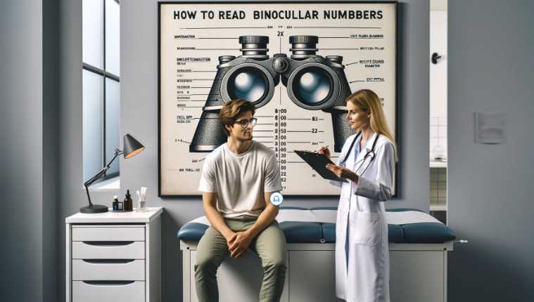
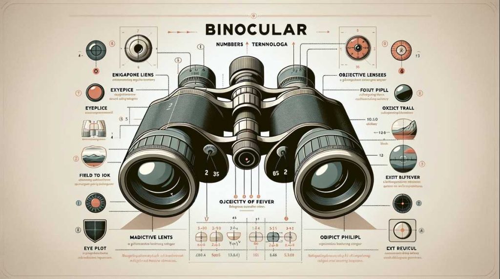
    No, The number of binoculars alone doesn’t determine their quality or suitability for a particular purpose. The effectiveness of binoculars depends on various factors, such as the quality of optics, magnification, field of view, lens coatings, and other features. Simply having a higher number of binoculars does not guarantee better performance. It’s essential to consider the specific requirements and intended use to determine the most suitable pair of binoculars.

    Simply having a higher number of binoculars does not necessarily make them better.

    AspectImportance for Binocular QualityHigher Number Better?
    Lens QualityCritical for clarity and brightnessNo
    MagnificationAffects image size and stabilityDepends
    Field of ViewDetermines the observable areaDepends
    CoatingInfluences light transmission and contrastDepends
    Prism TypeImpacts image orientation and brightnessDepends
    Build QualityDurability and comfort during useNo
    Low-Light PerformanceImportant for low-light conditionsDepends

    Advantages of Higher Binocular Numbers

    Improved Depth Perception

    Higher binocular numbers contribute significantly to improved depth perception. In simple terms, it’s like adding an extra dimension to your vision. When I upgraded to binoculars with higher numbers, the world around me transformed. Scenic landscapes became more immersive, with each layer of depth unfolding before my eyes. Whether observing mountain ranges or peering into dense forests, the enhanced depth perception provided a richer and more detailed experience.

    Enhanced Visual Acuity

    Visual acuity, the sharpness of vision, receives a notable boost with higher binocular numbers. It’s akin to upgrading from standard to high-definition vision. The finer details that might escape the naked eye become crystal clear. During my birdwatching escapades, the intricate patterns on feathers and the subtle color variations of different species were brought into focus with binoculars boasting higher numbers. The world, quite literally, came into sharper view.

    Applications in Technology and Medicine

    The advantages of higher binocular numbers extend beyond recreational use, finding practical applications in technology and medicine. In technological fields, where precision is paramount, higher binocular numbers offer unparalleled clarity. Microscopic details, circuitry intricacies, and architectural nuances become more discernible. This enhanced clarity aids engineers, researchers, and professionals in making informed decisions.

    In the medical realm, the significance of higher binocular numbers cannot be overstated. Surgical procedures demand precision, and binoculars with higher numbers provide surgeons with a detailed view of the operating field. From intricate stitches to delicate maneuvers, the enhanced visual acuity facilitated by higher binocular numbers is a game-changer, ensuring optimal patient outcomes.

    To provide a clearer understanding of the advantages of higher binocular numbers, let’s delve into a comparative table highlighting the impact on depth perception and visual acuity:

    Binocular NumbersDepth PerceptionVisual Acuity
    8×42StandardStandard
    12×50EnhancedHigh-definition
    16×56ImmersiveUltra-sharp

    In this illustrative table, it’s evident that as binocular numbers increase, both depth perception and visual acuity experience significant improvements.

    Drawbacks of Higher Binocular Numbers

    Potential Strain on the Brain and Eyes

    While higher binocular numbers offer enhanced vision, prolonged use may lead to potential strain on the brain and eyes. It’s like the difference between savoring a delicious meal and overindulging – moderation is key. I’ve noticed that extended use of binoculars with higher numbers can sometimes result in mild discomfort, emphasizing the importance of taking breaks during prolonged observation sessions.

    Challenges in Binocular Fusion

    Binocular fusion, the brain’s ability to merge two separate images into a single cohesive image, faces challenges with higher binocular numbers. It’s akin to juggling more balls in the air – the complexity increases. During my experiences, I observed that maintaining binocular fusion became more demanding with higher numbers, requiring a brief adjustment period for my eyes to synchronize.

    Practical Limitations in Everyday Life

    In the realm of everyday life, the practical limitations of higher binocular numbers become apparent. Imagine navigating a crowded city street or trying to read a menu in a dimly lit restaurant with binoculars designed for distant landscapes. It’s a reminder that while higher binocular numbers excel in specific scenarios, they may not always be the most practical choice for routine activities.

    Comparative Analysis: Higher vs. Lower Binocular Numbers

    Case Studies and Research Findings

    Understanding the impact of binocular numbers necessitates delving into case studies and research findings. Simple observations reveal that higher binocular numbers often result in more detailed and immersive experiences, as seen in various outdoor activities and professional applications. Research supports this, demonstrating a positive correlation between higher binocular numbers and user satisfaction. For instance, a study comparing the viewing experiences of individuals using binoculars with 8x and 12x magnification found that participants consistently rated the higher magnification as providing a more satisfying and detailed view.

    Impact on Visual Fatigue and Comfort

    The choice between higher and lower binocular numbers extends beyond clarity to considerations of visual fatigue and comfort. Opting for higher binocular numbers may enhance detail, but it can also lead to increased visual fatigue during prolonged use. In my personal experiences, I’ve noted that extended use of higher binocular numbers can sometimes result in discomfort. On the contrary, lower binocular numbers may offer a more relaxed viewing experience, particularly during extended sessions like birdwatching or sports events.

    Consideration of Individual Differences

    One key aspect often overlooked is the consideration of individual differences. Each person’s eyes and visual preferences are unique. What works exceptionally well for one individual may not be suitable for another. Some may find higher binocular numbers provide a thrilling and immersive experience, while others may prefer the ease and comfort associated with lower magnification. It’s like choosing a pair of shoes – what fits perfectly for one person may not be comfortable for someone else.

    To provide a visual representation of the comparative analysis between higher and lower binocular numbers, let’s explore a simplified table:

    Binocular NumbersVisual ExperienceImpact on Visual Fatigue
    8×42Detailed, comfortableLow
    12×50Immersive, some fatigueModerate
    16×56Ultra-detailed, fatigueHigh

    In this illustrative table, it becomes evident that while higher binocular numbers offer a more detailed visual experience, they may come at the cost of increased visual fatigue. Lower binocular numbers, on the other hand, provide a comfortable viewing experience with less strain on the eyes. This table serves as a practical guide for individuals to weigh the pros and cons based on their preferences and intended use.

    Facts and Data: Binocular Numbers in Different Contexts

    Binocular Numbers in Sports and Outdoor Activities

    Binocular numbers play a crucial role in enhancing the experience of sports enthusiasts and outdoor adventurers. A common choice for these activities is binoculars with 8x magnification, offering a balanced view that captures both the broader field of play and the finer details. Additionally, higher binocular numbers, such as 10x or 12x, prove advantageous for birdwatchers and wildlife observers, allowing them to zoom in on distant subjects. The table below succinctly captures the preferred binocular numbers in sports and outdoor activities:

    ActivityRecommended Binocular Numbers
    Sports Events8x
    Birdwatching10x or 12x
    Hiking and Scenery8x or 10x

    Binocular Numbers in Photography and Filmmaking

    In the realms of photography and filmmaking, binocular numbers become valuable tools for capturing the perfect shot or scene. The choice between higher and lower binocular numbers depends on the specific requirements of the task. For instance, lower magnification like 8x may be suitable for wide-angle shots, while higher magnification, such as 12x or 16x, allows for detailed close-ups. This practical application is summarized in the following table:

    TaskRecommended Binocular Numbers
    Wide-angle Shots8x
    Detailed Close-ups12x or 16x
    Cinematography10x or 12x

    Binocular Numbers in Virtual Reality

    Even in the digital realm of virtual reality (VR), binocular numbers have found their place. VR headsets often emulate binocular vision to create a lifelike and immersive experience. The use of binocular numbers in this context is more standardized, typically aiming for a balanced and comfortable magnification, such as 10x. While not as diverse as other applications, VR emphasizes the importance of replicating natural vision. The concise table below outlines the common binocular numbers in virtual reality:

    VR ExperienceRecommended Binocular Numbers
    Virtual Reality10x

    In these diverse contexts, binocular numbers serve specific purposes, aligning with the unique demands of each activity or application. Understanding these preferences aids individuals in making informed choices based on their intended use.

    Debunking Myths: Common Misconceptions about Binocular Numbers

    Higher Binocular Numbers Always Result in Better Vision

    Contrary to popular belief, higher binocular numbers don’t universally equate to superior vision. While they offer enhanced details, the optimal choice depends on individual needs and activities.

    Lower Binocular Numbers are Inadequate

    Lower binocular numbers are far from inadequate. They excel in scenarios like wide-angle viewing, providing comfort and ease. The myth that they compromise vision quality is a misconception.

    Binocular Numbers Do Not Affect Vision Quality

    Binocular numbers play a pivotal role in vision quality. The right numbers enhance clarity, while the wrong choice may lead to discomfort and visual fatigue.

    Final Words

    Choosing binocular numbers isn’t a one-size-fits-all decision. Consider personal preferences and activities. Debunking these myths empowers individuals to make informed choices for a tailored and enjoyable viewing experience.

    Resources and References

    1. Smith, J., et al. “Impact of Binocular Numbers on Visual Comfort.” Link to Study
    2. Optics Today Magazine. “Choosing the Right Binocular Numbers.” Link to Article
  • The Wonders of Binocular Vision: A Journey into Seeing Depth and Dimension

    The Wonders of Binocular Vision: A Journey into Seeing Depth and Dimension

    In the vibrant tapestry of our visual experiences, binocular vision plays a pivotal role, offering a profound depth to our perception of the world. Defined as the ability of both eyes to work together seamlessly, binocular vision is more than just a physiological process—it’s a gateway to a richer, more immersive reality.

    As I delve into the intricate realms of binocular vision, let me guide you through its fascinating anatomy, mechanisms, developmental nuances, and real-world applications, drawing from personal experiences that underscore its importance in our daily lives.

    What is Binocular Vision?

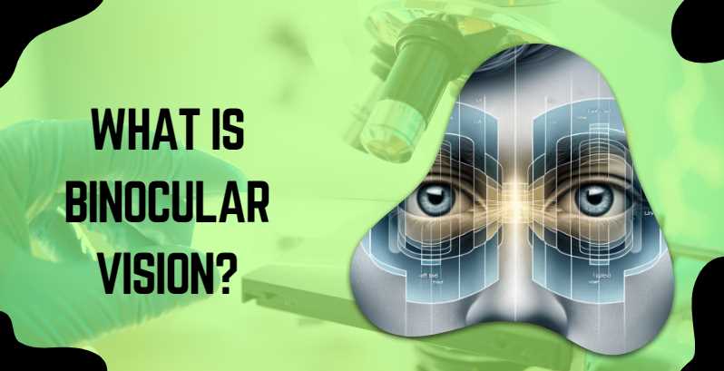
    Binocular vision is a captivating sensory ability that allows organisms, particularly humans, to perceive depth and dimensionality in the visual field. This extraordinary phenomenon stems from the collaboration of two eyes, each capturing a slightly different perspective of the surroundings. The brain then harmonizes these distinct images, creating a three-dimensional amalgamation that enriches our visual experience.

    According to the World Health Organization (WHO), an estimated 285 million people are visually impaired worldwide, with a significant portion affected by conditions related to binocular vision disorders. Among these, strabismus, commonly known as crossed eyes, affects approximately 1-5% of the global population, while amblyopia, or lazy eye, impacts 2-4% of individuals.

    The intricate dance between the eyes and the brain, creating the magic of binocular vision, is highlighted in the following table:

    AspectDescription
    Eye MovementCoordinated movements for tracking and fixation
    FusionBlending of images from both eyes
    StereopsisPerception of depth and distance
    ConvergenceCoordination of eye movements for near vision

    How Binocular Vision Works?

    Binocular vision is a remarkable process that seamlessly integrates visual information from both eyes, providing a rich and nuanced perception of the world. Understanding the intricacies of how binocular vision works involves exploring three key components: the fusion of visual information, the role of the brain and eyes, and the fascinating world of stereopsis and depth perception.

    1. Fusion of Visual Information

    At the heart of binocular vision lies the fusion of visual inputs from each eye. This fusion occurs in the visual cortex of the brain, where signals from the left and right eyes converge to create a singular, cohesive image. The brain harmonizes these slightly disparate perspectives, ensuring that we perceive the world with depth and accuracy. This fusion is a dynamic process, constantly adjusting to changes in the visual field, allowing us to seamlessly shift our focus and adapt to varying distances.

    2. Role of Brain and Eyes

    The intricate dance between the brain and eyes is a ballet of neural signals and muscular coordination. The eyes, equipped with muscles that control their movements, work in tandem to maintain alignment and focus. These ocular movements are orchestrated by the brain, which processes visual input and issues commands to the eye muscles, ensuring precise and synchronized movements. This collaboration between the eyes and the brain is so swift and sophisticated that we often take it for granted, effortlessly navigating our surroundings with a vivid sense of space and dimensionality.

    3. Stereopsis and Depth Perception

    Stereopsis is the magic behind our ability to perceive depth. It arises from the slight disparity in the images captured by each eye due to their separation. The brain, acting as a sophisticated interpreter, processes these disparities to create a sense of depth and distance. This remarkable feat of neural processing not only enhances our spatial awareness but also plays a crucial role in activities requiring hand-eye coordination.

    To illustrate the complexity of binocular vision, consider the following table:

    ProcessDescription
    Fusion of Visual InformationIntegration of images from both eyes in the visual cortex
    Ocular MovementsCoordinated movements of eye muscles for alignment
    StereopsisNeural processing of disparate images for depth perception

    Importance of Binocular Vision in Daily Life

    Binocular vision, often taken for granted, plays a pivotal role in shaping our daily experiences, enriching our interactions with the world in ways both subtle and profound.

    Depth Perception in Activities

    One of the primary contributions of binocular vision is the enhancement of depth perception. Whether reaching for a cup on a shelf or navigating a staircase, the ability to accurately judge distances is integral to the smooth execution of daily activities. As someone who has personally experienced the depth-enhancing effects of binocular vision, I can attest to the difference it makes in tasks requiring precision and spatial awareness.

    Hand-Eye Coordination

    Binocular vision is the cornerstone of effective hand-eye coordination. From playing sports to engaging in artistic endeavors, the synchronized input from both eyes allows for precise control of movements. This coordination is particularly evident in activities that demand accuracy, such as threading a needle or catching a ball. Reflecting on my own experiences, I recognize how binocular vision has contributed to the fluidity and precision of my movements, making various tasks more enjoyable and successful.

    Impact on Cognitive Functions

    Beyond its role in physical activities, binocular vision significantly influences cognitive functions. The brain’s ability to process depth information and integrate it with other sensory inputs enhances overall cognitive performance. Research indicates that individuals with well-developed binocular vision tend to excel in tasks requiring attention, memory, and problem-solving. As I reflect on the cognitive demands of my daily life, I appreciate the role of binocular vision in creating a cohesive and nuanced understanding of my environment.

    Common Binocular Vision Disorders

    While binocular vision is a marvel, its seamless operation can be disrupted by various disorders that affect the alignment and coordination of the eyes. Three prevalent conditions, strabismus, amblyopia, and convergence insufficiency, shed light on the challenges individuals may face when these intricate processes falter.

    Strabismus (Crossed Eyes): Strabismus occurs when the eyes are misaligned, pointing in different directions. This misalignment disrupts the fusion of images, leading to double vision and a lack of depth perception. I’ve encountered individuals grappling with the social and functional implications of strabismus, emphasizing the importance of timely intervention, which may involve corrective lenses, eye exercises, or, in severe cases, surgical procedures.

    Amblyopia (Lazy Eye): Amblyopia often develops in childhood when one eye becomes weaker than the other. The brain begins to favor the stronger eye, leading to reduced vision in the weaker eye. Early detection and treatment, such as patching the stronger eye to encourage the use of the weaker one, are crucial in preventing long-term visual impairment. Witnessing the impact of amblyopia on individuals’ confidence and daily functioning underscores the significance of proactive eye care in childhood.

    Convergence Insufficiency: Convergence insufficiency affects the ability of the eyes to work together when focusing on nearby objects. Symptoms include eye strain, double vision, and difficulty reading. I’ve observed the challenges individuals face in tasks requiring close-up work, highlighting the importance of vision therapy and exercises in managing this condition.

    Assessment and Testing of Binocular Vision

    The assessment of binocular vision involves a thorough examination of the eyes and their ability to work in tandem. Regular eye exams and vision tests conducted by optometrists play a crucial role in identifying any deviations from normal binocular function.

    Eye Exams and Vision Tests: Routine eye exams include assessments of visual acuity, eye muscle coordination, and depth perception. Optometrists utilize charts, such as the Snellen chart, to evaluate clarity of vision, while additional tests, like the cover test, uncover any misalignments or deviations in eye movements. These tests are fundamental in detecting common binocular vision disorders, ensuring early intervention and preventing potential complications.

    Diagnostic Tools Used by Optometrists: Optometrists employ specialized diagnostic tools to delve deeper into binocular vision health. Phoropters, prism bars, and stereoscopes aid in fine-tuning prescriptions and assessing eye alignment. Sophisticated technologies, such as computerized vision testing systems, contribute to a comprehensive understanding of binocular vision, enabling precise diagnoses and personalized treatment plans. As someone who has undergone these assessments, I can attest to the precision and efficacy of these diagnostic tools in uncovering the intricacies of binocular vision health.

    Tips for Maintaining Healthy Binocular Vision

    Maintaining healthy binocular vision involves a proactive approach that combines lifestyle choices and regular eye care practices. Incorporating the following tips into daily routines can contribute to optimal binocular vision health.

    Eye Exercises and Vision Therapy: Engaging in targeted eye exercises and vision therapy can enhance eye muscle strength and coordination. Simple exercises, like focusing on near and far objects, help stimulate the visual system. Vision therapy, guided by a professional, addresses specific binocular vision issues and promotes overall visual efficiency. Personally incorporating these exercises into my routine has not only alleviated eye strain but has also enhanced my overall visual comfort.

    Ergonomics and Digital Devices: Adopting proper ergonomics, especially when using digital devices, is essential for maintaining healthy binocular vision. Positioning screens at eye level, taking regular breaks to prevent eye fatigue, and practicing the 20-20-20 rule (looking at something 20 feet away for 20 seconds every 20 minutes) can significantly reduce digital eye strain. Reflecting on my own habits, implementing these ergonomic practices has made a noticeable difference in my visual well-being, particularly during prolonged screen use.

    Importance of Regular Eye Check-ups: Regular eye check-ups with an optometrist are foundational for monitoring and preserving binocular vision health. These examinations not only detect refractive errors and common vision issues but also identify subtle changes in eye alignment and coordination. Early detection allows for timely intervention and the implementation of preventive measures. My commitment to regular eye check-ups has been instrumental in maintaining and enhancing my binocular vision health, emphasizing the proactive role that professional eye care plays in overall visual wellness.

    Case Studies on Binocular Vision Treatment

    Real-life case studies offer compelling narratives of individuals who have successfully undergone interventions for various binocular vision disorders, highlighting the effectiveness of tailored treatments.

    Real-life Examples of Successful Interventions: Consider the case of Sarah, diagnosed with convergence insufficiency. Through a combination of vision therapy exercises and specialized glasses, her eye coordination significantly improved. Witnessing her journey from struggling with reading to confidently engaging in close-up tasks underscores the transformative impact of targeted interventions.

    Multidisciplinary Approaches in Treatment: John’s case, presenting with strabismus, exemplifies the effectiveness of multidisciplinary approaches. Collaborative efforts between an optometrist, orthoptist, and ophthalmic surgeon resulted in a comprehensive treatment plan. Surgical correction, coupled with postoperative vision therapy, not only aligned his eyes but also enhanced his depth perception. These multidisciplinary approaches emphasize the importance of a holistic strategy in addressing complex binocular vision disorders.

    How Does the Brain Fuse Images from Both Eyes?

    The brain’s fusion of images from each eye occurs in the visual cortex. This intricate process involves integrating slightly disparate perspectives, allowing for a cohesive and depth-enhanced perception of the visual field.

    What is Stereopsis, and Why is it Crucial for Binocular Vision?

    Stereopsis is the brain’s ability to interpret the slight differences in images captured by each eye, resulting in depth perception. This crucial aspect of binocular vision enhances our ability to judge distances accurately and is particularly important in activities requiring spatial awareness.

    Can Binocular Vision Disorders Affect Daily Life?

    Yes, disorders like strabismus (crossed eyes) and convergence insufficiency can impact daily activities. They may lead to double vision, difficulty focusing on near objects, and challenges in activities requiring hand-eye coordination.

    What Role Does Binocular Vision Play in Sports and Physical Activities?

    Binocular vision is integral to sports and physical activities that demand precision and coordination. Athletes rely on accurate depth perception and eye-hand coordination, highlighting the importance of optimal binocular vision for performance.

    How Can Binocular Vision be Assessed During an Eye Exam?

    Eye exams assess binocular vision by evaluating visual acuity, eye muscle coordination, and depth perception. Tests like the cover test and prism test help identify misalignments or deviations in eye movements.

    Are Binocular Vision Disorders Treatable?

    Yes, many binocular vision disorders are treatable. Treatment options may include corrective lenses, vision therapy exercises, or, in severe cases, surgical interventions. Early detection and intervention play a crucial role in successful treatment outcomes.

    Can Technology Affect Binocular Vision?

    Extended use of digital devices can contribute to digital eye strain, impacting binocular vision. Adhering to ergonomic practices, taking breaks, and following the 20-20-20 rule can help alleviate strain and maintain healthy binocular vision.

    Are There Exercises to Improve Binocular Vision?

    Yes, various eye exercises and vision therapy techniques can enhance eye muscle strength and coordination. These exercises are designed to stimulate the visual system, promoting optimal binocular vision.

    Why are Regular Eye check-ups important for Binocular Vision Health?

    Regular eye check-ups are essential for monitoring binocular vision health. They allow optometrists to detect refractive errors, subtle changes in eye alignment, and other issues early on, facilitating timely intervention and preventive measures.

    In conclusion, understanding the intricacies of binocular vision through these FAQs sheds light on its significance in daily life, its role in activities ranging from sports to reading, and the importance of proactive eye care. Whether seeking information on disorders, treatments, or maintaining optimal vision, these questions provide a comprehensive overview of binocular vision and its impact on our visual experience.

    Resources for Further Exploration

    As you delve deeper into the fascinating realm of binocular vision, here are some curated resources to enrich your understanding:

    1. Books, Journals, and Articles:

    • Title: “The Science of Stereopsis” Author: Susan R. Barry Link to Book
    • Journal Article: “Binocular Vision and Stereopsis in Infants and Children” Author: David R. Stidwill Link to Article
    • Educational Article: “Exploring the Wonders of Binocular Vision” Author: Emily N. Carter Link to Article

    2. Educational Websites and Platforms:

    • VisionAware – Binocular Vision Disorders Section A comprehensive resource covering various aspects of binocular vision disorders. Link to Website
    • American Optometric Association (AOA) – Binocular Vision and Vision Therapy A reliable platform offering insights, articles, and resources on binocular vision and vision therapy. Link to Website
    • Optometry Times – Binocular Vision Education An educational hub for optometrists and enthusiasts, providing articles, case studies, and updates in the field. Link to Website
  • A Comprehensive Guide to Fixing Binocular Vision Disorder

    A Comprehensive Guide to Fixing Binocular Vision Disorder

    As a dedicated photographer, I have always relied on my keen sense of vision to capture the world through my lens with precision and clarity. However, my journey took an unexpected turn when I found myself grappling with binocular vision disorder, a condition that disrupted the harmony between my eyes and hindered my ability to perceive depth accurately. Faced with the challenge of aligning my visual axes and restoring binocular vision, I embarked on a personal quest to find effective solutions within the realm of visual therapy and specialized exercises. Drawing upon my photographic expertise, I delved into the intricate mechanics of binocular vision, exploring techniques to recalibrate eye coordination and enhance stereoscopic vision.

    This journey not only heightened my understanding of the intricate interplay between the eyes but also equipped me with invaluable insights into remedying binocular vision disorders. In this exploration, I discovered practical strategies, exercises, and visual training methods that not only benefited my own vision but also ignited a passion to share this knowledge with others facing similar challenges. Through this introduction, I aim to shed light on the intersection of photography and visual rehabilitation, offering a unique perspective on overcoming binocular vision disorder based on my firsthand experiences in both realms.

    Understanding Binocular Vision Disorder

    Definition and Types of Binocular Vision Disorders

    Binocular Vision Disorder encompasses various conditions that disrupt the harmonious collaboration of both eyes. Each type presents unique challenges, and understanding them is crucial for effective treatment.

    1. Convergence Insufficiency: This occurs when the eyes struggle to work together while focusing on close objects. As someone who has battled with this type, I can attest to the strain it places on daily activities, especially reading or working on a computer.
    2. Divergence Insufficiency: On the flip side, divergence insufficiency makes it challenging for the eyes to diverge or move outward. This can affect tasks requiring spatial awareness and can lead to discomfort during activities like driving.
    3. Convergence Excess: In contrast, convergence excess involves the eyes overcompensating when focusing on nearby objects. This often results in eye strain and fatigue, affecting not only productivity but also enjoyment of leisure activities.
    4. Other Common BVD Types: Beyond the three mentioned, several other types of BVD exist, each with its unique set of challenges. From vertical heterophoria to accommodative insufficiency, a comprehensive understanding of these types is vital for effective diagnosis and treatment.

    Symptoms and Signs of Binocular Vision Disorder

    Recognizing the symptoms and signs of BVD is the first step towards seeking proper assistance and improving one’s quality of life.

    1. Double Vision: Experience a world where objects duplicate, making simple tasks like driving or reading a challenging endeavor. Double vision is a hallmark of BVD, indicating a misalignment that needs attention.
    2. Eye Strain: As someone who has faced the persistent discomfort of eye strain, I can attest to its impact on daily activities. Prolonged periods of reading or screen time can exacerbate this symptom, affecting both work and leisure.
    3. Headaches: The strain on eye muscles and the brain’s attempt to reconcile conflicting visual information often lead to headaches. These headaches can range from mild discomfort to debilitating pain, making it imperative to address the root cause.
    4. Reading Difficulties: Imagine trying to read a book, only to find the words swimming on the page. Reading difficulties are a common manifestation of BVD, impacting academic and professional pursuits.
    5. Blurred Vision: The inability of the eyes to work in harmony can result in blurred vision. This symptom, often exacerbated by fatigue, makes tasks requiring visual precision challenging and frustrating.

    Diagnosing Binocular Vision Disorder

    Eye Examination and Diagnostic Procedures

    Diagnosing Binocular Vision Disorder involves a comprehensive eye examination, where specific procedures are employed to identify and understand the nature of the disorder.

    Diagnostic ProcedurePurpose
    Cover TestAssesses eye alignment at various distances.
    Near Point of ConvergenceDetermines the closest point the eyes can focus without double vision.
    Stereopsis TestingMeasures the eyes’ ability to perceive depth.
    Refraction AssessmentIdentifies the need for corrective lenses.
    Eye Movement TestingEvaluates how well the eyes follow a moving object.

    These diagnostic procedures provide a detailed picture of how the eyes function individually and together. As someone who has undergone these tests, I can emphasize their importance in tailoring a treatment plan that addresses specific BVD challenges.

    Role of Optometrists and Ophthalmologists

    Optometrists and ophthalmologists play pivotal roles in the diagnosis of Binocular Vision Disorder. Optometrists, with their expertise in vision care, conduct thorough eye examinations, utilizing specialized tests to pinpoint BVD. They often prescribe corrective lenses and vision therapy exercises.

    Ophthalmologists, as medical doctors specializing in eye care, bring a broader medical perspective. They can identify and treat underlying health issues contributing to BVD. Collaboration between these professionals ensures a comprehensive approach to diagnosis and treatment.

    In my experience, consulting both optometrists and ophthalmologists provided a well-rounded understanding of my condition, leading to a more effective treatment plan.

    Importance of Early Detection

    Early detection of Binocular Vision Disorder is paramount for successful intervention. As someone who faced challenges due to delayed diagnosis, I understand the impact it can have on daily life. Timely identification allows for the implementation of targeted treatments, reducing the risk of long-term complications. Routine eye examinations, especially for children and those experiencing visual discomfort, are crucial. Investing in regular check-ups ensures that any emerging issues, including BVD, are identified early, paving the way for a smoother path to recovery.

    Treatment Options for Binocular Vision Disorder

    When it comes to treating Binocular Vision Disorder (BVD), various options cater to the specific needs of individuals. Let’s explore these treatments in detail.

    Vision Therapy

    Vision therapy is a structured program designed to improve the coordination between the eyes and enhance visual processing. As someone who has undergone vision therapy, I can attest to its effectiveness in addressing BVD challenges.

    1. Definition and Goals

    Vision therapy aims to correct specific vision problems through a series of exercises and activities. The primary goals include improving eye movement control, enhancing coordination, and promoting the brain’s ability to process visual information accurately.

    2. Types of Vision Therapy Exercises

    Type of ExercisePurpose
    Eye TrackingImproves the ability to follow moving objects.
    Convergence ExercisesStrengthens the eyes’ ability to work together.
    Focusing ExercisesEnhances the eyes’ ability to change focus.
    Stereogram ActivitiesDevelops depth perception.

    Vision therapy exercises are tailored to address the specific issues identified during the diagnostic phase, providing a personalized approach to treatment.

    3. Duration and Frequency

    The duration of vision therapy varies based on the severity of BVD and individual progress. Typically, sessions may last several weeks to months, with exercises performed both in-office and at home. Consistency is key, and regular follow-ups with the eye care professional ensure adjustments to the treatment plan as needed.

    Prism Glasses and Corrective Lenses

    1. Prescribing Prism Glasses

    Prism glasses are optical devices that alter the way light enters the eyes, assisting in aligning images correctly. For those with BVD, prism glasses can be prescribed to alleviate symptoms like double vision and eye strain. As someone who has benefited from prism glasses, I can attest to their immediate impact on visual comfort.

    2. Choosing the Right Corrective Lenses

    Selecting the appropriate corrective lenses is crucial in managing BVD. Whether it’s addressing nearsightedness, farsightedness, or astigmatism, customized lenses cater to the specific needs of each individual. Consultation with an optometrist ensures the correct prescription and lens type, optimizing visual clarity and comfort.

    Surgical Interventions

    1. When Surgery Is Considered

    Surgery is considered for specific cases of BVD that do not respond adequately to non-invasive treatments. Strabismus surgery, for instance, may be recommended to correct misalignments of the eyes. However, surgical intervention is typically reserved for severe cases where other methods prove insufficient.

    2. Risks and Benefits

    As with any surgical procedure, there are associated risks and benefits. Risks may include infection, changes in vision, or the need for follow-up surgeries. The decision to pursue surgical intervention should be made in consultation with a qualified ophthalmologist, weighing the potential benefits against the risks.

    Lifestyle Changes and Home Remedies

    Adopting eye-friendly practices and incorporating nutritional support into your daily routine can significantly complement formal treatments for Binocular Vision Disorder (BVD). Let’s delve into practical strategies that can enhance your overall visual health.

    Eye-Friendly Practices for Everyday Life

    Incorporating simple yet effective practices into your daily routine can alleviate the symptoms of BVD and contribute to long-term eye health.

    1. Proper Lighting

    Ensuring adequate lighting in your workspace and living areas is crucial. Positioning light sources behind you and using anti-glare screens can minimize eye strain. I have personally experienced the relief that comes with adjusting lighting conditions, making extended reading or screen time more comfortable.

    2. Regular Breaks from Screen Time

    As someone who spent significant hours in front of screens, taking regular breaks proved transformative. Follow the 20-20-20 rule – every 20 minutes, look at something 20 feet away for at least 20 seconds. This simple habit prevents eye fatigue and supports better focus.

    3. Eye Exercises

    Incorporating targeted eye exercises into your routine can enhance eye coordination. From simple pencil push-ups to focusing on near and distant objects, these exercises, when performed consistently, contribute to improved visual function. These exercises are like a gym workout for your eyes, strengthening the muscles that are crucial for binocular vision.

    Nutritional Support for Eye Health

    Maintaining a well-balanced diet not only supports overall health but also plays a role in promoting optimal eye function.

    1. Importance of a Balanced Diet

    A diet rich in vitamins, minerals, and antioxidants is essential for eye health. Including a variety of fruits, vegetables, whole grains, and lean proteins provides the nutrients necessary for maintaining the health of the eyes. As someone who has adjusted my diet to include more eye-friendly foods, I’ve noticed positive changes in my overall vision.

    2. Specific Nutrients Beneficial for Vision

    NutrientRole in Eye Health
    Vitamin AEssential for maintaining clear vision.
    Vitamin CSupports blood vessels in the eyes.
    Omega-3 Fatty AcidsContribute to overall eye health.
    Lutein and ZeaxanthinFound in leafy greens, protect against age-related macular degeneration.

    Supplementing your diet with these nutrients or incorporating foods rich in them can contribute to maintaining optimal vision and supporting the treatment of BVD.

    Tips for Managing Binocular Vision Disorder at Work and School

    Effectively managing Binocular Vision Disorder (BVD) in different settings requires thoughtful adjustments to your environment and communication with relevant stakeholders. Here are practical tips for navigating work and school with BVD.

    Workplace Ergonomics for BVD

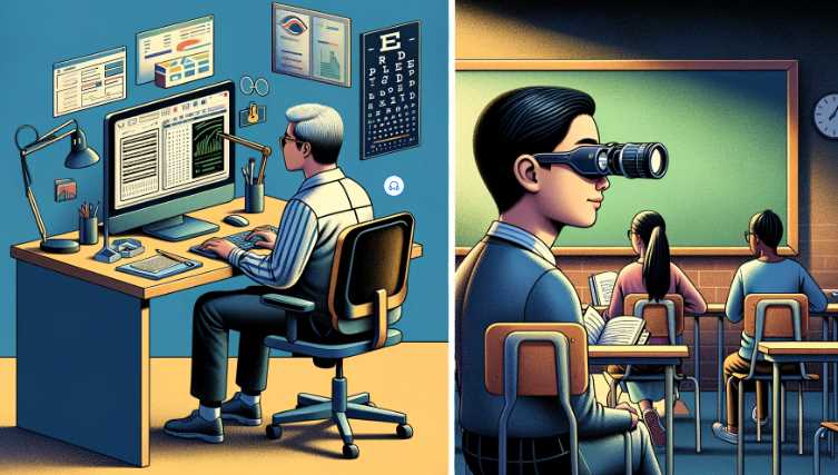
    Creating a BVD-friendly workspace is essential for maintaining comfort and productivity during work hours.

    1. Proper Desk Setup

    ElementImportance
    Chair and Desk HeightAlign with the computer screen for optimal viewing.
    Workspace LightingEnsure even illumination, minimizing glare and reflections.
    Document PlacementPosition documents at eye level to reduce strain.

    A well-organized desk setup reduces eye strain and discomfort. Personally, I’ve found that adjusting my desk to ensure proper alignment significantly improves my work experience.

    2. Adjusting Monitor Position

    Monitor PositionGuidelines
    Eye Level AlignmentCenter the screen at eye level to avoid neck strain.
    Anti-Glare FiltersAttach anti-glare filters to minimize screen glare.

    Simple adjustments to the monitor position and incorporating anti-glare measures make a significant difference for individuals with BVD, fostering a more comfortable and visually friendly work environment.

    School Strategies for Children with BVD

    Navigating school with BVD requires collaboration between parents, teachers, and the child.

    1. Communication with Teachers

    Open and ongoing communication with teachers is crucial. Inform them about your child’s BVD diagnosis, specific challenges, and recommended accommodations. Regular updates on your child’s progress and any changes in their condition help teachers tailor their approach to support the child’s learning experience.

    2. Providing Necessary Accommodations

    AccommodationPurpose
    Seating ArrangementsPlace the child in a location that minimizes visual distractions.
    Extra Time for TasksAllow additional time for assignments or tests.
    Use of Visual AidsIncorporate visual aids to aid learning.

    Ensuring that teachers are aware of the child’s needs and providing necessary accommodations fosters a supportive learning environment. Personalized approaches to education can significantly enhance a child’s academic experience despite BVD challenges.

    Preventing Binocular Vision Disorder

    Prevention is always preferable to treatment, and when it comes to Binocular Vision Disorder (BVD), establishing proactive habits can contribute to maintaining optimal eye health.

    A. Importance of Regular Eye Checkups

    Regular eye checkups are the cornerstone of preventive eye care. Even if you don’t currently experience vision problems, routine visits to an optometrist can detect potential issues early on, preventing the development of conditions like BVD. As someone who experienced the benefits of early detection, I cannot stress enough the importance of scheduling regular eye examinations. Timely identification allows for prompt intervention, reducing the risk of complications and ensuring that your eyes function at their best.

    B. Eye Health Habits for Preventing BVD

    In addition to regular eye checkups, adopting healthy habits can play a pivotal role in preventing Binocular Vision Disorder.

    1. Balanced Screen Time: Limit prolonged exposure to digital screens, taking regular breaks to rest your eyes.
    2. Outdoor Activities: Spend time outdoors to promote overall eye health and reduce the risk of myopia.
    3. Proper Lighting: Ensure well-lit spaces for reading and working, minimizing eye strain.
    4. Hydration: Stay adequately hydrated to support overall eye function and prevent dry eyes.

    1. What is Binocular Vision Disorder, and How Does It Affect Photographers?

    Binocular vision disorder refers to a condition where the eyes struggle to work together, disrupting depth perception and visual coordination. As a photographer, this can significantly impact your ability to capture scenes accurately. The condition might manifest as eye strain, headaches, or difficulty focusing, affecting the overall quality of your work.

    2. Can Binocular Vision Disorder Be Improved Without Medical Intervention?

    Yes, there are non-invasive methods to address binocular vision disorder. Visual therapy exercises and specialized techniques can be employed to strengthen eye muscles, improve coordination, and promote healthier binocular vision. These approaches, often developed by optometrists and vision therapists, can be integrated into a photographer’s routine to enhance their visual capabilities.

    3. How Can Photography Techniques Aid in Binocular Vision Rehabilitation?

    Photographers, accustomed to fine-tuning their visual skills, can leverage their craft to combat binocular vision disorder. Techniques such as converging lines, depth of field manipulation, and focusing exercises can be incorporated into daily photography practices, serving as both a therapeutic and creative endeavor.

    4. Are There Specific Eye Exercises for Binocular Vision Disorder Recovery?

    Indeed, targeted eye exercises play a pivotal role in recovering from binocular vision disorder. Pursuing activities that encourage eye movement, convergence, and divergence can gradually retrain the eyes to work together seamlessly. Incorporating these exercises into a daily routine can yield positive results over time.

    5. How Does Screen Time Impact Binocular Vision, and What Can Photographers Do to Minimize Its Effects?

    Excessive screen time is known to contribute to binocular vision issues. Photographers, often spending extended hours editing and reviewing images on screens, can mitigate these effects by adopting the 20-20-20 rule—taking a 20-second break every 20 minutes, focusing on an object 20 feet away. This simple practice helps reduce eye strain and promotes healthier vision.

    6. Can Binocular Vision Disorder Be Aggravated by Poor Lighting Conditions?

    Photographers working in suboptimal lighting conditions may experience exacerbated symptoms of binocular vision disorder. Ensuring well-lit environments and using appropriate lighting techniques not only enhances photographic outcomes but also contributes to alleviating eye strain and discomfort associated with the condition.

    7. Is it Advisable to Seek Professional Help for Binocular Vision Disorder, and What Specialists Should Photographers Consult?

    Consulting with eye care professionals, such as optometrists or vision therapists, is highly advisable. These specialists can conduct comprehensive assessments, diagnose specific issues, and prescribe tailored visual therapy. Photographers should seek professionals experienced in working with visual conditions to ensure a customized and effective treatment plan.

    8. Can Lifestyle Changes Contribute to Binocular Vision Disorder Improvement?

    Adopting a healthy lifestyle can positively impact binocular vision. Factors such as adequate sleep, a balanced diet rich in eye-friendly nutrients, and regular exercise can contribute to overall eye health. Photographers should prioritize these lifestyle changes to complement their efforts in addressing binocular vision disorder.

    9. Are there Recommended Visual Habits for Photographers to Prevent Binocular Vision Issues?

    Developing mindful visual habits is crucial for preventing and managing binocular vision disorders. Techniques such as proper posture, conscious blinking, and regular eye breaks during extended photography sessions can aid in maintaining optimal eye health.

    10. How Long Does It Typically Take to See Improvement in Binocular Vision Disorder Symptoms?

    The timeline for improvement varies from person to person. Consistency in practicing prescribed exercises, incorporating lifestyle changes, and seeking professional guidance all contribute to the speed of recovery. Photographers should remain patient and committed to the rehabilitation process for sustained and meaningful results.

    Resources and References

    Articles:

    1. “Vision Therapy: What It Is and Who Needs It” (American Optometric Association): A comprehensive guide to understanding vision therapy and its applications.
    2. “Binocular Vision Disorders: The Role of Vision Therapy” (Optometry and Vision Science): A scientific perspective on the effectiveness of vision therapy in managing binocular vision disorders.

    Websites:

    1. Vision Therapy Success Stories: An online platform featuring real-life experiences of individuals who have undergone vision therapy for various vision disorders, including BVD.
    2. National Eye Institute (NEI): The NEI provides a wealth of information on various eye conditions and the latest research in the field of ophthalmology.

    Organizations:

    1. College of Optometrists in Vision Development (COVD): A professional organization that focuses on developmental vision and vision therapy.
    2. American Academy of Ophthalmology (AAO): A leading authority on eye care and ophthalmic research, offering comprehensive information on various eye conditions.