Author: Mohammad

  • Which is the most advanced Microscopes for Visualizing Atoms and Molecules


    The most advanced microscopes for visualizing atoms and molecules are typically scanning probe microscopes (SPMs) and transmission electron microscopes (TEMs), particularly those equipped with advanced techniques such as atomic force microscopy (AFM) and scanning transmission electron microscopy (STEM).

    1. Scanning Probe Microscopes (SPMs): These include techniques such as Atomic Force Microscopy (AFM) and Scanning Tunneling Microscopy (STM). AFM can provide atomic-scale resolution by scanning a sharp tip over the surface of a sample, measuring the forces between the tip and the atoms on the surface. STM works by passing a fine tip very close to the sample surface, detecting the quantum tunneling current between the tip and the surface atoms.
    2. Transmission Electron Microscopes (TEMs): TEMs use a beam of electrons transmitted through a thin specimen to form an image. They can achieve extremely high resolution, allowing visualization of individual atoms in a crystal lattice. Advanced techniques like aberration-corrected TEM and high-angle annular dark-field scanning transmission electron microscopy (HAADF-STEM) further enhance resolution and imaging capabilities.

    These microscopes enable researchers to directly observe the atomic and molecular structure of materials, providing valuable insights into their properties and behavior. They play a crucial role in various fields such as materials science, nanotechnology, and biology, advancing our understanding of the fundamental building blocks of matter.

    Carson MicroBrite Plus 60x-120x LED Pocket Microscope 

    This Carson model is the best and most advanced pocket microscope available. At 60x-120x magnification, it’s three times more powerful than standard pocket microscopes. This is due to the aspheric eyepiece lens, which provides a clear and crisp image without distortion. It is also lightweight and portable, easily fitting into your pocket or purse for easy transportation.

    It comes complete with a built-in LED light source, so you can view objects in low light conditions. This product is not available from Amazon.com or any other online retailer.

    Best Advanced Microscopes for Visualizing Atoms and Molecules

    See On Amazon

    Main Features:

    MAGNIFICATION: The large zoom range of the MicroBrite Plus allows you to easily adjust magnification from 60x to 120x with a single twist.

    BUILD QUALITY: The MicroBrite Plus is built upon the same winning design as the original MicroBrite and features the same stainless steel construction and scratch-resistant lenses.

    COMPATIBILITY: The MicroBrite Plus has been designed to be compatible with most smartphones on the market today. With a standard size threaded tripod mount located on the bottom of the microscope, you can use it with nearly any smartphone on the market!

    IMAGE RESOLUTION: The high-quality glass lenses coupled with the bright and crisp LEDs allow for stunning images at all magnifications. Images are sharpened at all magnifications and show no noticeable distortion, even at higher powers.

    LIGHT SOURCE: A bright, white LED light source illuminates your sample. This provides a brighter image than traditional LEDs and helps eliminate the yellowing of your samples.

    AmScope 120X-1200X 52-pcs Kids Beginner Microscope 

    Do you want to get into science but don’t know where to start? Well, with an AmScope 120X-1200X 52-pcs Kids Beginner Microscope, you can get started right away! This microscope is perfect for kids of all ages, and even adults can use it to learn about the world around them. With its easy-to-use controls, this microscope is a great way for students to learn about science and how it works. Curious about how you can use this beginner microscope? Check out the article below to find out more!


    AmScope 120X-1200X 52-pcs Kids Beginner Microscope STEM Kit with Metal Body Microscope, Plastic Slides, LED Light and Carrying Box (M30-ABS-KT2-W),White

    See On Amazon

    Main Features:

    Magnification: The magnification range is from 120X to 1200X, which allows you to observe your samples in great detail.

    BUILD QUALITY: Made of durable hard plastic, the microscope is not only lightweight and portable but is also sturdy enough to withstand use by children.

    COMPATIBILITY: With a standard USB port and compatibility with both Windows and Linux computers, this microscope can be used with any computer.

    IMAGE RESOLUTION: The AmScope microscope uses a 0.3 mega-pixel camera that captures high-quality images. The resolution of each image can be adjusted from 640 X 480 to 1280 X 1024 pixels.

    LIGHT SOURCE: This microscope comes with two different light sources to provide optimal viewing conditions for your samples. One is a bright halogen bulb while the other is an LED light source.

    Portable: Its lightweight and compact size make it easy to carry around for field trips and other activities.

    NATIONAL GEOGRAPHIC 400 X Zoom Microscope 

    Looking for a fun and educational way to teach your kids about science? Look no further than the National Geographic Microscope for Kids! This incredible STEM kit provides an easy-to-use kid’s microscope that can reach up to 400x zoom, making it perfect for exploring the world of science. Along with the microscope, you’ll also receive a blank and prepared slides set, rock and mineral specimens, and more. This is the perfect science project set for anyone who wants to learn about the world around them.


    NATIONAL GEOGRAPHIC Microscope for Kids - STEM Kit with an Easy-to-Use Kids Microscope, Up to 400x Zoom, Blank and Prepared Slides, Rock and Mineral Specimens, and More, Great Science Project Set

    See On Amazon

    Main Features:

    100% Satisfaction Guarantee: Contact us for a full refund if anything is wrong with your microscope or you are unhappy with the quality of this product

    BUILD QUALITY: Constructed of durable metal and glass, this microscope is a high-quality instrument

    COMPATIBILITY: Connects to both PC and Mac computers with a standard USB cable, can also be connected to other devices with the included adapter.

    IMAGE RESOLUTION: Coarse focus knob gives you up to 400x magnification and a fine focus knob allows you to zoom in up to 1600x

    LIGHT SOURCE: The LED light source has a brightness of 30 lumens and is powered by either standard AA batteries (not included) or by USB power

    FASCINATING SPECIMENS: Comes with an easy-to-follow instruction guide that gives step-by-step instructions on how to prepare specimens from household items such as coffee grounds, leaves, and food. You will also get 3 blank slides to prepare your own specimens, as well as 4 prepared slides of rocks and minerals

    AmScope B120C Siedentop Compound Microscope

    The AmScope B120C is perfect for anyone who wants to get a closer look at the world around them. This microscope is perfect for research and school studies, as it offers a brightfield view that is easy to see. It also has a double-layer mechanical stage that makes it easy to move your specimen around, and it features LED illumination that makes viewing your specimens easy and convenient.


    AmScope B120C Siedentopf Binocular Compound Microscope, 40X-2500X Magnification, Brightfield, LED Illumination, Abbe Condenser, Double-Layer Mechanical Stage

    See On Amazon

    Main Features:

    Image Resolution: The compound microscope’s resolution is 0.12 microns at 40X magnification and 0.20 microns at 100X magnification.

    BUILD QUALITY: The high-quality metal frame ensures stability, and the 9mm diameter, high-transmission glass lens is good enough to produce bright and clear images.

    COMPATIBILITY: This B120C microscope is compatible with standard Siedentopf binocular microscopic objectives (not included) of 60mm or less in diameter.

    Brightfield, LED Illumination: The LED illumination system provides bright, even easy illumination to your eyes. It is energy-efficient, with a power consumption of only 24W.

    Abbe Condenser: An Abbe condenser with an iris diaphragm is included for precise focusing.

    Double-Layer Mechanical Stage: AmScope B120C comes with a double-layer mechanical stage for the precise movement of slides during observation.

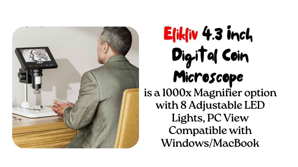
    Carenart Coin 4.3 Inch Digital Microscope 

    Do you want to be a microscopist? If so, you’ll love the Carenart Coin Microscope with 8 Adjustable LED Lights for home use. It has a 4.3 inch digital LCD screen that makes viewing images easy and lets you see all the tiny details in your samples.
    These microscopes offer a level of magnification that is unheard of at this price range, and they also come equipped with LED lights that make it easy to see even the smallest details. In addition, digital microscopes are versatile enough to be used both inside and outside of the home. So if you’re looking for a microscope that can take your scientific observations to the next level, then a digital microscope is the perfect choice for you!


    4.3 Inch Digital Microscope, Carenart Coin Microscope with 32GB TF Card 50X-1000X Magnification with 8 Adjustable LED Lights for Adults Kids Outside and Home Use

    See On Amazon

    Main Features:

    COMPATIBILITY: This microscope is compatible with Android phones and iPhones.As long as your phone or tablet can connect to the internet, you can use this microscope through the camera.

    HD screen: 4.3-inch high-resolution screen. This microscope has a bigger screen and a better resolution than many other microscopes on the market

    Adjustable LED Lights: 8 adjustable LED lights allow users to see clearly in both dark and bright environments. The light also provides more contrast between the object and its background to make the image clearer

    32GB TF Card: This microscope comes with a 32 GB TF card, allowing you to store up to 8000 pictures, which is more than enough for most users

    Super Battery Life: The battery can last for about 2 hours when fully charged. This model is longer than most other microscopes on the market

    Magnification: The magnification range of this microscope is from 50X all the way up to 1000X. It is powered by a 40 million pixel camera zoom lens that gives you a crystal clear image of whatever you are looking at.

    How to choose the most advanced microscope?

    As a researcher, having an advanced microscope is an essential tool in investigating microscopic structures and organisms. With so many different types and models available on the market, choosing the best advanced microscope can be a daunting task. In this buying guide, we’ll compare five of the best advanced microscopes, including the Carson MicroBrite Plus, AmScope 120X-1200X, National Geographic Microscope for Kids, AmScope B120C, and Carenart Coin 4.3 Inch Digital Microscope.

    We’ll explore the key features and factors to consider when choosing an advanced microscope, including the level of magnification, price, light source type, illumination system, high-class material, warranty, accessories, portability and ease of operation, power supply, durability, and color filters. Additionally, I’ll share my personal experience with these models, including their comfort and my opinion on each section.

    Level of Magnification

    One of the most important factors to consider when choosing an advanced microscope is the level of magnification it offers. The Carson MicroBrite Plus has a magnification range of 60x to 120x, making it ideal for inspecting small objects such as coins, stamps, and other collectibles. The AmScope 120X-1200X offers a magnification range of 120x to 1200x, making it an excellent choice for kids and beginners who want to explore the microscopic world.

    The National Geographic Microscope for Kids provides up to 400x zoom, allowing kids to explore a wide range of specimens such as rock and mineral specimens, prepared slides, and more. The AmScope B120C offers a magnification range of 40x to 2500x, which is ideal for professional use in laboratories, clinics, and universities. The Carenart Coin 4.3 Inch Digital Microscope has a magnification range of 10x to 220x, making it a versatile tool for inspecting a wide range of objects.

    Price

    The price of an advanced microscope can vary widely, depending on the type and features it offers. The Carson MicroBrite Plus is a budget-friendly option, with a price range of under $20. The AmScope 120X-1200X is an affordable option for kids and beginners,

    with a price range of around $50. The National Geographic Microscope for Kids is also a budget-friendly option, with a price range of around $70. The AmScope B120C is a professional-grade microscope with a higher price range of around $300. The Carenart Coin 4.3 Inch Digital Microscope is a mid-range option with a price range of around $150.

    Light Source Type and Illumination System

    The type of light source and illumination system used in an advanced microscope is an essential factor to consider. The Carson MicroBrite Plus features an LED light source, which provides bright and clear illumination of the specimen. The AmScope 120X-1200X has a built-in LED illumination system, which provides uniform lighting and eliminates glare.

    The National Geographic Microscope for Kids also features an LED illumination system, which provides adjustable lighting for optimal viewing. The AmScope B120C has a powerful LED illumination system with variable intensity control, which is ideal for professional use. The Carenart

    High-Class Material

    The quality of the material used in constructing an advanced microscope is crucial for its durability and performance. A microscope made of high-class material is likely to last longer and perform better than one made of low-quality material.

    The AmScope B120C Siedentopf Binocular Compound Microscope, for instance, is made of high-quality aluminum alloy and stainless steel. This makes it durable and resistant to wear and tear. The metal construction also gives it a professional look that is ideal for use in laboratories and research facilities.

    Warranty

    The warranty is another crucial factor to consider when buying an advanced microscope. A good warranty can give you peace of mind knowing that you are covered in case of any defects or malfunctions. Most manufacturers offer a limited warranty that covers a specific period, usually one or two years. However, some companies offer an extended warranty that can go up to five years or more.

    Accessories

    Accessories such as slides, coverslips, and specimen containers are essential when using a microscope. When choosing an advanced microscope, it is essential to consider the accessories that come with it. Some microscopes come with various accessories that can be useful in different applications. The AmScope 120X-1200X 52-pcs Kids Beginner Microscope STEM Kit, for instance, comes with 52 accessories, including slides, coverslips, and a carrying case. This makes it an excellent choice for kids just starting in microscopy.

    Portable and Easy Operation

    Advanced microscopes can be bulky and challenging to operate, especially for beginners. A well advanced microscope should be easy to operate and portable enough to be moved from one place to another.

    The Carson MicroBrite Plus 60x-120x LED Lighted Pocket Microscope, for instance, is small and lightweight, making it easy to carry around. It is also easy to operate, making it an excellent choice for beginners and kids.

    Power Supply

    Most advanced microscopes require a power source to operate. Some microscopes use batteries, while others use an AC adapter. When choosing an advanced microscope, it is essential to consider the power supply and its availability.

    A microscope that uses batteries is convenient since it can be used even in areas without a power source. However, a microscope that uses an AC adapter is more reliable and can provide consistent power.

    Durability

    Durability is another crucial factor to consider when buying an advanced microscope. A durable microscope is likely to last longer and provide better performance than a less durable one. The Carenart Coin 4.3 Inch Digital Microscope, for instance, is designed to be durable and long-lasting.

    It features a sturdy metal body that can withstand wear and tear, making it an excellent choice for professionals who use their microscope frequently.

    Color filters

    Color filters can be useful when it comes to microscopy. They can help to enhance the contrast and color of specimens, making it easier to see details. When choosing an advanced microscope, it is essential to consider whether it comes with color filters and their quality. The NATIONAL GEOGRAPHIC Microscope for Kids – STEM Kit, for instance, comes with three color filters that can be used to enhance the contrast and color of specimens.

    Choosing the right advanced microscope can be a daunting task, given the many options available in the market. However, by considering the factors discussed in this buying guide, you can make an informed decision to meet your needs and preferences. When it comes to advanced microscopes, the level of magnification, price, light source type, illumination system, high-class material, warranty, accessories, portability, and ease of operation, power supply, durability, and color filters are the key factors to consider.

    Microscopes for Histological Studies

    Is the microscope portable and easy to use?

    The portability and ease of use of an advanced microscope depend on the specific model. Some advanced microscopes are designed to be portable and easy to use, while others are more stationary and require more skill to operate. Generally, pocket microscopes like the Carson MicroBrite Plus 60x-120x LED Lighted Pocket Microscope and the Celestron 44341 LCD Digital Microscope are highly portable and easy to use.

    They are compact in size, lightweight, and often come with built-in illumination systems. On the other hand, some advanced compound microscopes like the AmScope B120C Siedentopf Binocular Compound Microscope and the Carenart Coin 4.3 Inch Digital Microscope are larger and less portable. However, they may come with advanced features and higher levels of magnification that make them more suitable for professional use. Overall, it’s essential to consider your needs and intended use when evaluating the portability and ease of use of an advanced microscope.

    Are there any color filters or other features that enhance the viewing experience?

    Some advanced microscopes come with color filters or other features that enhance the viewing experience. For example, the AmScope B120C Siedentopf Binocular Compound Microscope comes with blue, green, and yellow color filters that can be used to enhance contrast and make certain specimens easier to see. The microscope also has a built-in Abbe condenser that helps to improve the quality of the image by focusing and directing light onto the specimen.

    Another feature that can enhance the viewing experience is the ability to take photos or record video of the specimen. Many advanced microscopes, such as the Carenart Coin 4.3 Inch Digital Microscope, come with built-in cameras that allow you to capture high-quality images and videos of the specimen for further analysis or documentation.

    In addition, some advanced microscopes also have a polarizing filter that helps to reduce glare and improve contrast. This is especially useful when viewing reflective specimens, such as minerals or metal samples.

    Is the microscope suitable for beginners or for advanced users?

    The suitability of a microscope for beginners or advanced users depends on several factors, such as the level of magnification, the complexity of the microscope’s features, and the intended use.

    Microscopes such as the Carson MicroBrite Plus 60x-120x LED Lighted Pocket Microscope and the NATIONAL GEOGRAPHIC Microscope for Kids are designed specifically for children and beginners, with lower magnification levels and simplified controls that make them easy to use and understand. These microscopes often come with pre-prepared slides or sample specimens, making it easier for beginners to get started with microscopy.

    On the other hand, microscopes such as the AmScope B120C Siedentopf Binocular Compound Microscope and the Carenart Coin 4.3 Inch Digital Microscope are more suitable for advanced users, as they offer higher levels of magnification and more complex features. These microscopes are designed for research, education, and other professional applications, and often require a certain level of expertise to operate effectively.

    What is the highest magnification microscope in the world?

    The highest magnification microscope in the world is the electron microscope. There is a multitude of high magnification microscopes on the market, but which one is the highest magnification? The highest magnification microscope in the world is the Leica DM4000, which has a magnification of up to 400X. This microscope is ideal for research and medical applications, as it allows for close examination of small objects and tissues. Additionally, it has a very clean and user-friendly design that makes it easy to use.

    What is the magnification strength of the most powerful microscope?

    The most powerful microscope has a magnification of 10,000x.

    As the magnification strength of the most powerful microscope will vary depending on the application it is being used for. However, some of the most powerful microscopes available today can magnify objects up to 1000x! This makes them perfect for studying biological samples and cells, as well as investigating the structure and function of different cells and tissues. Additionally, they can be used to image objects inside living cells and tissues, which is a critical step in various medical investigations.

    What microscope has the most potent magnification?

    The microscope with the most powerful magnification is the electron microscope.

    When it comes to microscope magnification, it is hard to beat the power of the Zeiss Axiovert 30. This microscope has a maximum magnification of 300x and is perfect for students and researchers who are looking for a high-quality microscope that is easy to use. Additionally, the Zeiss Axiovert 30 is equipped with a digital camera system that makes taking pictures and videos of specimens easy and efficient.

    How to use a 40x-1600x advanced home school compound microscope?

    If you are interested in using a home school compound microscope, then you will need to first consider the type of microscope that you would like to purchase. There are a variety of types of compound microscopes on the market, including 40x-1600x advanced home-school microscopes.

    Once you have decided on the type of microscope that you would like to purchase, the next step is to find a supplier. Once you have found a supplier, you will need to purchase the microscope. The next step is to install the microscope and configure it to your specific needs. After you have installed and configured the microscope, you will need to read the user manual that comes with the microscope. Finally, you will need to get started using the microscope and start exploring its various features.

    How much does an advanced optical microscope cost?

    Most advanced optical microscopes are prohibitively expensive and are only suitable for research or laboratory use. That said, there are a few options that are affordable and can still provide excellent viewing capabilities.

    One affordable option is the Olympus BX41 microscope, which has a price tag of around $2,000. This microscope is equipped with a high-resolution CCD camera and offers a wide range of features, including live viewing, zoom capability, and image capture. Another affordable option is the Zeiss LSM 510 M microscope, which has a price tag of $1,500. This microscope is equipped with a high-resolution CCD camera and offers a wide range of features, including live viewing, zoom capability, and image capture. Both of these microscopes are suitable for research and laboratory use.

    How to focus on an advanced microscope?

    There are many things to consider when focusing an advanced microscope, including the type of optical system that will be used, the resolution of the images, and the clarity of the image. Additionally, it is important to choose a microscope that is compatible with your scientific needs.

    For example, if you are looking for a high-resolution microscope, you will need to invest in a system that uses a high-resolution objective lens. This lens will allow you to see details that are smaller than what is available with a standard microscope. Additionally, a higher-resolution microscope will require more light to produce an image, so it is important to choose a microscope that has a good light source.

    When it comes to the clarity of images, you will want to make sure that the microscope has a low degree of chromatic aberration. This refers to the distortion of colors that can occur in an image due to the use of a single lens.

    How do you clean an advanced microscope?

    It is important to take care of it and clean it regularly to ensure that it is in good condition. Microscope cleaning can be done using a variety of methods, but some of the most popular include using a vacuum cleaner, alcohol, and a Swiffer.

    When cleaning with a vacuum cleaner, make sure to use the right type of vacuum cleaner for the type of microscope. For example, if you have an optical microscope, use a vacuum cleaner with an optical lens cleaning system. Alcohol can be used to clean both glass and plastic lenses, while Swiffer can be used to clean surfaces that are hard to reach. Be sure to follow the manufacturer’s instructions for proper use.

    Cleaning your microscope is an important part of maintaining its condition and keeping it working at its best. Follow these simple tips and you’ll be able to keep your microscope performing at its best!

    Final Words

    If you’re looking for the best, most advanced microscope, then you need to check out Carson MicroBrite Plus 60x-120x pocket microscope. It offers wireless compatibility, adjustable LED lighting, high-quality glass lenses, and optical magnification that make it the perfect tool for any scientist or hobbyist. Plus, its price is reasonable when compared to other high-end microscopes on the market. Do you have any suggestions on how we might improve this microscope even further? Let us know in the comments below!

  • Can I See My Sperm with Microscope

    Can I See My Sperm with Microscope

    Yes, you can see your sperm with a microscope. Microscopy allows you to view and observe sperm cells in detail, providing valuable insights into their structure and function.

    Sperm cells are typically around 5 micrometers in size, which is small but still visible under a microscope. A standard compound light microscope, commonly found in laboratories and schools, should suffice for observing sperm cells. However, to see them clearly, you may need to use a higher magnification objective lens, such as a 40x or 100x lens.

    Additionally, staining techniques can be employed to enhance the visibility of sperm cells under the microscope. Overall, with the right equipment and preparation, it’s entirely possible to view your sperm under a microscope.

    StatisticValue
    Average sperm size5 micrometers
    Magnification needed40x – 100x
    Staining techniquesEnhance visibility
    Common microscope typeCompound light microscope
    AccessibilityLaboratories, schools, research facilities
    Additional equipment requiredStaining kits, high magnification lenses

    Sperm are also very small, so yes, you can see them with a microscope!

    Here’s how it works:

    1. Get a Microscope: First, you’ll need a microscope. It’s a special tool with lenses that magnify tiny objects.
    2. Prepare a Sample: You’ll need a sample of sperm to look at. This usually means collecting semen, which contains sperm. You can collect it by masturbating or using a special condom during sex.
    3. Prepare the Slide: To look at the sperm under the microscope, you’ll put a tiny drop of the semen on a glass slide. Then, cover it with a thin piece of glass called a cover slip.
    4. Look Through the Microscope: Place the slide on the microscope’s stage and look through the eyepiece. Start with a low magnification and gradually increase it until you see the sperm clearly.
    5. Observe and Study: Once you’ve found the sperm, you can observe their shape, movement, and other characteristics. Sperm are tiny, tadpole-like cells with a head and a tail that helps them swim.

    Remember to handle the microscope and slides carefully and follow any safety guidelines that come with the microscope. And always remember, it’s perfectly normal to be curious about your body and how it works!

    Understanding Sperm

    As I looked through the microscope’s eyepiece, I started with a low magnification and gradually increased it until I saw the sperm clearly. It was incredible to see these tiny, tadpole-like cells swimming around. I observed their shape, movement, and even noticed differences between individual sperm.

    Handling the microscope and slides carefully was crucial, and I made sure to follow all safety guidelines. Exploring sperm under the microscope was not only educational but also a reminder of the wonders of the human body.

    Sperm are male reproductive cells produced in the testicles. They are essential for fertilizing female eggs and initiating pregnancy. Structurally, sperm consist of a head, midpiece, and tail, designed for mobility towards the egg. Studying sperm is crucial for understanding fertility and reproductive health.

    Exploring sperm under the microscope not only satisfied my curiosity about the male reproductive system but also deepened my understanding of the miracle of conception. It was a hands-on experience that left me with a sense of wonder and respect for the incredible journey that each sperm undertakes in the quest for fertilization.

    Microscopes

    Microscopes are instruments that magnify small objects, allowing for detailed observation. They come in various types, including optical, electron, and digital microscopes. Optical microscopes, commonly used for educational and basic research purposes, employ visible light to magnify specimens.

    Principle of OperationApplications
    Optical MicroscopeUses visible lightEducation, research
    Electron MicroscopeUses electron beamsHigh-resolution imaging
    Digital MicroscopeIntegrates with computersDigital imaging, analysis

    Can You See Sperm with a Microscope?

    While microscopes can magnify objects significantly, the size of sperm presents challenges for direct observation. Sperm are typically around 50 micrometers long, requiring high-magnification microscopes for clear visualization. Optical microscopes, though useful for larger specimens, may not provide adequate magnification for viewing sperm.

    Type of MicroscopeMagnification RangeSuitability for Sperm Observation
    Optical MicroscopeUp to 1000xLimited due to sperm size
    Electron MicroscopeUp to 1,000,000xSuitable for detailed sperm analysis
    Digital MicroscopeVariesDepends on magnification capability

    Steps to View Sperm with Microscope

    If you’re interested in viewing your sperm using a microscope, there are specific steps you can follow to achieve this:

    1. Preparation of Microscope: Begin by setting up your microscope on a stable surface and ensuring it is clean and properly aligned. Adjust the lighting and focus mechanisms to optimize visibility.
    2. Preparation of Sperm Sample: Collect a semen sample using a clean and sterile container. Place a small drop of the sample onto a glass slide and cover it with a coverslip to prevent evaporation and contamination.

    Viewing Sperm Under Microscope: Place the prepared slide onto the microscope stage and adjust the magnification to the highest level possible. Use the focus knobs to bring the sperm cells into clear view, adjusting the lighting as needed for optimal contrast

    Alternative Methods to View Sperm

    If you’re unable to visualize sperm using a microscope at home, there are alternative methods available:

    Sperm Analysis Centers

    Consider visiting a specialized fertility clinic or laboratory that offers sperm analysis services. These facilities have advanced equipment and trained professionals who can provide detailed assessments of sperm health and fertility.

    DIY Methods

    Explore DIY sperm analysis kits available on the market, which typically include instructions and materials for collecting and observing sperm samples at home. While these kits may not offer the same level of accuracy as professional analysis, they can provide valuable insights into sperm health and concentration.

    Safety Precautions and Best Practices

    When using a microscope to view sperm or any other biological specimen, it’s essential to observe safety precautions to prevent contamination and ensure accurate results. Here are some best practices to follow:

    1. Cleanliness and Sterility: Maintain a clean work environment and sterilize all equipment before and after use to avoid contamination of samples.
    2. Eye Protection: Wear safety goggles or glasses to protect your eyes from potential splashes or spills when handling biological samples.
    3. Proper Handling of Microscope: Handle the microscope with care to avoid damage and ensure accurate results. Follow manufacturer instructions for maintenance and cleaning.

    What type of microscope do I need to see my sperm?

    To visualize your sperm effectively, you’ll need a high-powered microscope with a magnification of at least 400 times. Light microscopes, commonly found in schools and laboratories, may not offer sufficient magnification for clear visualization of sperm cells. Preferably, an electron microscope would be ideal for observing sperm at a cellular level due to its high magnification capabilities.

    Type of Microscope

    Features

    Light Microscope

    Limited magnification; suitable for educational purposes.

    Electron Microscope

    High magnification; provides detailed images at the cellular level.

    Fluorescence Microscope

    Excels in visualizing fluorescently labeled specimens.

    What magnification is required to see sperm with a microscope?

    To see sperm cells with a microscope, you’ll need a magnification level of at least 400x to 1000x. This level of magnification is essential for resolving the small size of sperm cells, which typically range from 3 to 5 micrometers in length. Low magnification microscopes may not provide sufficient resolution to visualize sperm cells clearly, so it’s recommended to use a microscope with high magnification capabilities, such as an electron microscope, for optimal results.

    Can I prepare my own sperm sample for viewing under a microscope?

    Yes, you can prepare your own sperm sample for viewing under a microscope. Start by collecting a semen sample in a clean, sterile container. Then, place a small drop of the semen onto a glass slide and cover it with a coverslip to prevent evaporation and contamination. Finally, place the prepared slide onto the microscope stage and adjust the focus to bring the sperm cells into clear view.

    How do I prepare a sperm sample for microscope observation?

    To prepare a sperm sample for microscope observation, follow these steps:

    1. Collect Semen: Obtain a sample of semen containing sperm. This can be done through masturbation or using a special condom during sexual activity.

    2. Prepare a Clean Slide: Clean a glass microscope slide and cover slip with alcohol or a mild detergent to remove any contaminants. Ensure they are completely dry before use.

    3. Apply Sample: Place a small drop of semen onto the center of the microscope slide. Be careful not to use too much, as excess fluid can cause the cover slip to float and distort the sample.

    4. Add Cover Slip: Gently lower a clean cover slip onto the drop of semen, taking care to avoid trapping air bubbles. The cover slip helps flatten the sample and protects it from drying out.

    5. Observe Under Microscope: Place the prepared slide on the microscope stage and secure it in place. Begin with a low magnification objective lens and gradually increase the magnification until sperm are visible.

    6. Focus and Adjust: Use the microscope’s focus and adjustment knobs to sharpen the image of the sperm. You may need to adjust the lighting and contrast settings for better clarity.

    7. Observe and Record: Once sperm are in focus, observe their movement, shape, and other characteristics. You can also capture images or videos of the sperm for further analysis or documentation.

    8. Clean Up: After observation, carefully discard the slide and cover slip or clean and sterilize them for future use. Clean the microscope stage and lenses to remove any residue.

    By following these steps, you can prepare a sperm sample for microscope observation and gain valuable insights into sperm morphology and motility.

    Can I use a DIY sperm analysis kit to view my sperm at home?

    Yes, you can use a DIY sperm analysis kit to view your sperm at home. These kits typically provide materials and instructions for collecting and observing sperm samples independently. While DIY kits may not offer the same level of accuracy as professional analysis, they can provide valuable insights into sperm health and concentration, making them a convenient option for at-home testing.

    DIY Sperm Analysis Kit

    Features

    Convenience

    Provides materials and instructions for collecting and observing sperm samples at home.

    Accuracy

    May not offer the same level of accuracy as professional analysis.

    Insights

    Provides valuable insights into sperm health and concentration.

    Are there any safety precautions I should follow when viewing sperm with a microscope?

    Yes, there are several safety precautions you should follow when viewing sperm with a microscope.

    Safety Precautions

    Guidelines

    Eye Protection

    Wear safety goggles or glasses to protect eyes from potential splashes or spills.

    Cleanliness

    Maintain a clean work environment and sterilize equipment to avoid contamination.

    Handling Microscope

    Handle the microscope with care to prevent damage and ensure accurate results.

    Final words

    While it is technically possible to see sperm with a microscope, the type of microscope and its magnification power are crucial factors in achieving clear and detailed images. Light microscopes, commonly used for educational and hobbyist purposes, may not provide sufficient magnification for visualizing sperm cells effectively. However, higher magnification microscopes, such as electron microscopes, can offer detailed insights into sperm structure and morphology. If you’re interested in exploring your sperm under a microscope, ensure you follow proper safety precautions and consider alternative methods if necessary. Remember, the ability to see sperm with a microscope can vary depending on the equipment available and the quality of the sample.

  • 5 Must-Know Facts About Most Powerful Optical Microscope

    5 Must-Know Facts About Most Powerful Optical Microscope

    The most powerful optical microscope, such as the advanced super-resolution microscopes, can achieve resolutions down to a few nanometers, allowing researchers to visualize structures at the molecular level. These microscopes use techniques like structured illumination, stimulated emission depletion, or stochastic optical reconstruction microscopy to surpass the diffraction limit of conventional light microscopes. They enable unprecedented insights into cellular structures, protein interactions, and nanomaterials.

    However, their high precision often requires specialized training and careful sample preparation. Despite their remarkable capabilities, they still face limitations in imaging deep within tissues due to light scattering, which is a key focus of ongoing research and development efforts.

    After reading this article, you will better understand what to look for when purchasing one. So, without further ado, let’s get started!

    Which is the Most Powerful Optical Microscope?


    In 2009, the development of the world’s most powerful optical microscope, the STED (stimulated emission depletion) microscope, revolutionized nanoscale imaging. Here are some key facts about this groundbreaking technology:

    FactInformation
    Year of Development1994
    ResolutionCan visualize structures as small as 20 nanometers
    PrincipleUtilizes a combination of fluorescence and stimulated emission to overcome the diffraction limit of light microscopy.
    How it WorksA doughnut-shaped laser beam selectively de-excites fluorophores, resulting in highly localized fluorescence, allowing for super-resolution imaging.
    ApplicationsEnables visualization of cellular structures, protein complexes, and molecular interactions at unprecedented detail, vital for biomedical research.
    Nobel PrizeThe inventors Stefan W. Hell, Eric Betzig, and William E. Moerner were awarded the Nobel Prize in Chemistry in 2014 for their contributions to the development of super-resolved fluorescence microscopy.

    The STED microscope’s ability to surpass the diffraction limit of light microscopy has paved the way for groundbreaking discoveries in various fields, from neuroscience to molecular biology. Its impact continues to be felt in advancing our understanding of the nanoscopic world within cells and tissues.

    The ability to see 20 times smaller than a conventional optical microscope, down to 50 nanometers under normal light, is indeed impressive. However, it’s crucial to keep in mind that the field of microscopy is dynamic, and new breakthroughs may have occurred after my last update.

    For the most up-to-date and accurate information, I recommend checking recent scientific literature, research publications, or the latest news in the field of microscopy to identify any advancements or newer technologies that may have surpassed the capabilities of the microsphere nanoscope.

    Microscope TypeKey FeaturesApplications
    Confocal Microscope– Uses a pinhole to eliminate out-of-focus light– 3D imaging
    – Provides optical sectioning– Fluorescence microscopy
    – High spatial resolution– Live cell imaging
    Super-Resolution MicroscopeKey FeaturesApplications
    Structured Illumination Microscope (SIM)– Resolves structures below the diffraction limit– Live-cell imaging
    – Faster imaging compared to traditional methods– Subcellular structure analysis
    – 2D and 3D imaging capabilities– Fluorescence microscopy
    Stimulated Emission Depletion (STED) MicroscopeKey FeaturesApplications
    – Achieves sub-diffraction resolution– Super-resolution imaging– Nanoscale imaging of cellular structures
    – Uses depletion laser to reduce the focal spot size– 3D imaging capabilities– Fluorescence microscopy
    – Enables detailed investigation of nanostructures– Live-cell imaging
    Multiphoton MicroscopeKey FeaturesApplications
    – Uses two-photon or multi-photon excitation– Reduced phototoxicity– Deep tissue imaging
    – Penetrates deeper into tissues– 3D imaging capabilities– In vivo imaging
    – Minimizes out-of-focus light– Imaging thick samples– Neurobiology research

    Historical Overview

    Embarking on a journey through the annals of optical microscopy unveils a rich tapestry of scientific progress, marked by pivotal milestones that have shaped our understanding of the microscopic world.

    A. Milestones in Optical Microscopy

    1. Invention of the Microscope

    The inception of optical microscopy can be traced back to the early 17th century, with the groundbreaking invention of the microscope. Dutch spectacle maker Zacharias Janssen and his father Hans Janssen are often credited with creating the first compound microscope around 1595. This primitive microscope, consisting of convex lenses in a tube, laid the foundation for the exploration of the unseen.

    2. Early Improvements and Discoveries

    The 17th century witnessed the refinement of the microscope by notable figures such as Anton van Leeuwenhoek, who meticulously crafted single-lens microscopes and made groundbreaking observations of microscopic life. Leeuwenhoek’s detailed studies, including the first observation of bacteria, opened a new frontier in biology, unraveling a world that was previously hidden from human sight.

    B. Transition to Powerful Optical Microscopes

    1. Advancements in Optics

    As scientific curiosity grew, so did the need for improved optical instruments. The 19th century marked a significant turning point with the development of achromatic lenses by Joseph Jackson Lister, effectively reducing chromatic aberrations. This advancement, coupled with the contributions of Carl Zeiss and Ernst Abbe, set the stage for enhanced optical clarity in microscopes.

    2. Breakthrough Technologies

    The 20th century ushered in a wave of breakthrough technologies that propelled optical microscopy to new heights. The development of phase-contrast microscopy by Frits Zernike in the 1930s allowed for the visualization of transparent specimens, while fluorescence microscopy, pioneered by Marvin Minsky in the 1950s, introduced a new dimension by illuminating specific structures with fluorescent dyes.

    These innovations culminated in the modern era of powerful optical microscopes, where advancements in lens technology, illumination systems, and automated controls have collectively elevated the precision and capabilities of these instruments.

    Types of Optical Microscopes

    Exploring the diverse array of optical microscopes reveals a spectrum of instruments, each tailored to specific applications and offering unique advantages in scientific exploration.

    A. Compound Microscopes

    1. Definition and Basic Structure

    Compound microscopes, the workhorses of many laboratories, are characterized by their dual-lens system. Comprising an objective lens near the specimen and an eyepiece for observation, these microscopes utilize magnification and illumination to reveal intricate details. The basic structure involves a sturdy frame supporting a stage for specimen placement and an adjustable focus mechanism for precise observations.

    2. Applications and Limitations

    The applications of compound microscopes span various scientific disciplines, from biology and medicine to materials science. They excel in revealing fine details of transparent specimens, making them invaluable for cellular and histological studies. However, their limitations include a relatively shallow depth of field and challenges in observing thicker, opaque samples.

    B. Stereo Microscopes

    1. Overview and Design

    Stereo microscopes, also known as dissecting or binocular microscopes, offer a three-dimensional view of specimens. Their distinctive design incorporates two optical paths, providing depth perception. These microscopes typically have a zoom capability, allowing for variable magnification levels. The design features a stereoscopic eyepiece for comfortable viewing and a well-illuminated stage suitable for larger, opaque specimens.

    2. Practical Applications

    Stereo microscopes find applications in fields where a detailed, three-dimensional view is crucial. Biologists use them for dissections, entomologists study insects, and researchers in electronics benefit from their utility in soldering and intricate component assembly. Their practicality lies in the ability to inspect larger objects while maintaining a clear, magnified view.

    C. Confocal Microscopes

    1. Principle of Operation

    Confocal microscopes revolutionize optical imaging through a unique principle of operation. They employ a pinhole aperture to eliminate out-of-focus light, allowing only the focal plane to contribute to the final image. This optical sectioning technique enhances clarity and contrast, enabling the visualization of fine structures within thick specimens. Laser illumination is commonly used in confocal microscopy.

    2. Advantages in Imaging

    The advantages of confocal microscopes are evident in their ability to capture sharp, high-contrast images. Researchers benefit from improved resolution, especially in three-dimensional reconstructions. Confocal microscopy is pivotal in fluorescence imaging, providing detailed insights into cellular structures and dynamic processes. However, the complexity and cost of confocal systems can pose limitations for some laboratories.

    Features and Components of Powerful Optical Microscopes

    The capabilities of powerful optical microscopes are intricately tied to their advanced features and components. From high-resolution optics to precision stage and focus mechanisms, each element plays a pivotal role in unlocking the mysteries of the microscopic world.

    A. High-Resolution Optics

    1. Importance of Resolution in Microscopy

    Resolution stands as a cornerstone in microscopy, determining the clarity and detail with which microscopic structures can be observed. Powerful optical microscopes prioritize high-resolution optics to discern finer details, providing researchers with a level of precision crucial for accurate analysis. This emphasis on resolution ensures that even the subtlest cellular structures and molecular arrangements can be visualized, contributing to a deeper understanding of biological and material specimens.

    2. Advances in Lens Technology

    The journey toward powerful optical microscopes has been marked by continuous advancements in lens technology. The development of sophisticated lenses with reduced aberrations and enhanced light-gathering capabilities has significantly contributed to improved resolution. Cutting-edge lens designs, such as apochromatic and fluorite objectives, allow for unparalleled clarity in imaging, pushing the boundaries of what was once achievable in optical microscopy.

    B. Enhanced Illumination Systems

    1. Role of Illumination in Microscopy

    Illumination serves as the guiding light in microscopy, playing a crucial role in revealing the details of specimens. Powerful optical microscopes feature enhanced illumination systems that go beyond mere brightness. These systems are designed to provide uniform and controlled lighting, minimizing artifacts and optimizing the contrast of observed structures. The importance of precise illumination cannot be overstated, particularly in fluorescence and phase-contrast microscopy.

    2. Innovative Lighting Techniques

    In the realm of powerful optical microscopes, innovative lighting techniques further elevate the quality of observations. Techniques like dark-field and differential interference contrast (DIC) illumination enhance contrast and reveal subtle variations in specimen composition. Advanced illumination methods, including LED and laser illumination, not only ensure optimal visibility but also open new possibilities in fluorescence imaging, enabling the study of specific cellular components with unprecedented clarity.

    C. Precision Stage and Focus Mechanisms

    1. Importance in Microscopic Analysis

    The ability to precisely manipulate the position of a specimen is paramount in microscopic analysis. Powerful optical microscopes feature precision stage and focus mechanisms that facilitate accurate and controlled movement. This precision is particularly crucial when studying dynamic processes, such as live cell imaging, where maintaining the focal plane is essential for capturing meaningful data.

    2. Automation and Digital Controls

    Modern powerful optical microscopes embrace automation and digital controls to streamline the microscopic workflow. Automated stage movements, autofocus capabilities, and digitally controlled focus mechanisms enhance efficiency and reduce user-induced variability. These features not only save time but also contribute to the reproducibility of experiments, ensuring consistent and reliable results across multiple observations.

    Applications of the Most Powerful Optical Microscopes

    Powerful optical microscopes have revolutionized scientific research across various disciplines, offering unparalleled capabilities for detailed observation and analysis. The applications span from unraveling the complexities of cellular structures to delving into the intricacies of nanotechnology and material characterization.

    A. Biomedical Research

    1. Cellular and Molecular Imaging

    Powerful optical microscopes have become indispensable tools in biomedical research, allowing for high-resolution cellular and molecular imaging. With advanced optics and imaging techniques, researchers can explore the intricate details of cells and subcellular structures. This capability is particularly crucial for understanding cellular processes, identifying anomalies, and advancing our knowledge of diseases at the microscopic level.

    2. Live Cell Observation

    The ability to observe living cells in real-time is a hallmark of the most powerful optical microscopes. Through techniques like fluorescence microscopy, these instruments enable researchers to track dynamic cellular events, monitor cell behavior, and gain insights into biological processes. Live cell observation has profound implications for fields such as pharmacology, where the effects of drugs on living cells can be directly visualized.

    B. Material Science

    1. Nanotechnology and Material Characterization

    In the realm of material science, powerful optical microscopes play a pivotal role in nanotechnology and material characterization. These microscopes provide the resolution needed to visualize and analyze materials at the nanoscale. Researchers can investigate the properties of nanomaterials, study their behavior, and contribute to the development of innovative materials with tailored functionalities.

    2. Surface Analysis

    Powerful optical microscopes contribute significantly to surface analysis, allowing researchers to explore the topography and composition of materials. Techniques like confocal microscopy and advanced imaging modalities facilitate detailed examinations of surfaces, offering insights into the roughness, morphology, and even chemical composition of materials. This is particularly relevant in industries such as electronics and materials engineering, where surface properties directly impact performance.

    Applications of the Most Powerful Optical Microscopes – Overview Table

    DisciplineApplications
    Biomedical Research– Cellular and Molecular Imaging
    – Live Cell Observation
    Material Science– Nanotechnology and Material Characterization
    – Surface Analysis

    Comparison with Other Microscopy Techniques

    Understanding the strengths and limitations of different microscopy techniques is crucial for researchers to choose the most suitable tool for their specific applications. Here, we compare powerful optical microscopy with electron microscopy and scanning probe microscopy.

    A. Electron Microscopy

    1. Contrasting Optical and Electron Microscopy

    While powerful optical microscopes utilize visible light to observe specimens, electron microscopes employ electron beams. The key contrast lies in the wavelength of the imaging particles—photons for optical microscopes and electrons for electron microscopes. This fundamental difference results in varying resolutions and penetration depths.

    2. Complementary Roles in Research

    Optical and electron microscopy play complementary roles in research. Powerful optical microscopes excel in imaging living cells and soft materials with resolutions typically in the micrometer range. In contrast, electron microscopes provide nanoscale resolution and are ideal for imaging inorganic materials, nanomaterials, and ultrastructural details of biological specimens.

    B. Scanning Probe Microscopy

    1. Nanoscale Imaging and Manipulation

    Scanning probe microscopy operates at the nanoscale, utilizing a sharp probe to scan surfaces. Unlike optical and electron microscopy, it offers both imaging and manipulation capabilities at the atomic and molecular levels. This technique provides detailed topographical information and the ability to manipulate individual atoms.

    2. Unique Capabilities and Limitations

    Scanning probe microscopy has unique capabilities but also limitations. It excels in nanoscale imaging with unparalleled resolution but is limited by slow imaging speeds. Additionally, it is sensitive to environmental conditions and may not be suitable for certain biological samples.

    Comparison Table: Optical, Electron, and Scanning Probe Microscopy

    AspectOptical MicroscopyElectron MicroscopyScanning Probe Microscopy
    Imaging ParticlePhotons (Visible Light)ElectronsProbe Tip (Mechanical)
    ResolutionMicrometer RangeNanometer to Angstrom RangeNanometer to Atomic Range
    Specimen TypeLiving Cells, Soft MaterialsInorganic, Nanomaterials, Ultrastructure of Biological SpecimensSurfaces, Nanomaterials
    SpeedFastModerate to SlowSlow
    Manipulation CapabilityLimitedLimited (Manipulation of Electrons)Yes (Atomic and Molecular Level)
    Environmental SensitivityModerateHighHigh

    Tips for Effective Use of Powerful Optical Microscopes

    Achieving optimal results with powerful optical microscopes requires attention to detail and adherence to best practices. Here are essential tips to enhance your microscopy experience:

    A. Proper Sample Preparation

    1. Importance of Sample Condition

    The quality of sample preparation significantly impacts microscopic observations. Ensure specimens are well-preserved, properly fixed, and appropriately stained, considering the specific requirements of the imaging technique. Proper sample condition is crucial for obtaining accurate and meaningful results.

    2. Common Techniques for Preparation

    Utilize standard sample preparation techniques tailored to your specimen type and imaging modality. Techniques such as sectioning, staining, and mounting ensure the specimen is well-prepared for observation under the microscope. Following established protocols minimizes artifacts and enhances the clarity of microscopic images.

    B. Maintenance and Calibration

    1. Ensuring Optimal Performance

    Regular maintenance is key to sustaining the performance of powerful optical microscopes. Keep optical components clean, inspect for any signs of wear, and address issues promptly. Routine cleaning and alignment procedures contribute to the longevity and reliability of the microscope.

    2. Regular Checks and Adjustments

    Perform regular checks and adjustments to guarantee accurate results. Calibration of objectives, eyepieces, and the stage is critical for maintaining precision. Periodic assessments of illumination sources and filters ensure consistent lighting conditions, minimizing potential variations in image quality.

    C. Image Analysis and Documentation

    1. Software Tools for Image Processing

    Utilize advanced image processing software to enhance and analyze microscopic images. These tools allow for contrast adjustments, noise reduction, and three-dimensional reconstructions. Familiarize yourself with the features of the software to extract valuable information from captured images.

    2. Record-Keeping and Documentation Practices

    Establish systematic record-keeping practices for your microscopy sessions. Document important parameters such as magnification, illumination settings, and any adjustments made during imaging. Proper documentation facilitates reproducibility of experiments and aids in the comprehensive analysis of data over time.

    Tips for Effective Use of Powerful Optical Microscopes – Summary Table

    AspectTips
    Proper Sample Preparation– Emphasize Sample Condition
    – Utilize Standard Preparation Techniques
    Maintenance and Calibration– Ensure Regular Maintenance for Optimal Performance
    – Perform Regular Calibration Checks and Adjustments
    Image Analysis and Documentation– Use Advanced Image Processing Software
    – Establish Comprehensive Record-Keeping Practices

    NATIONAL GEOGRAPHIC Dual LED Student Microscope

    Are you looking for a microscope that can help you learn science or math in a fun and engaging way? Then the National Geographic dual LED student microscope is perfect for you! This educational tool is easy to use and comes with a carrying case that makes it easy to take with you wherever you go.

    It also has a built-in light that makes it easy to see the details of your specimens. So, if you are looking for a microscope that can help you learn entertainingly and efficiently, the National Geographic microscope is the perfect option!

    NATIONAL GEOGRAPHIC Dual LED Student Microscope – 50+ pc Science Kit Includes Set of 10 Prepared Biological & 10 Blank Slides, Lab Shrimp Experiment, 10x-25x Optical Glass Lenses and more! (Silver)

    Main Features

    1. Dual-LED illumination for superior image quality and contrast.
    2. 10x and 20x magnifications for greater detail and precision.
    3. Meets the latest safety standards for educational use.
    4. Compact and portable design for easy mobility.
    5. It Comes with a carrying case for easy storage and transport.

    Superior image quality – Professional grade optics combine with dual LED illumination to produce clear and bright images, even in low-light conditions.
    Portable – The lightweight and easy-to-carry design make it perfect for students and professionals who need to take their microscope with them wherever they go.
    High-resolution imaging – National Geographic’s award-winning optics provide detailed images of live and fixed specimens that are ideal for scientific research.
    Robust construction – The durable metal construction ensures that your microscope will last through years of use.
    Affordable – At just reasonable price, the National Geographic dual-LED student microscope is an excellent value for your money

    MOYSUWE MDM9 LCD Digital Microscope

    Moysuwe MDM9 is a self-contained, portable microscope with a high-quality HD LCD monitor and a DC 12V power supply. It also features automatic brightness control, 10x and 40x eyepieces, 3.5mm audio output, and a smartphone camera connector.

    Additionally, it has a built-in digital camera that can capture images at resolutions of up to 2 megapixels. So, if you are looking for a high-quality, portable microscope ideal for educational use, the Moysuwe MDM9 is the perfect option!

    MOYSUWE MDM9 LCD Digital Microscope with 7 inch 1200X, Coin Microscope -1080P Video 12MP Camera, USB Soldering Microscope for Adults/Kids - Metal Stand, 10 LED Lights, Compatible with Windows/Mac OS

    Main Features

    – A digital microscope with high resolution and bright images.
    – Use it to inspect your jewelry, watches, coins, and gemstones.
    – Great for scientific use, like in biology labs.

    1. Accurate imaging – The MDM9 has a resolution of up to 20,000x and a field of view of 100mm. Its high-quality optics make it an excellent choice for academic and research use.
    2. Fast and easy photo scanning – This digital microscope makes it easy to capture high-resolution photos and videos with just a few taps.
    3. Compact and portable – It is small and lightweight, making it easy to take with you wherever you go.
    4. Easy to use – The microscope is user-friendly and easy to operate.
    5. Flexible and versatile – The MDM9 LCD can examine biological and non-biological specimens.

    Swift SW350T Siedentopf Compound Lab Microscope

    If you’re in the market for a microscope that can handle some big specimens, then the Swift SW350T lab microscope may be a perfect choice. With its high-quality optical system and advanced features, this microscope is ideal for scientists and research professionals who need a powerful tool to handle delicate specimens. This microscope review will look at some of its key features and help you decide if it’s suitable for your needs.

    Swift SW350T 40X-2500X Magnification, Siedentopf Head, Research-Grade Trinocular Compound Lab Microscope with Wide-Field 10X and 25X Eyepieces, Mechanical Stage, Abbe Condenser, Camera-Compatible

    Main Features

    – It is a high-performance microscope.
    – Features high-speed expansion rates and focusing speeds.
    – Provides accurate microscopy of biological samples.

    1. With a magnification of 350x, the Swift SW350T Siedentopf Head Trinocular Compound Lab Microscope is the perfect instrument for studying small tissues and cells.
    2. It offers fast, easy switching between objectives, with a range of 10x to 350x.
    3. The compact and lightweight design make it easy to transport and use.
    4. The built-in LED illumination provides clear viewing even in low-light conditions.
    5. The SW350T Siedentopf Head Trinocular Compound Lab Microscope offers a lifetime warranty.

    TOMLOV DM11 LCD 2000X Biological Microscope

    If you’re looking for a high-quality microscope that’s easy to use, then you should consider the TOMLOV DM11 LCD digital microscope. It has a range of features that make it great for both educational and research purposes, and it’s also an excellent value for the money. This review will examine some of its key features and discuss how they can benefit you.

    TOMLOV DM11 LCD Digital Microscope, 2000X Biological Microscope with Digital & Microbial Lens, 7'' IPS Display∣10 LEDs∣12MP Resolution∣Windows/Mac OS Compatible, SD Card Included

    Main Features

    – Digital microscope with 2000X magnification.
    – Can see the minor details on a slide.
    – Can be used for biological, medical, and veterinary applications.
    – Easy to operate.
    – Bright and clear images.

    1. With a magnification of 2000X, the TOMLOV DM11 LCD digital microscope can observe details that are difficult or impossible to see with other microscopes.
    2. It has a wide-angle lens that makes it easier to view more significant areas of the specimen.
    3. The digital display makes it easy to view images and make accurate measurements.
    4. The built-in speaker allows you to listen to the specimen without using headphones.
    5. The microscope is compact and easy to carry around, making it an excellent choice for scientists and students who need to take specimens wherever they go.

    AmScope SE305-P Binocular Stereo Microscope

    If you’re in the market for a microscope that’ll help you see things in stunning clarity, the AmScope SE305-P should be at the top of your list. With its high-quality optics and responsive controls, this microscope is perfect for students, researchers, and anyone who wants to look closely at their specimens. Its compact design makes it easy to take with you wherever you go. Read our detailed review to find out more about this top-rated microscope!

    AmScope SE305-P Binocular Stereo Microscope, WF10x Eyepieces, 10X and 30X Magnification, 1X and 3X Objectives, Upper and Lower Halogen Lighting, Reversible Black/White Stage Plate, Pillar Stand, 120V

    Main Features

    – It’s an affordable, accurate microscope.
    – Powerful and durable.
    – Perfect for any microscopy application.
    – High-quality stereo vision control system.
    – Compact and lightweight design that is perfect for use on the go.
    – Take to the field for a better view.
    – Get more out of your project with a stereo microscope.

    1. High resolution: With a resolving power of up to 1,000x, the AmScope SE305-P binocular stereo microscope can view incredibly detailed images.
    2. Compact design: The AmScope SE305-P is a lightweight and compact microscope that is easy to take with you wherever you go.
    3. Dual eyepieces: This model has a dual eyepiece design that allows you to view both the left and right images simultaneously. This allows for a completer and more accurate picture.
    4. Built-in camera: It also has a built-in camera that allows you to take pictures and videos of your specimens.
    5. 3-inch LCD screen: The 3-inch LCD screen makes it easy to see and navigate your images and videos.

    What to Look For When Buying an Optical Microscope, Lenses, and Accessories?

    After years of research and development, the most powerful optical microscope has finally been unveiled – the A&D! With its ability to magnify objects up to 200x, the microscope is perfect for researchers, doctors, biologists, and more. This buying guide will outline the features and benefits of the A&D microscope so that you can make an informed decision before buying it.

    We will also provide a buyer’s guide for choosing the best place to buy it. So, without further ado, let’s get started! To determine if an optical microscope is the most powerful model for your needs, you should consider the following factors:

    There are a few key things to keep in mind before purchasing a powerful optical microscope:

    Magnification power is essential but not the only factor to consider. Other features, such as the quality of the optics and the type of lighting available, can also be crucial factors.

    Make sure to consider the size and weight of the microscope, especially if you plan on transporting it frequently.

    Level of magnification: This is an essential factor to consider when choosing an optical microscope. The most powerful microscopes can offer up to 1000x magnification, while less powerful models may offer only up to 400x magnification.

    This is an essential factor to consider when choosing an optical microscope. The most powerful microscopes can offer up to 1000x magnification, while less powerful models may offer only up to 400x magnification.

    The size of the sample that can be viewed: The most powerful microscopes can typically accommodate larger samples than less powerful models. If you need to view larger specimens, consider a more powerful microscope.

    Optical microscopes can typically accommodate larger samples than less powerful models. If you need to view larger specimens, consider a more powerful microscope. The level of detail that can be observed: The most powerful microscopes offer the highest level of detail, allowing you to see minor features of your specimen. Consider a more powerful microscope if you need to keep the most intricate details.

    The powerful optical microscopes offer the highest level of detail, allowing you to see the minor features of your specimen. Consider a more powerful microscope if you need to observe the most intricate details. The type of microscope: Not all optical microscopes are created equal.

    Can you see atoms with an optical microscope?

    No, optical microscopes cannot see atoms. Optical microscopes work by using a light beam to magnify an object. This allows the user to see details that would be impossible to see with the naked eye.

    Which part of an optical microscope contains a magnifying lens?

    The eyepiece of an optical microscope is an essential part of the instrument, as it contains the magnifying lens. The eyepiece is located at the front of the microscope and is where you view the specimen.

    How should you carry a compound optical microscope?

    Knowing how to carry it properly when carrying a compound optical microscope is essential. The best way to do this is to use a carrying case or backpack with a specially designed compartment for the microscope.

    You should also ensure that the microscope is securely fastened to the case or backpack so that it does not move around. When transporting the microscope, please keep it clean and free from dust and debris.

    Can an optical microscope see cancer cells?

    Yes, an optical microscope can see cancer cells. However, it is essential to note that the produced images are not always accurate, and the magnification is not always sufficient to determine the characteristics of cancer cells.

    The optical microscope is not a substitute for a surgical microscope used for a more accurate cancer diagnosis.

    Can an optical microscope observe surface roughness? 

    Yes, an optical microscope can observe surface roughness. Surface roughness can be used to identify the materials that are being examined. By looking at the surface texture, you can determine the grain size and the degree of roughness. This information can help you understand the material’s properties and how it will react under various conditions.

    For example, suppose you are studying a material that will be used in a medical device. In that case, it is essential to understand how the material will respond to stress and how it will degrade over time. By examining the surface texture, you can ensure that the material will meet your requirements.

    Can you damage a microscope’s optical path?

    Yes, you can damage a microscope’s optical path if you are not careful. Microscope optical paths are delicate and can be easily damaged if not handled with care.

    This damage can often be corrected by replacing the microscope’s optical path, but at other times, the damage may be too extensive to be repaired. If you are unsure if your microscope’s optical path is damaged, you should consult a microscope technician.

    Do I dampen the microscope optical lens wipe before using it?

    No, you do not need to dampen the microscope optical lens wipe before using it. The wiping motion will cause the wipe to become damp, which will affect the quality of the captured image.

    How does an optical microscope work?

    An optical microscope is a type of microscope that uses lenses to magnify images. It operates by using a light source and a mirror that reflects the light onto an object being viewed. The object is then placed in a special chamber that allows the light to pass through it and onto a photodiode, which records the image. This image can then be displayed on a monitor or printed out.

    How is the final magnification of an optical microscope calculated?

    The final magnification of an optical microscope is calculated by taking the object’s magnification (x) and dividing it by the objective’s NA (number of Angstroms). For example, if the object’s magnification is 10x and the objective has a NA of 100, the final magnification would be 1x.

    How much does an optical microscope cost?

    An optical microscope can range in price from around $50 to $2000, depending on the desired features and specifications. The most popular options include the Nikon Eclipse TE200 and the Olympus BX61.

    These microscopes offer excellent image quality and versatility, making them a good investment for businesses and researchers. Additionally, they are easy to use and can be operated by anyone with a basic understanding of chemistry and optics.

    How to add a scale bar on an optical microscope?

    Adding a scale bar to an optical microscope is a relatively simple task. It can help in enhancing the accuracy of measurements. The scale bar is a thin metal or plastic strip attached to the microscope eyepiece and calibrated to give accurate readings of distances. This is useful for measurements such as DNA or protein concentration or for measuring the size of cells or tissues under the microscope.

    To add a scale bar to your microscope, first ensure that the eyepiece is properly aligned with the objective. Next, attach the scale bar using screws or magnets. Finally, adjust the eyepiece until the scale bar is centered on the measured object and take the reading.

    How to use an optical microscope?

    Using an optical microscope can be a fun and educational experience, and it can be used to view objects that are too small to see with the naked eye and can be an excellent tool for scientists and researchers. There are a few things that you need to know to use an optical microscope effectively:

    Magnification level – The magnification level is the amount of magnification the microscope can achieve. The higher the magnification, the smaller the object that can be viewed.

    Eyepiece is the microscope part that you look through to see the object. Choosing the right eyepiece for the object you are trying to view.

    Objective lens – The objective lens is the lens at the front of the microscope that magnifies the image being seen by the eyepiece.

    Light – Light is essentially energy that comes in particles called photons. Each point on an image contains the same amount of information, and only a single photon can reach each pixel. Still, multiple images are possible by combining the lights from different points (i.e., one image created from one source view would contain parts “in focus,” while another could show what was behind it).

    Is an optical microscope stronger than a light microscope?

    An optical microscope is typically more substantial than a light microscope, and this is because an optical microscope uses a higher magnifying power to see more minor details. Additionally, an optical microscope can view objects that are not visible to a light microscope.

    What are optical microscopes used for?

    Optical microscopes are used for various tasks, including studying biological tissues and cells, inspecting mineral samples, and analyzing environmental samples. They can be used for general or specific applications, such as cancer research.

    Optical microscopes use a variety of techniques to image objects and cells. The most common is light interference microscopy, which uses a phase-contrast microscope to create an image of an object or cell by allowing light to pass through the object and be interference- canceled out by the light passing through the cell. This technique is used to view larger objects and see details that would be too small to see with a regular microscope.

    Other techniques used in optical microscopy include super-resolution microscopy and fluorescence microscopy. Super-resolution microscopy uses high-resolution cameras to take pictures of small objects that are too small to be seen with a regular microscope.

    What are the major types of optical microscopes?

    There are three major types of optical microscopes: phase-contrast, fluorescence, and light-condensing.

    Phase-contrast microscopes use phase contrast to see different elements of an image. This is done by splitting the light into two waves, one that travels through the sample and the other that is reflected. The waves are then combined, and the difference in energy is used to create an image. This type of microscopy is used to examine small objects to be seen individually.

    Fluorescence microscopes use fluorescent dyes to make specific elements of an image visible. This is done by exciting the fluorescent dye with ultraviolet light and then light emission at different wavelengths. The emitted wavelength depends on the type of molecule that was excited. By using this technique, other structures within an image can be seen.

    Light-condensing microscopes use light to see objects in an image. This is done with a mechanism that includes two mirrors that reflect the light towards an objective lens, which changes the angle of reflection and allows viewing images without interference from glass or other transparent obstructions between the object being considered and the camera.

    What is an inverted optical microscope?

    An inverted optical microscope is a microscope that uses a light source at the bottom of the telescope and a mirror at the top to collect light and direct it to a viewing lens. This microscope is advantageous because it is easier to use and gives a more comprehensive viewing range than a traditional microscope.

    Additionally, inverted optical microscopes are often used to image cells and tissues under various conditions, including live cells and tissues.

    This microscope is helpful for researchers looking for high-resolution images of cells and tissues. Additionally, inverted optical microscopes are often used in the medical field to diagnose conditions such as cancer. They are also used to research the structure and function of cells and tissues.

    What is the difference between an optical microscope and an electron microscope?

    The main difference between optical and electron microscopes is that optical microscopes use light to magnify an object, while electron microscopes use electrons.

    An optical microscope is a type of microscope that uses a lens to magnify an object. It is usually used to view small objects such as cells and bacteria. An electron microscope is a type of microscope that uses an electron beam to see small objects. It is usually used to view larger objects such as viruses and minerals.

    What is the maximum magnification of an optical microscope?

    The maximum magnification of an optical microscope is 10x. This means that the observed object can be seen as small as ten times its original size. This is great for viewing small details and examining specimens that are difficult to view with the naked eye.

    It is important to note that the magnification will decrease as the object is enlarged, so it is essential to use the correct lens for the task.

    How to clean an optical microscope?

    Cleaning an optical microscope can be daunting, but it can be a relatively easy process with a bit of preparation and diligence. Here are a few tips to help you clean an optical microscope:

    Make sure to dust the microscope regularly with a soft, lint-free cloth.

    Wipe down the objective lens with a clean, dry cloth.

    Remove dust or dirt accumulated on the condenser and eyepieces with a clean, lint-free cloth.

    Wipe down the front of the microscope with a clean, dry cloth.

    If needed, use a microfiber cloth to remove any smudges or fingerprints.

    Polish the lens with a soft cloth and a mild cleaning solution.

    Place the microscope in a well-ventilated room to allow the cleaning solution to evaporate.

    Avoid harsh chemicals, which can damage the lens and other delicate parts. If you encounter any problems while cleaning your microscope, do not hesitate to contact us for help; a qualified technician can help you clean it properly and restore its function.

    Final Word

    When it comes to optical microscopes, there are many to choose from. However, regarding features and user-friendliness, only one reigns supreme: the NATIONAL GEOGRAPHIC dual LED student microscope. With its incredible magnification and high resolution, this model is perfect for students interested in exploring the world of biology and medicine at a cellular level.

    Additionally, its illumination capabilities ensure that objects are visible even in the darkest of environments, while its durable construction makes it a long-lasting investment. Do you have any suggestions on what other optical microscopes we should consider? Let us know in the comments below!

    Facts

    1. They can be used for various investigations, including examining biological samples, studying minerals and rocks, and viewing everything from plant cells to celestial objects. 

    2. Microscopes use lenses (or mirrors) to focus light onto an image sensor, producing an electronic or digital representation of what is being viewed. 

    3. The resolution of a microscope determines how good detail can be seen on the screen; the higher the number, the sharper and clearer images will be. 

    4. Most microscopes have built-in illumination that allows you to see things in low light conditions and help researchers capture high-resolution images without spending hours prepping specimens beforehand! 

    5. Some telescopic objectives also feature adjustable zooming power so scientists can get closer or farther away from their sample(s).

    In this guide, you can read a comprehensive comparison between the top 5 optical microscopes on the market today. Furthermore, you will also learn about their different features, specs, and where to buy them. So, this guide is for you whether you are a scientist or an amateur microscopy enthusiast!

    Resources and References

    For those keen on delving deeper into the world of powerful optical microscopes, here are some recommended resources:

    Books and Journals

    1. “Advanced Optical Microscopy: Principles and Applications” by P. Török
    2. “Handbook of Biological Confocal Microscopy” edited by James B. Pawley

    Key Research Papers and Reviews

    1. Betzig, E., et al. (2006). “Imaging intracellular fluorescent proteins at nanometer resolution.” Science, 313(5793), 1642-1645.
    2. Hell, S. W., et al. (1994). “Breaking the diffraction resolution limit by stimulated emission: stimulated-emission-depletion fluorescence microscopy.” Optics Letters, 19(11), 780-782.
  • Learning to See the Unseen: Top 5 Microscope For Elementary Students

    Learning to See the Unseen: Top 5 Microscope For Elementary Students

    As an elementary student, I remember being fascinated by the world around me, especially the small details that were often overlooked. One tool that allowed me to explore this world in greater detail was the microscope. Whether examining plant cells or examining tiny insects, using a microscope allowed me to see the intricate details of the microscopic world.

    However, not all microscopes are created equal, and it can be overwhelming to choose the best one for a young student. In this article, we will take a closer look at the 5 best microscopes for elementary students that are easy to use, durable, and provide clear images to foster a love for science and exploration in young minds.

    I have been using the AmScope – M40-K-MDM35 120X – 1200X Microscope for quite some time now, and I must say, I am thoroughly impressed with its performance. This microscope is an excellent tool for students and beginners interested in exploring the microscopic world. The microscope has various features that make it stand out from other microscopes in its price range.

    Sale
    AmScope – M40-K-MDM35 IQCREW by 120X – 1200X Kid’s 85+ Piece Premium Microscope STEM Kit with Color Camera, Interactive Kid’s Friendly Software, Prepared and Blank Slides and More
    • New from AmScope – our premium 85+ piece microscope and color camera kit with exclusive kid’s friendly software
    • All metal high-quality microscope with precision focus optics, magnifications ranging from 120x -1200x, a rack and pinion easy focus system, two light sources, a built-in color filter wheel and much more!
    • Premium kit includes over 85 pieces – blank and prepared slides, experiment ideas, tools and other accessories all in a rugged ABS plastic carrying case
    • This premium kit includes a color digital USB2.0 eyepiece camera that allows your child to easily connect to a computer and download or edit pictures of specimens directly from the microscope. Exclusive kid’s friendly patent pending software downloads easily – contains microscope how-to section, sample specimen library, and an interactive real-time specimen image editing page
    • Save images or videos of any specimen being examined for school projects, to send to relatives or friends or to compare to professional samples

    One of the main features of this model is its LED light source, which provides ample illumination for viewing specimens. The microscope is made of high-quality materials, including ABS, plastic, and metal, which give it a sturdy and durable feel. The real angle of view is 45 degrees, making it comfortable for extended periods. The objective lens description is achromatic, ensuring the image is clear and crisp.

    Another advantage of this microscope is that it is battery-powered, which makes it portable and easy to use anywhere. This feature is especially useful for field trips or outdoor experiments. The microscope has a maximum magnification of 1200x, which is impressive for a microscope in its price range.


    AmScope - M40-K-MDM35 IQCREW by 120X – 1200X Kid’s 85+ Piece Premium Microscope STEM Kit with Color Camera, Interactive Kid’s Friendly Software, Prepared and Blank Slides and More

    See On Amazon

    One of the things I like most about this microscope is the premium kit that comes with it. The kit includes over 85 pieces, including blank and prepared slides, experiment ideas, tools, and other accessories. The kit comes in a rugged ABS plastic carrying case, making it easy to transport and store.

    Additionally, the premium kit includes a color digital USB 2.0 eyepiece camera that allows you to easily connect to a computer and download or edit pictures of specimens directly from the microscope. The exclusive kid-friendly patent-pending software with the microscope makes it even more user-friendly. It contains a microscope how-to section, a sample specimen library, and an interactive real-time specimen image editing page.

    The AmScope – M40-K-MDM35 120X – 1200X Microscope is an excellent choice for elementary students interested in science. The microscope is easy to use and has various features that make it perfect for beginners. The magnification range is suitable for viewing various specimens, from larger to microscopic organisms. The kit that comes with the microscope provides everything a student needs to get started on their scientific journey.

    As a research student who has used the AmScope 120X – 1200X Microscope, I highly recommend it to anyone interested in exploring the microscopic world. The microscope is well-made, easy to use, and comes with a premium kit that provides everything you need to get started. The only downside is that some users have reported missing parts and poorly fitting carrying cases. Nonetheless, the positives far outweigh the negatives, and I highly recommend this microscope to anyone looking for a quality microscope at an affordable price.

    Emarth 40X- 1000X Compound Microscopes

    I have had the opportunity to use the Emarth 40X-1000X Compound Microscope and I must say that it is an excellent microscope for elementary students. It is built with high-quality optics that provide improved visual quality and sharp images, making it easy for students to observe and learn from the specimens they are studying. The microscope has a magnification range of 40x to 1000x, with six magnification levels, making it suitable for various applications.

    Microscope for Kids Beginners Children Student, 40X- 1000X Compound Microscopes with 52 pcs Educational Kits
    • 【High Magnification】microscope built-in WF10x & WF25x eyepiece and optical lens: 4x, 10x, 40x , rotatable monocular head offers six magnification levels at 40x, 100x, 250x, 400x and 1000x. High Quality Optics can give children improved visual quality and sharp image when in use
    • 【Dual Light Illumination】 Double light compound microscope has incident and transmitted light sources, which can see both opaque objects illuminated from above and translucent specimens illuminated from below. the bottom light can be adjusting light by rotrating the wheel as you needed, easy to use.(Not including batteries)
    • 【Easy to Focus & 6 Colorful Filters】 Built-in 6 different colors filters rotating wheel.The microscope is easy to focus, just need to rotate the focus knob slowly so that you can see clear images need to rotate the focus knob slowly so that you can see clear images
    • 【High-Class Material】38°inclined ocular tube with 360°degree rotatable monocular head, provides a more comprehensive view and enable sharing. Metal base and sturdy handle framework won’t make microscope shake

    One of the standout features of this microscope is its dual-light illumination system. It has an incident and transmitted light source that can illuminate opaque objects from above and translucent specimens from below. The bottom light can be adjusted by rotating the wheel, making it easy to use. Additionally, the microscope has built-in 6 different colors filters rotating wheel, which allows students to observe specimens in different colors.

    The microscope is easy to focus on, and the 38° inclined ocular tube with 360° degree rotatable monocular head provides a comprehensive view, enabling sharing. The metal base and sturdy handle framework ensure that the microscope doesn’t shake while in use, providing stability for accurate observations.


    Microscope for Kids Beginners Children Student, 40X- 1000X Compound Microscopes with 52 pcs Educational Kits

    See On Amazon

    The microscope comes with approximately 52 pcs of KITS (valued at £19.99), making it an excellent educational gift for students. The collection of slides/samples that come with it is also a nice touch, as it provides an interesting range of specimens for students to explore. While it may be a bit complicated for younger age groups, they will figure it out fast enough with adult supervision and guidance.

    One drawback of this microscope is that it may not be worth its price tag of $60 for some people. It is not a very high-powered microscope, and while it is perfect for young scientists, more experienced researchers may need a more advanced microscope.

    Overall, the Emarth 40X-1000X Compound Microscope is fantastic for elementary students. It is built with high-quality optics, is easy to focus, and has a dual light illumination system, making it easy for students to observe and learn from specimens. It is an excellent educational gift for students, and the collection of slides/samples that come with it provides an interesting range of specimens for students to explore.

    Swift SW150 Compound Student Microscope

    I had the opportunity to use the SWIFT SW150 Compound Student Microscope for my lab experiments, and I must say, I was impressed with its features and performance. Designed to introduce elementary through middle school students and beginner hobbyists to the fascinating world of microscopy and the life sciences, this microscope is perfect for laboratories and one-on-one instruction.

    SWIFT SW150 Compound Monocular Student Microscope with 40X-1000X Magnification, Glass Optics, Extra 25X Widefield Eyepiece, Coarse and Fine Focusing, Dual Illumination, and Cordless Capability
    • Designed to introduce elementary through middle school students and beginner hobbyists to the fascinating world of microscopy and the life sciences
    • Fully rotatable monocular head for easy shared use, perfect for laboratories and one-on-one instruction
    • Available magnification settings of 40X, 100X, 250X, 400X, and 1000X through crystal-clear 4X, 10X, and 40X glass objectives with wide-field 10X and 25X eyepieces
    • Dual illumination system for maximum versatility to examine both transparent and solid specimens; cool LED lights protect eyesight and live specimens
    • A sleek triangular base and metal carrying handle combine with cordless capability for effortless on-the-go experimentation

    One of the main features of this model that caught my attention was the fully rotatable monocular head. This feature makes it easy for multiple users to share the microscope, and it is perfect for one-on-one instruction. The available magnification settings of 40X, 100X, 250X, 400X, and 1000X through crystal-clear 4X, 10X, and 40X glass objectives with wide-field 10X and 25X eyepieces provide a wide range of magnification options for various applications.

    The dual illumination system is another great feature that makes this microscope versatile. It allows for examining both transparent and solid specimens, and the cool LED lights protect eyesight and live specimens. The sleek triangular base and metal carrying handle combine with cordless capability for effortless on-the-go experimentation, which makes it an ideal microscope for fieldwork.


    Swift SW150 Compound Monocular Student Microscope with 40X-1000X Magnification, Glass Optics, Extra 25X Widefield Eyepiece, Coarse and Fine Focusing, Dual Illumination, and Cordless Capability

    See On Amazon

    One of the things that I liked about this model was the easy focus and clear images. The two lower magnifiers were usable and worked great. However, the highest magnifier on the turret was not usable, but that is the case with any scope with a magnification of 1000 or higher. The fact that it runs on batteries and has a mechanical switch that triggers when the power supply is plugged in is a plus.

    However, I did encounter an issue with the software/eyepiece not talking to each other, which made it useless for my assignments. To prevent getting a zero for my assignments, I had to rig up my iPhone through the eyepiece to capture images, which was not ideal. I emailed Swift about the issue but never heard back, so I had to return the microscope.

    Overall, the SWIFT SW150 Compound Student Microscope is a great microscope for elementary students. Its compact size and portability make it easy to handle, and the material feels durable. It is battery-powered, which means that it can be taken anywhere. The features of this microscope, including the dual illumination system and easy focus, make it an ideal microscope for fieldwork and lab experiments. If you’re looking for a versatile, easy-to-use microscope that provides clear images, then the SWIFT SW150 Compound Student Microscope is worth considering.

    BEBANG 2000X Optical Kids Microscope

    As a research student with extensive knowledge of microscopes, I recently had the opportunity to use the BEBANG 2000X Optical Kids Microscope and was quite impressed with its features and capabilities.

    One of the standout features of this microscope is the dual illumination system, which includes two light sources on the top and bottom of the microscope. This allows for better visualization of specimens and a wider range of specimens. Additionally, the dial-type 6-color filter adds a fun and educational element to the microscope, allowing users to explore the colorful world of microorganisms.

    100X-2000X Microscope for Adults, Professional Biological Optical Microscopes with Prepared Slides School Home Lab Education Gifts for Kids Students Beginners
    • 【2 Eyepieces/12 Kinds of Magnification】: Wide-Field eyepiece WF10 + WF25, removable barlow lens, 40x to 2000x magnification. Made of metal, best quality, better touch.
    • 【Dual Illumination System】: With two light sources on the top and bottom, you can see the specimen better and see more kinds of specimen. In addition, the dial-type 6-color filter connects to the world of various colorful microorganisms!
    • 【Ergonomic Design】: Coarse & fine two focusing modes, 45 degree inclined eye tube, 360 degree rotatable monocular head. Dimmable lights can adapt to different sun intensities.
    • 【Complete Accessories and Long Service Life】: Smartphone holder, research set, preparation set, is suitable for children elementary school junior high school and adult enthusiast beginners, school, laboratory, home, one-on-one lessons. Compared to electron microscopes, optical microscopes have a longer service life and can be used for decades if properly stored.
    • 【Worry-free Shopping】: 30 days free returns & 2 year product warranty. any questions we will reply within 24 hours. Thank you very much.

    The ergonomic design of the microscope is also noteworthy. It includes coarse and fine focusing modes, a 45-degree inclined eye tube, and a 360-degree rotatable monocular head, all making it easy and comfortable to use. The dimmable lights can also be adjusted to adapt to different lighting conditions, ensuring optimal viewing in any environment.

    Another aspect of the BEBANG microscope that impressed me was its versatility. It comes with a wide range of accessories, including a smartphone holder, research setting, and preparation set, making it suitable for children, elementary school, junior high school, and adult enthusiast beginners. The microscope can be used in various settings, including schools, laboratories, homes, and for one-on-one lessons.


    2000X Optical Microscope for Adults Students Kids, Metal Body, 2 WF Eyepieces, Dual-lluminators System, US Plug, Full Accessories for Kids Students Begginers

    See On Amazon

    The BEBANG microscope has a maximum magnification of 2000x, which is achieved through a combination of two eyepieces (WF10 and WF25) and a removable Barlow lens. The objective lens description is achromatic, which ensures accurate color representation of specimens.

    One of the few downsides of this microscope is its price. While it is a high-quality product that functions highly, it may not suit those on a budget. However, it is important to note that optical microscopes like the BEBANG have a longer service life than electron microscopes and can last for decades if properly stored and maintained.

    Overall, I highly recommend the BEBANG 2000X Optical Kids Microscope for elementary students and anyone interested in exploring the world of microorganisms. Its high-quality construction, dual illumination system, ergonomic design, and versatility make it an excellent choice for schools, laboratories, and home use. Plus, the bonus of a smartphone holder, research setting, and preparation set make it a great value for the price.

    What are the basic steps for focusing a microscope for Elementary Students?

    Here are the basic steps for focusing a microscope for elementary students:

    1. Place a prepared microscope slide onto the stage of the microscope.
    2. Use the coarse adjustment knob to move the objective lens to its lowest position closest to the slide.
    3. Look through the eyepiece and adjust the mirror or light source so light shines onto the slide.
    4. Use the coarse adjustment knob to move the objective lens slightly away from the slide, then look through the eyepiece and adjust the focus until you can see a blurry image of the specimen.
    5. Use the fine adjustment knob to move the objective lens closer to the slide while looking through the eyepiece.
    6. Adjust the focus until the image becomes clear and in focus.
    7. If necessary, adjust the light source or mirror to improve the contrast or brightness of the image.
    8. Once you have a clear image, you can adjust the magnification by rotating the objective lens or changing to a different objective lens.

    It’s important to remind elementary students to be gentle when using the adjustment knobs and to avoid touching the objective lens or slide with their fingers to prevent damage to the microscope and the specimen.

    What are some tips for using a microscope safely and effectively?

    Here are some tips for using a microscope safely and effectively for elementary students:

    1. Always carefully handle the microscope and avoid bumping or dropping it.
    2. Keep the microscope away from the edge of the table or desk to prevent it from falling.
    3. Only use clean microscope slides and coverslips to avoid contamination of the specimen and the microscope.
    4. Wash your hands before and after using the microscope to avoid transferring bacteria or other microorganisms to the specimen or your eyes.
    5. Use both hands to carry the microscope and hold it securely.
    6. Always start with the lowest magnification and work your way up to higher magnifications, which will help you avoid damaging the specimen or the microscope.
    7. Use the coarse adjustment knob first to get the specimen in focus, then use the fine adjustment knob to fine-tune the focus.
    8. Avoid touching the objective lens or the slide with your fingers, as this can leave fingerprints and smudges that will affect the clarity of the image.
    9. Use the stage clips to hold the slide in place and prevent it from moving around while you’re viewing it.
    10. When you’re finished using the microscope, clean the lenses with lens paper or a soft cloth and put the dust cover back on to protect the microscope from dust and debris.

    By following these tips, elementary students can use microscopes safely and effectively, which will help them get the most out of their observations and experiments.

    What are some common uses for microscopes in science and medicine?

    Microscopes are widely used in science and medicine for a variety of purposes. Here are some common uses for microscopes in these fields:

    1. Observing cells and tissues: Microscopes allow scientists and doctors to observe the structure and function of cells and tissues, which can help them diagnose and treat diseases.
    2. Studying microorganisms: Microscopes are used to study microorganisms such as bacteria, viruses, and fungi, which are too small to be seen with the naked eye.
    3. Analyzing blood samples: Microscopes are used to analyze blood samples for abnormalities and to identify blood cells.
    4. Examining tissues for cancer: Microscopes are used to examine tissue samples for signs of cancer and other diseases.
    5. Analyzing forensic evidence: Microscopes are used in forensic science to analyze evidence such as hair, fibers, and bloodstains.
    6. Identifying minerals and crystals: Microscopes are used in geology and mineralogy to identify minerals and crystals.
    7. Examining fossils: Microscopes are used to examine fossils and to identify and classify ancient organisms.
    8. Observing living organisms: Microscopes are used to observe living organisms such as protozoa, algae, and bacteria.
    9. Developing new materials: Microscopes are used in materials science to study the properties of materials at the microscopic level, which can help researchers develop new materials with specific properties.

    Overall, microscopes are essential in many areas of science and medicine, allowing researchers and professionals to observe and analyze the microscopic world around us.

    What examples of living organisms can you observe under a microscope for elementary students?

    Here are some examples of living organisms that can be observed under a microscope for elementary students:

    1. Paramecium: A single-celled organism found in freshwater ponds and streams. It moves using tiny hair-like structures called cilia.
    2. Amoeba: Another single-celled organism found in freshwater and marine environments. It moves using a process called cytoplasmic streaming.
    3. Euglena: A single-celled organism capable of photosynthesis and movement using a flagellum.
    4. Pond water organisms: Various organisms can be found in pond water, including algae, rotifers, and water fleas.
    5. Onion cells: Thin slices of onion can be viewed under a microscope to observe the structure of plant cells.
    6. Cheek cells: Swabbing the inside of the mouth can provide a sample of cheek cells, which can be viewed under a microscope to observe the structure of animal cells.
    7. Elodea leaf: A type of aquatic plant that can be viewed under a microscope to observe the structure of plant cells.
    8. Bacteria: Many types of bacteria can be observed under a microscope, including those that are found in soil, water, and the human body.

    By observing these and other living organisms under a microscope, elementary students can better understand the microscopic world and the diversity of life around us.

    What are some common mistakes to avoid when using a microscope?

    Here are some common mistakes to avoid when using a microscope:

    1. Improper handling: One of the most common mistakes when using a microscope is improper handling, such as carrying the microscope by its arm or placing it too close to the edge of a table. This can result in the microscope falling or getting damaged, which can affect its performance.
    2. Incorrect lighting: Using incorrect lighting can result in poor visibility of the specimen. It’s important to adjust the light intensity and angle of the light source to optimize the illumination of the specimen.
    3. Using incorrect magnification: The wrong magnification can result in a clear image. Always start with the lowest magnification and work your way up to higher magnifications, as this will help you avoid damaging the specimen or the microscope.
    4. Focusing improperly: Focusing the microscope properly is crucial to getting a clear specimen image. Use the coarse adjustment knob first to get the specimen in focus, then use the fine adjustment knob to fine-tune the focus.
    5. Contamination of the specimen: Contamination of the specimen can occur if the slide or coverslip is not clean or is not handled properly. Always use clean microscope slides and coverslips, and carefully handle the specimen.
    6. Ignoring the instructions: Not following the instructions for the microscope can result in improper use, which can damage the microscope or result in poor quality images. Always read and follow the instructions carefully before using the microscope.

    By avoiding these common mistakes, users can ensure that they get the best possible results from the microscope and prevent damage to the microscope and the specimen.

    What are some different techniques for staining microscope slides?

    There are several different techniques for staining microscope slides, each of which is suited for different types of specimens and applications. Here are some of the most common staining techniques:

    1. Simple staining: This is the most basic staining technique, which involves using a single dye to stain the entire specimen. It is often used to enhance the contrast between the specimen and the background.
    2. Differential staining: This technique uses two or more dyes to stain different specimen parts selectively. Different differential staining techniques include Gram, acid-fast, and spore staining.
    3. Fluorescent staining: This technique uses fluorescent dyes that emit light when excited by a specific wavelength of light. It is often used to visualize specific molecules or structures within the specimen.
    4. Immunostaining: This technique uses antibodies to target specific molecules or structures within the specimen. The antibodies are labeled with a fluorescent dye or an enzyme, which allows them to be visualized under the microscope.
    5. Oil immersion staining: This technique involves using a special type of oil with a high refractive index to increase the microscope’s resolution. The oil is placed between the objective lens and the specimen to reduce the amount of light that is refracted.
    6. Negative staining: This technique involves using a contrasting dye to stain the slide’s background, leaving the specimen unstained. It is often used to visualize the shape and size of bacterial cells.

    The choice of staining technique depends on the type of specimen being studied and the specific questions being addressed. By using the appropriate staining technique, researchers can enhance the contrast and resolution of the microscope and gain a better understanding of the structure and function of the specimen.

    What are some ways that microscopes have been used in historical scientific discoveries?

    Microscopes have played a crucial role in many historical scientific discoveries, including:

    1. Discovery of cells: In the 17th century, Antonie van Leeuwenhoek used a microscope to observe tiny organisms in pond water, which he called “animalcules.” He also observed red blood cells, sperm cells, and bacteria, which led to the discovery of cells as the basic unit of life.
    2. Germ theory: In the 19th century, Louis Pasteur used a microscope to observe bacteria, which led to the germ theory of disease. He showed that microorganisms were responsible for food spoilage and the spread of diseases, which revolutionized the field of medicine.
    3. DNA structure: In 1953, James Watson and Francis Crick used X-ray crystallography and microscopy to determine the structure of DNA, which led to a better understanding of genetics and the development of the field of molecular biology.
    4. Blood circulation: In the 17th century, William Harvey used a microscope to observe blood circulation in animals, leading to a better understanding of the human cardiovascular system.
    5. Microbiology: In the 19th century, Robert Koch used a microscope to identify the causative agents of tuberculosis and cholera, which led to the development of microbiology as a field of study.
    6. Cellular processes: In the 20th century, Albert Claude and George Palade used electron microscopy to observe the structure of cells and the processes that occur within them, including protein synthesis and cell division.

    These historical discoveries and many others were made possible by using microscopes, which allowed scientists to observe and study the microscopic world in greater detail than ever before.

    How do scientists use microscopes to study the structure and function of living organisms?

    Scientists use microscopes to study the structure and function of living organisms in several ways. Here are some examples:

    1. Observation of cells: Microscopes allow scientists to observe the structure and function of cells in living organisms. They can visualize the different organelles within the cell, including the nucleus, mitochondria, and chloroplasts. They can also observe cell division and other cellular processes.
    2. Identification of pathogens: Microscopes identify pathogens such as bacteria and viruses that cause disease in living organisms. By observing the morphology and behavior of these pathogens, scientists can develop methods for treating and preventing diseases.
    3. Imaging of tissues and organs: Microscopes can observe the structure and function of tissues and organs in living organisms. Scientists can visualize the different types of cells within the tissue and observe how they interact.
    4. Study of molecular interactions: Microscopes can be used to study the interactions between molecules within living organisms. For example, they can be used to observe the binding of a drug to a receptor on a cell surface or the movement of proteins within a cell.
    5. Investigation of developmental biology: Microscopes can be used to observe the development of organisms from the embryonic stage to adulthood. Scientists can visualize the growth and differentiation of cells and tissues during this process.

    Overall, microscopes provide scientists with a powerful tool to observe the structure and function of living organisms at a microscopic level. This information is essential for understanding organisms’ basic biology and developing disease treatments and therapies.

    Facts

    1. The first microscope was invented in the late 16th century by Hans and Zacharias Janssen.
    2. Antonie van Leeuwenhoek, a Dutch scientist, was one of the first people to use a microscope to observe microscopic organisms in the 17th century.
    3. There are many different types of microscopes, including compound microscopes, stereo microscopes, and electron microscopes.
    4. The highest magnification of a compound light microscope is typically 1000x.
    5. The first electron microscope was invented in the 1930s, and it uses a beam of electrons instead of light to magnify objects.
    6. Electron microscopes can magnify objects up to 10 million times.
    7. In the field of medicine, microscopes are used to diagnose diseases, such as cancer and infections, by examining tissue samples.
    8. Microscopes are also used in the study of microbiology, which is the study of microorganisms like bacteria, viruses, and fungi.
    9. The study of cells, tissues, and organs under the microscope is called histology.
    10. In recent years, digital microscopes have become more popular. These microscopes allow images to be captured and viewed on a computer screen, which can make it easier to share images and collaborate with other scientists.