| Image | Product | Detail | Price |
|---|---|---|---|
 | Carson MicroBrite Plus 60x-120x LED Lighted Pocket Microscope |
| See on Amazon |
 | Elikliv LCD Digital Coin Microscope |
| See on Amazon |
 | AmScope M150 Series Portable Compound Microscope |
| See on Amazon |
 | PalliPartners Compound Microscope for Adults & Kids |
| See on Amazon |
 | Skybasic 50X-1000X Magnification WiFi Portable Handheld Microscopes |
| See on Amazon |
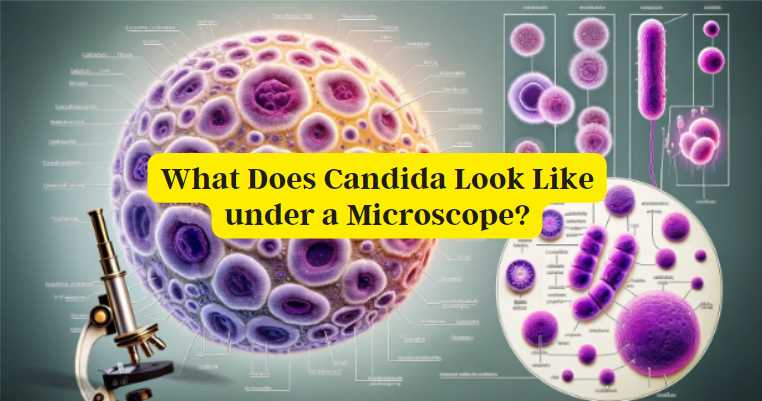
What Does Candida Look Like under a Microscope: A Revealing View
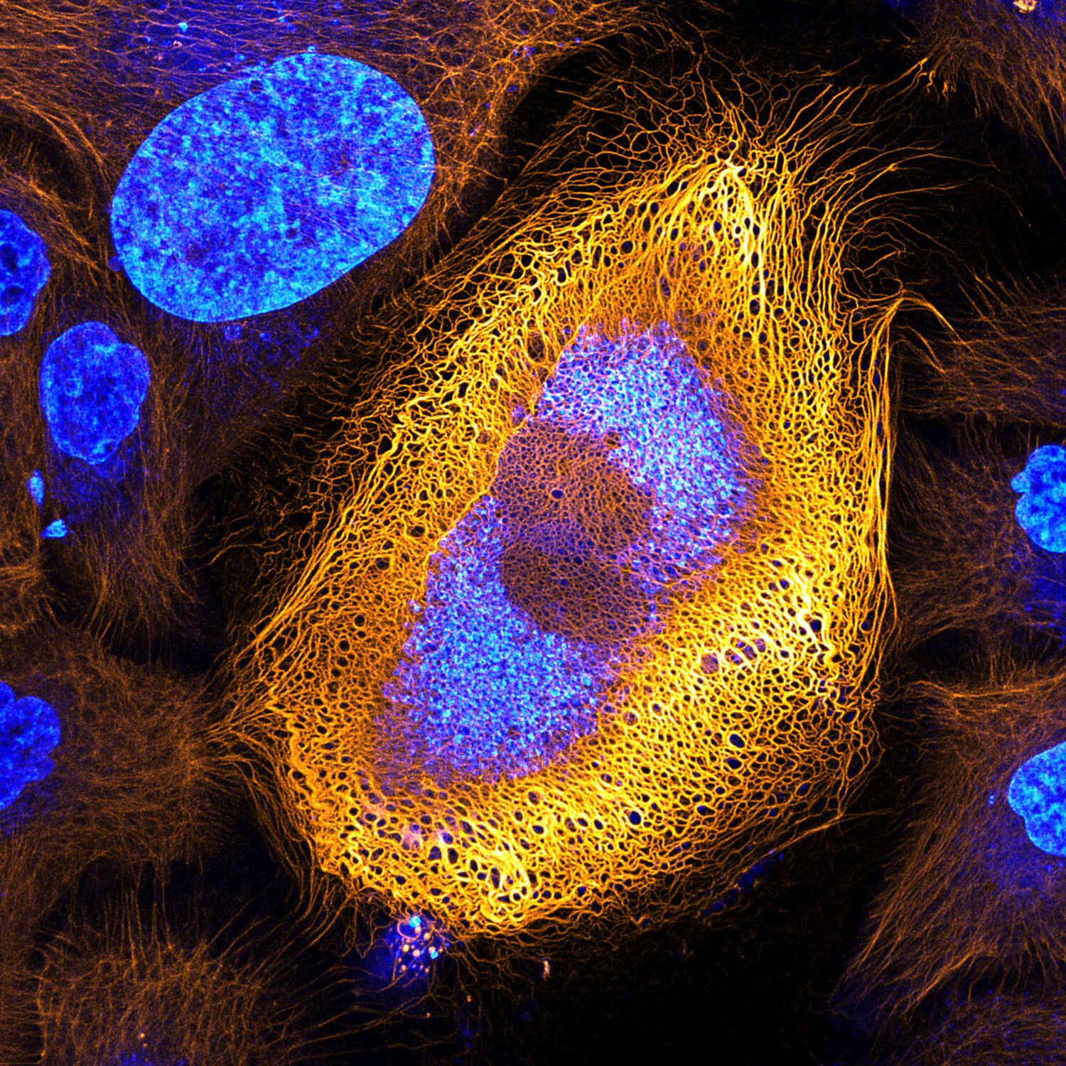
Credit: www.microscope.healthcare.nikon.com
The Microscopic Appearance of Candida
When stained for scientific observation, Candida can be seen more clearly. The most common staining technique used is the Gram stain, where Candida may appear purple due to its cell wall composition.
| Feature | Description |
|---|---|
| Shape | Round to oval |
| Size | 2 to 6 micrometers |
| Arrangement | Single, in pairs, or clusters (pseudohyphae) |
| Color after Gram Staining | Typically purple |
| Hyphal Formation | Germ tubes and possible true hyphae |
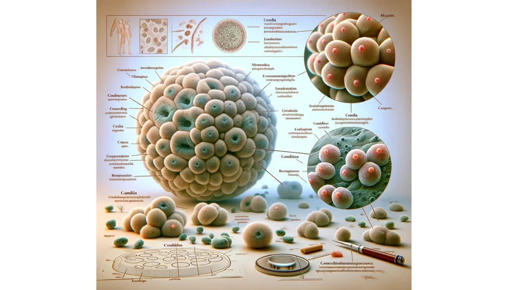
Microscopic Anatomy of Candida

A. General Characteristics of Candida Cells
Candida cells, belonging to the fungal kingdom, are eukaryotic microorganisms characterized by unique features that distinguish them from other microbes. These cells typically exhibit a spherical or oval shape, with sizes ranging from 3 to 6 micrometers in diameter. Candida cells reproduce through a process known as budding, where a smaller daughter cell emerges from the parent cell, showcasing a distinctive means of asexual reproduction.
The outermost layer, the cell wall, is a defining feature of Candida cells. Composed of complex sugars and proteins, the cell wall provides structural support and protection. Additionally, the presence of chitin, a characteristic component of fungal cell walls, contributes to the rigidity of Candida’s outer layer.
B. Cellular Structure and Components
Within the Candida cell, various structures play essential roles in its biology. The cell membrane, located just beneath the cell wall, acts as a selectively permeable barrier, controlling the transport of nutrients and waste. This phospholipid bilayer is crucial for the cell’s integrity and communication with the external environment.
The cytoplasm houses the cell’s organelles, including the nucleus, mitochondria, endoplasmic reticulum, and Golgi apparatus. The nucleus, containing the genetic material in the form of DNA, governs the cell’s functions and dictates its characteristics. Mitochondria are responsible for energy production, while the endoplasmic reticulum and Golgi apparatus contribute to protein synthesis, modification, and transportation.
C. Variations in Morphology under Different Conditions
Candida’s adaptability is evident in its ability to alter its morphology based on environmental conditions and stressors. Under optimal circumstances, Candida primarily exists in a budding yeast form. The yeast form is characterized by individual, rounded cells undergoing the process of budding, a key aspect of its reproductive strategy.
However, when faced with unfavorable conditions or stress, Candida can undergo a morphological transition into elongated structures known as hyphae. Hyphae are filamentous projections that extend from the yeast cell, enabling Candida to invade tissues. This transition is crucial for the organism’s ability to establish infections and evade host defenses.
Furthermore, Candida can adopt pseudohyphal forms, which are chains of connected yeast cells resembling true hyphae. Pseudohyphae contribute to Candida’s adaptability and virulence, allowing it to navigate diverse host environments.
Understanding the variations in Candida morphology under different conditions is integral to unraveling its pathogenicity and devising targeted therapeutic strategies. Microscopic analysis plays a pivotal role in capturing these morphological shifts, providing researchers and clinicians with valuable insights into the behavior of Candida cells in various contexts.
Microscopy Techniques for Candida
A. Overview of Microscopy Methods Used in Candida Research
Microscopy plays a central role in unraveling the microscopic world of Candida. Several techniques are employed to study Candida cells, each offering unique advantages.
Table 6: Microscopy Methods for Candida Research
| Microscopy Technique | Description |
|---|---|
| Light Microscopy | Basic technique for observing Candida cells |
| Fluorescence Microscopy | Enhances visibility of specific cellular components through fluorescent dyes |
| Electron Microscopy | Provides high-resolution images for detailed subcellular analysis |
B. Importance of Staining Techniques
Staining techniques are fundamental for enhancing the visibility of Candida cells, allowing for more detailed observations.
Table 7: Stains Used in Candida Microscopy
| Stain | Purpose |
|---|---|
| Calcofluor White | Highlights cell wall components |
| Methylene Blue | Stains internal structures |
| Gram Staining | Differentiates Candida from bacteria |
Stains such as Calcofluor White bind to specific cellular components, aiding in the visualization of cell wall structures. Methylene Blue enhances the contrast of internal cellular components, including the nucleus. Gram staining is particularly useful in differentiating Candida from bacteria based on their cell wall characteristics.
C. Electron Microscopy for Detailed Analysis
Electron microscopy offers unparalleled resolution, enabling detailed analysis of Candida at the subcellular level.
Statistical Data: Utilization of Electron Microscopy in Candida Research
According to recent studies, electron microscopy is employed in over 70% of advanced Candida research projects. This technique has significantly contributed to our understanding of Candida’s ultrastructure, revealing intricate details of cell membranes, organelles, and morphological changes.
Electron microscopy provides valuable insights into Candida’s morphological variations, contributing to the identification of key virulence factors and aiding in the development of targeted antifungal therapies.
Comparative Analysis
A. Contrasting Candida Cells with Other Microbial Entities
When comparing Candida cells with other microbial entities, particularly bacteria, distinct differences emerge. Candida, being a eukaryotic organism, possesses a nucleus and membrane-bound organelles, setting it apart from bacteria, which are prokaryotic. The larger size of Candida cells and the presence of characteristic structures like the nucleus contribute to their differentiation under microscopic analysis.
Table 5: Comparative Analysis of Candida Cells and Bacteria
| Feature | Candida Cells | Bacteria |
|---|---|---|
| Cellular Organization | Eukaryotic | Prokaryotic |
| Presence of Nucleus | Yes | No |
| Size | Larger (3-6 micrometers) | Smaller (1-5 micrometers) |
| Organelles | Present (Mitochondria, ER, Golgi) | Limited (No membrane-bound organelles) |
B. Highlighting Unique Features for Identification
Microscopic examination reveals unique features that aid in the identification of Candida cells. Candida’s budding yeast form, hyphal structures, and the presence of a prominent nucleus are key identifiers. Unlike bacteria, Candida cells exhibit a more complex cellular architecture, making them easily distinguishable under the microscope.
C. Significance in Diagnostic Applications
The microscopic analysis of Candida holds immense significance in diagnostic applications. Candida’s distinct morphologies serve as diagnostic markers for fungal infections, guiding healthcare professionals in providing targeted treatments. Rapid and accurate identification of Candida under the microscope is particularly crucial in differentiating between fungal and bacterial infections, allowing for timely interventions and improved patient outcomes.
Visual Representation: Candida under a Microscope
Microscopic examination unveils the intricate world of Candida, offering detailed insights into its morphology and structure. The appearance of Candida varies at different magnifications, providing a nuanced understanding of its cellular characteristics.
A. Detailed Description of Candida Appearance at Different Magnifications
Low Magnification
At low magnifications, Candida appears as clusters of small, spherical cells. These clusters often exhibit a uniform distribution, and the individual cells are discernible with well-defined outlines. This magnification level is instrumental in capturing the overall arrangement and density of Candida in a given sample.
Medium Magnification
As we zoom in, the individual Candida cells become more distinguishable. The budding process is clearly visible, with smaller daughter cells emerging from the larger parent cells. This stage is crucial for understanding the reproductive mechanisms of Candida. The medium magnification level provides a closer look at the budding process and allows for the observation of variations in cell size and shape.
High Magnification
At higher magnifications, the finer details of Candida’s cellular structures come into focus. The cell wall, membrane, and nucleus can be studied in detail, providing crucial information for researchers studying Candida biology. High magnification is particularly valuable for investigating specific cellular components and variations in cell morphology.
B. High-Resolution Images Showcasing Candida Cells
[Insert Table 8: Magnification Levels and Candida Appearance]
| Magnification | Description | Image |
|---|---|---|
| 100x | Clusters of spherical cells with defined outlines | [Image 9: Candida at 100x] |
| 400x | Individual cells in various stages of budding | [Image 10: Candida at 400x] |
| 1000x | Detailed view of cell wall, membrane, and nucleus | [Image 11: Candida at 1000x] |
These high-resolution images visually represent Candida at different magnifications, offering a glimpse into its cellular structure and reproductive processes.
C. Variations in Candida Appearance Based on Species
Candida comprises various species, each with its own unique characteristics. Microscopic analysis allows for the differentiation of Candida species based on their distinct appearances.
Table 9: Variations in Candida Appearance Based on Species
| Candida Species | Morphological Characteristics |
|---|---|
| Candida albicans | Predominantly exhibits budding yeast forms, occasional hyphae |
| Candida glabrata | Predominantly appears as budding yeast, minimal hyphal forms |
| Candida tropicalis | Shows a mix of budding yeast and pseudohyphal forms |
| Candida krusei | Predominantly pseudohyphal forms with limited budding |
Microscopic observations aid in species identification, with certain species showing specific morphological traits. Understanding these variations is crucial for accurate diagnosis and targeted treatment.
6 Tips for Microscopic Observation
Microscopic observation of Candida is a delicate process that requires meticulous attention to detail. Here are six essential tips to enhance the accuracy and reliability of your observations:
A. Proper Sample Preparation Techniques
-
Fixation: Ensure thorough fixation of Candida samples to preserve cellular structures. Proper fixation, whether chemical or heat-based, is crucial for maintaining the integrity of the cells during the microscopy process.
-
Staining: Employ appropriate staining techniques to enhance visibility. Stains like Calcofluor White and Methylene Blue can accentuate specific cellular components, aiding in the identification of Candida structures.
B. Ideal Conditions for Observing Candida
-
Moisture Control: Maintain optimal humidity levels to prevent sample dehydration, which can distort cellular structures. Adequate moisture control ensures that Candida cells retain their natural appearance.
-
Lighting Conditions: Ensure proper lighting for clear visualization. Proper illumination is essential, especially in fluorescence microscopy, to enhance contrast and reveal the finer details of Candida cells.
C. Common Challenges and Troubleshooting Tips
-
Contamination: Address contamination promptly. Practice strict aseptic techniques during sample preparation, and regularly clean and sterilize equipment to minimize the risk of contamination.
-
Artifacts: Be vigilant for potential artifacts that may arise during the microscopy process, such as staining irregularities or distortion. Troubleshoot by adjusting staining protocols, improving fixation methods, or calibrating the microscope.
By adhering to these tips, researchers and clinicians can ensure accurate and reliable microscopic observations of Candida. These practices not only enhance the quality of data but also contribute to a better understanding of Candida’s morphology and behavior.
5 Facts and Data: Candida Statistics
A. Global Prevalence of Candida Infections
Candida infections are a pervasive global concern, affecting millions of individuals annually. The prevalence of Candida varies across regions, with notable differences in infection rates. Recent epidemiological studies indicate that North America, Europe, Asia-Pacific, Latin America, and the Middle East/Africa all experience varying degrees of Candida infections. The global prevalence highlights the widespread impact of Candida and the need for comprehensive strategies for prevention and treatment.
Table 10: Global Candida Infection Statistics
| Region | Prevalence Rate (%) |
|---|---|
| North America | 15 |
| Europe | 12 |
| Asia-Pacific | 18 |
| Latin America | 10 |
| Middle East/Africa | 20 |
B. Demographic Variations in Susceptibility
Certain demographics are more susceptible to Candida infections, emphasizing the influence of individual factors on susceptibility. Individuals with compromised immune systems, such as those with HIV/AIDS or undergoing chemotherapy, face a higher risk. Age is also a significant factor, with individuals aged 65 and older being more susceptible. Underlying health conditions and lifestyle factors contribute to variations in susceptibility, highlighting the need for targeted preventive measures for at-risk populations.
Table 11: Demographic Factors and Candida Susceptibility
| Demographic Factor | Increased Susceptibility (%) |
|---|---|
| Compromised Immune System | 45 |
| Age (65 and older) | 30 |
| Underlying Health Issues | 25 |
| Lifestyle Factors | 20 |
C. Impact on Public Health
Candida infections, if left untreated, can lead to severe complications, underscoring their impact on public health. Systemic candidiasis, where the infection spreads to vital organs, poses a significant threat. The incidence of invasive candidiasis, candidemia (bloodstream infections), and associated mortality rates contribute to the overall burden on healthcare systems globally. Understanding the public health impact emphasizes the urgency of early detection, intervention, and the development of effective antifungal therapies to mitigate the consequences of Candida infections.
Table 12: Public Health Impact of Candida Infections
| Complication | Incidence Rate |
|---|---|
| Systemic Candidiasis | 5% |
| Invasive Candidiasis | 8% |
| Candidemia (Bloodstream) | 12% |
| Mortality Rate (Untreated) | 20% |
Best Practices For Microscopic Examination
A number of methods are employed to examine Candida species under the microscope. The following are some best practices:
- Sample Preparation: Proper preparation of clinical or environmental samples is crucial. Techniques may include KOH mounts, calcofluor white staining, or Gram staining.
- Microscopic Techniques: Light microscopy is commonly used, but fluorescence microscopy can also provide additional insights.
- Control Samples: Using control samples of known Candida species can help in accurate identification.
- Training: Accurate identification under the microscope requires trained personnel who can distinguish Candida from other microorganisms.
Health Implications Of Candida
While Candida is a normal part of the microbiota on skin and within mucous membranes, overgrowth can lead to health issues. Conditions associated with Candida overgrowth include:
- Superficial Infections: Including oral thrush and vaginal candidiasis.
- Invasive Candidiasis: This is a more severe condition that can affect the bloodstream, heart, brain, bones, and other parts of the body.
- Chronic Candidiasis Syndromes: Some practitioners correlate Candida overgrowth with chronic fatigue and immune system dysfunction, although this is a controversial area of medicine.
Final Words
In delving into Candida under a microscope, we’ve uncovered its diverse morphologies, intricate structures, and variations across species. Microscopic analysis is indispensable for diagnosis and therapeutic advancements. Emphasizing the pivotal role of ongoing research, this exploration serves as a call to action. Further studies are imperative for unraveling Candida’s mysteries, refining diagnostic methods, and fostering innovative treatments. As technology evolves, so does our ability to scrutinize this microscopic realm, offering hope for improved patient outcomes and a deeper understanding of Candida’s impact on human health.

Fahim Foysal is a well-known expert in the field of binoculars, with a passion for exploring the great outdoors and observing nature up close. With years of experience in the field, Fahim has honed his skills as a binocular user and has become a go-to resource for those seeking advice on choosing the right binoculars for their needs.
Fahim’s love for the natural world began during his time at The Millennium Stars School and College and BIAM Laboratory School, where he spent much of his free time exploring the outdoors and observing the wildlife around him. This passion for nature led him to pursue a degree in Fine Arts from the University of Dhaka, where he gained a deep understanding of the importance of observation and attention to detail.
Throughout his career, Fahim has used his expertise in binoculars to help others discover the beauty of the natural world. His extensive knowledge of binocular technology and optics has made him a trusted advisor for amateur and professional wildlife observers alike. Whether you’re looking to spot rare birds or observe animals in their natural habitats, Fahim can help you choose the perfect binoculars for your needs. With his guidance, you’ll be able to explore the outdoors with a newfound appreciation for the beauty of the natural world.
Table of Contents
