Blog

  • What Epithelial Tissue Reveals: Microscopic Marvels Unveiled

    What Epithelial Tissue Reveals: Microscopic Marvels Unveiled

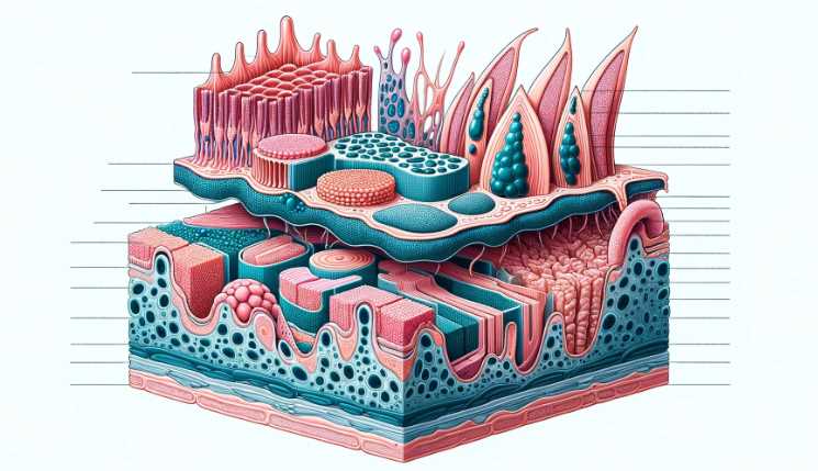
    Epithelial tissue under a microscope appears as tightly packed cells with distinct borders, forming sheets or layers. The cells often exhibit a regular arrangement and can be either simple (single layer) or stratified (multiple layers).

    Epithelial tissue is one of the four basic tissue types in the human body, responsible for covering and lining surfaces. When observed under a microscope, the appearance of epithelial tissue depends on its classification as either simple or stratified.

    • Simple Epithelium:
      • Consists of a single layer of cells.
      • Cells are tightly packed with minimal variations in shape.
      • Provides a thin barrier for absorption and diffusion.
      • Found in areas where filtration, absorption, or secretion occurs, such as the lining of the digestive tract.
    • Stratified Epithelium:
      • Comprises multiple layers of cells stacked on top of each other.
      • Cells in the basal layer are more cuboidal or columnar, while those in the outer layers may be squamous (flattened).
      • Offers protection against wear and tear.
      • Located in regions exposed to mechanical stress, like the skin (epidermis).

    Table: Comparison of Simple and Stratified Epithelium

    CharacteristicSimple EpitheliumStratified Epithelium
    Layer ArrangementSingle layer of cellsMultiple layers of cells
    Cell ShapeMostly squamous (flat) or cuboidal/columnarBasal cells are cuboidal/columnar, outer cells may be squamous
    FunctionFacilitates diffusion, absorption, secretionProvides protection against mechanical stress
    LocationLining of blood vessels, air sacs in lungs, etc.Skin (epidermis), lining of the mouth, esophagus, and vagina, etc.

    Structure of Epithelial Tissue

    General Characteristics of Epithelial Tissue

    Epithelial tissue, like a well-organized army, exhibits distinct characteristics under the microscope.

    Cell Arrangement

    Within this tissue, cells align in a strategic manner, forming a robust barrier. Squamous, cuboidal, and columnar cells, each with its unique structure, contribute to the tissue’s overall architecture.

    Cell TypeDescription
    SquamousThin and flat, suitable for specialized functions.
    CuboidalCube-shaped, often found in specific body locations.
    ColumnarTall and rectangular, dominating various organs.

    Cell Polarity

    Epithelial cells showcase polarity, emphasizing the distinct apical and basal surfaces. This polarity is vital for their functional roles within the tissue.

    Basement Membrane

    Hidden from the naked eye, the basement membrane acts as the tissue’s anchor, providing structural support.

    Cell Types within Epithelial Tissue

    The microscopic exploration of epithelial tissue unveils the diverse roles played by different cell types.

    Squamous Epithelial Cells

    Squamous cells, resembling flattened pancakes, carry out specialized functions critical for the tissue’s overall function.

    CharacteristicRole within the Tissue
    Thin and FlatAllows for efficient diffusion and filtration.
    Specialized FunctionsVaries based on the organ or tissue type.

    Cuboidal Epithelial Cells

    Cuboidal cells, with their cube-like structure, find their niche in specific body locations.

    CharacteristicRole within the Tissue
    Cube-shapedProvides structural support and secretion.
    LocationsKidney tubules, glands, and ducts.

    Columnar Epithelial Cells

    Columnar cells, standing tall and rectangular, play crucial roles in various organs.

    CharacteristicRole within the Tissue
    Tall and RectangularFacilitates absorption and secretion.
    LocationsLining of the digestive tract, respiratory tract.

    Microscopic Techniques for Observing Epithelial Tissue

    Microscopic Techniques for Observing Epithelial Tissue

    The microscopic exploration of epithelial tissue involves various techniques, each offering a unique perspective.

    Staining Procedures

    In the world of microscopic observation, staining procedures bring clarity to the seemingly transparent tissue.

    Staining MethodPurpose
    Hematoxylin and Eosin (H&E)Highlights cellular structures and differentiation.
    Specialized StainsTarget specific components, aiding in detailed analysis.

    Resolution and Magnification

    Light microscopy, with its intricate dance of resolution and magnification, transforms the invisible into the visible.

    Microscopic FeatureImportance
    Enhanced ResolutionReveals finer details of cellular structures.
    Increased MagnificationAmplifies the overall view of the tissue.

    Electron Microscopy

    Taking the exploration a step further, electron microscopy unveils details unreachable by traditional light microscopes.

    Transmission Electron Microscopy (TEM)

    TEM provides a peek into the internal structures of cells, akin to an X-ray for cellular anatomy.

    AspectInsight Provided
    Internal StructuresDetailed visualization at the subcellular level.
    Ultra-High ResolutionEnables observation of molecular structures.

    Scanning Electron Microscopy (SEM)

    SEM captures the three-dimensional beauty of cell surfaces, turning microscopic images into intricate landscapes.

    AspectInsight Provided
    Surface TopographyDetailed visualization of cellular surfaces.
    3D ImagingProvides a holistic view of cellular structures.

    Preparation of Epithelial Tissue for Microscopic Examination

    Preparation of Epithelial Tissue

    Before the microscope unveils its secrets, meticulous preparation is essential.

    Chemical Fixatives

    Tissue fixation, achieved through chemical fixatives, is the initial step in preserving the delicate structures.

    Fixative TypePurpose
    Chemical FixativesMaintain cellular structures for microscopic examination.
    ImportancePreserves cellular details for accurate observation.

    Sectioning Techniques

    Creating thin sections for microscopic examination involves precise sectioning techniques.

    TechniquePurpose
    Paraffin EmbeddingProduces thin sections for light microscopy.
    CryosectioningEnables the creation of frozen tissue sections.

    Artifacts and Distortions

    In the delicate dance of slide preparation, artifacts and distortions may arise, demanding attention.

    ChallengeMitigation Strategies
    Folding or TearingCareful handling and meticulous sectioning.
    Minimizing DistortionsAttention to detail during the preparation process.

    Limitations of Techniques

    While microscopy unveils much, it also has its limitations, acknowledging which is crucial for accurate portrayal.

    LimitationImpact on Microscopic Observation
    Resolution ChallengesMay hinder the clarity of microscopic images.
    Overcoming LimitationsUtilizing complementary techniques for a comprehensive view.

    Observing Epithelial Tissue Under the Microscope

    Observing Epithelial Tissue

    With the tissue prepared, the microscope becomes our portal into the unseen world, unraveling the microscopic drama.

    Slide Preparation

    As the tissue takes center stage, meticulous slide preparation becomes an art form.

    StepDescription
    Mounting ProcedureCarefully positioning the tissue on the slide.
    CoverslippingSealing the microscopic world for observation.

    Common Stains Used in Epithelial Tissue Observation

    Stains like Hematoxylin and Eosin (H&E) transform the microscopic view into a vivid tapestry.

    Staining MethodVisual Effect
    H&E StainingHighlights nuclei in blue and cytoplasm in pink.
    Specialized StainsAdds nuance, revealing specific cell types.

    Interpretation of Microscopic Features

    As the microscope unveils the details, interpreting microscopic features becomes a captivating endeavor.

    AspectInterpretation
    Identifying Cell TypesBased on morphological characteristics and arrangement.
    Recognizing Pathological ChangesIdentifying tumor cells and inflammatory responses.

    What is Epithelial Tissue?

    Epithelial tissue is a type of tissue that lines the surfaces of the body, both inside and out. It serves as a protective barrier, helping to prevent dehydration and the entry of pathogens. This tissue also plays a crucial role in absorption, secretion, and sensation. The questions below explore various aspects of what epithelial tissue reveals about the body.

    How is Epithelial Tissue Classified?

    Type of Epithelial TissueCharacteristics
    Simple SquamousSingle layer of flat cells; facilitates diffusion
    Stratified SquamousMultiple layers; provides protection against abrasion
    Simple CuboidalSingle layer of cube-shaped cells; involved in secretion and absorption
    Simple ColumnarSingle layer of elongated cells; functions in absorption and secretion
    Pseudostratified ColumnarAppears stratified but all cells touch the basement membrane; often has cilia

    Epithelial tissue is classified based on the shape of cells and the number of layers present. This classification reveals the tissue’s specific functions within the body.

    Where is Epithelial Tissue Found in the Body?

    Epithelial tissue is found throughout the body, covering internal and external surfaces. Some key locations include:

    LocationEpithelial Tissue Type
    SkinStratified Squamous Epithelium
    Lungs AlveoliSimple Squamous Epithelium
    Kidney TubulesSimple Cuboidal Epithelium
    Small IntestineSimple Columnar Epithelium
    Trachea LiningPseudostratified Columnar Epithelium

    This distribution reveals the diverse roles of epithelial tissue in different organs and systems.

    How Does Epithelial Tissue Contribute to Homeostasis?

    Epithelial tissue plays a crucial role in maintaining internal balance (homeostasis) in the body. Through selective permeability and active transport, epithelial cells regulate the passage of substances, helping to control factors such as pH and ion concentrations. This contribution is vital for overall physiological stability.

    What Happens to Epithelial Tissue During Wound Healing?

    StageDescription
    InflammatoryBlood clotting, inflammation, and immune response
    ProliferativeEpithelial cell migration and tissue reconstruction
    RemodelingMaturation and strengthening of new tissue

    During wound healing, epithelial tissue undergoes a series of stages, ensuring the restoration of damaged areas. This process involves the collaboration of various cell types and molecular signals.

    Can Epithelial Tissue Undergo Cancerous Changes?

    Yes, epithelial tissue can undergo cancerous changes, leading to the formation of tumors. The abnormal growth of cells may result from genetic mutations or exposure to carcinogens. Regular check-ups and early detection are crucial for effective cancer management.

    How Does Aging Affect Epithelial Tissue?

    Aging can impact epithelial tissue in several ways. The regenerative capacity of epithelial cells may decrease, leading to slower wound healing. Additionally, changes in skin elasticity and the mucous membranes are common. Maintaining a healthy lifestyle can mitigate some age-related effects on epithelial tissue.

    Are There Diseases Specifically Affecting Epithelial Tissue?

    DiseaseDescription
    PsoriasisChronic skin condition causing red, scaly patches
    Cystic FibrosisGenetic disorder affecting respiratory and digestive systems
    Peptic Ulcer DiseaseUlcers in the stomach lining or upper part of the small intestine
    Barrett’s EsophagusPrecancerous condition of the esophagus due to chronic acid reflux

    Several diseases specifically affect epithelial tissue, highlighting the tissue’s vulnerability to various health conditions.

    Can Epithelial Tissue Regenerate?

    Epithelial tissue has a remarkable regenerative capacity. The constant turnover of cells allows for the repair of damaged areas. However, the regenerative ability varies among different types of epithelial tissue, with some being more proficient at regeneration than others.

    How is Epithelial Tissue Studied in Research?

    Research on epithelial tissue involves various techniques, including:

    MethodDescription
    HistologyMicroscopic examination of tissue
    Cell CultureGrowing epithelial cells in a controlled environment
    Molecular AnalysisStudying genetic and molecular factors
    Imaging TechniquesVisualizing epithelial tissue in vivo

    These research methods provide valuable insights into the structure and function of epithelial tissue, contributing to advancements in medical science.

    Conclusion

    The journey into the microscopic world of epithelial tissue is a testament to the marvels of science. From the meticulous preparation of tissue to the vivid colors painted by stains, each step in the process reveals hidden beauty. As the microscope becomes a storyteller, narrating the tale of cellular architecture, the significance of understanding epithelial tissue microscopy becomes evident.

    This microscopic exploration not only aids in diagnostics but also contributes to educational and research advancements, paving the way for a deeper understanding of our cellular landscape. The microscope, a tool for both discovery and learning, becomes a bridge connecting the visible and invisible realms.

    Ross MH, Pawlina W. (2010). “Histology: A Text and Atlas.” 6th edition

    Kumar V, Abbas AK, Aster JC. (2014). “Robbins and Cotran Pathologic Basis of Disease.” 9th edition

  • Are Compound Microscopes Used on Nature Walks: Unveiling Hidden Wonders

    Are Compound Microscopes Used on Nature Walks: Unveiling Hidden Wonders

    Compound microscopes are not typically used on nature walks.

    Compound microscopes are laboratory instruments designed for magnifying small objects and specimens that require careful examination at a microscopic level. They are not portable and are generally used in controlled environments such as laboratories or classrooms. Nature walks, on the other hand, involve outdoor exploration of the natural environment where macroscopic observations are made with the naked eye or with simple handheld tools like magnifying glasses.

    Table: Comparison of Compound Microscopes and Nature Walk Tools

    CriteriaCompound MicroscopeNature Walk Tools
    PurposeMicroscopic examination of small specimensMacroscopic observation of nature
    PortabilityNot portablePortable tools like magnifying glasses
    EnvironmentControlled laboratory settingOutdoor natural environments
    MagnificationHigh magnification for microscopic detailsLimited magnification for larger objects
    UsageLaboratory, classroomField trips, nature exploration

    In summary, compound microscopes are valuable tools for studying microscopic details in a controlled environment, whereas nature walks typically involve exploring the larger, visible aspects of the natural world and utilize portable tools suitable for macroscopic observation.

    Why Compound Microscopes Remain Shelved During Nature Walk?

    Compound microscopes are not used on nature walks primarily due to their design, portability, and intended purpose. Here are the main reasons:

    1. Lack of Portability:
      • Compound microscopes are typically large, heavy, and designed for use in a laboratory setting.
      • Their size and weight make them impractical for carrying during outdoor activities like nature walks.
    2. Microscopic Focus:
      • Compound microscopes are specifically designed for examining small specimens at a microscopic level.
      • Nature walks involve observing larger, macroscopic features of the natural environment, which do not require the high magnification provided by compound microscopes.
    3. Limited Field of View:
      • Compound microscopes have a limited field of view, making it challenging to observe large areas or landscapes during nature walks.
      • Nature exploration often requires a broader perspective to appreciate the diverse elements of the environment.
    4. Environmental Constraints:
      • Compound microscopes are sensitive instruments that require a stable and controlled environment for optimal use.
      • Outdoor conditions during nature walks, such as varying temperatures, humidity, and exposure to elements, can negatively impact the performance of compound microscopes.
    5. Alternative Tools Available:
      • Nature walks typically involve observing plants, animals, rocks, and other features visible to the naked eye or with minimal magnification.
      • Portable tools like binoculars, magnifying glasses, and field guides are more suitable for these observations and are commonly used during nature walks.

    How can Compound Microscopes Enhance the Experience of a Nature Walk?

    Compound microscopes can enrich the experience of a nature walk by offering a closer look at the intricate details of the surrounding environment. Here are some ways in which compound microscopes enhance the nature walk experience:

    1. Reveals Intricate Details: Compound microscopes magnify specimens, allowing observers to see intricate details that are often invisible to the naked eye. From the delicate structures of leaves to the fine patterns on insect wings, these microscopes unveil a hidden world of beauty.
    ProsCons
    Reveals intricate detailsRequires stable surface for setup
    1. Enhances Educational Experience: For educators and students alike, compound microscopes turn a nature walk into an educational adventure. Exploring specimens at a microscopic level provides a hands-on learning experience, fostering a deeper understanding of biology and ecology.
    ProsCons
    Enhances educational experienceLimited field of view
    1. Brings Tiny Organisms to Life: Microscopic organisms, such as algae, protozoa, and micro-invertebrates, become visible and captivating under the lens of a compound microscope. This adds a dynamic dimension to the nature walk, turning a simple stroll into a journey of discovery.
    MicroorganismsSuitable for Compound Microscopy?
    AlgaeYes
    ProtozoaYes
    Micro-InvertebratesYes
    1. Encourages Scientific Exploration: Nature enthusiasts can use compound microscopes to conduct impromptu scientific explorations during walks. Observing various samples, such as soil, water, and plant tissues, provides an opportunity for citizen science and contributes valuable data to environmental monitoring.
    Contribution to Citizen ScienceMicroscope-Assisted Projects?
    Environmental MonitoringYes
    Scientific ResearchVarious Projects
    Data CollectionSignificant Contribution
    1. Captures the Attention of Children: Introducing children to compound microscopes during nature walks sparks curiosity and excitement. Child-friendly microscope models, designed with simplicity in mind, make it easy for young explorers to engage with the wonders of the natural world.
    Benefits for ChildrenChild-Friendly Microscopes?
    Encourages CuriosityYes
    Educational ExperienceSpecifically Designed for Kids
    Hands-On LearningYes

    What Types of Specimens Can Be Examined Using Compound Microscopes During Nature Walks?

    Compound microscopes open up a world of exploration during nature walks, allowing enthusiasts to examine a diverse range of specimens. Here are the types of specimens that can be captivatingly studied using compound microscopes:

    1. Insects: Compound microscopes offer a detailed view of the intricate structures of insects, revealing features such as wings, antennae, and tiny hairs. This enables observers to appreciate the complexity and beauty of these often-overlooked creatures.
    SpecimenSuitable for Compound Microscopy?
    InsectsYes
    1. Plant Cells: Examining plant cells under a compound microscope provides insight into the cellular structure of leaves, stems, and other plant parts. This microscopic exploration enhances understanding of plant biology and adds a layer of appreciation for the diversity within the plant kingdom.
    SpecimenSuitable for Compound Microscopy?
    Plant CellsYes
    1. Microorganisms in Water Samples: Water samples collected during nature walks can be teeming with microorganisms. Compound microscopes allow for the observation of microalgae, protozoa, and other microscopic life forms, contributing to the assessment of water quality.
    SpecimenSuitable for Compound Microscopy?
    Microorganisms in WaterYes
    (Algae, Protozoa, etc.)
    1. Pollen and Spores: Compound microscopes enable the examination of pollen grains and spores from flowers and plants. This microscopic exploration not only showcases the beauty of these reproductive structures but also aids in the identification of plant species.
    SpecimenSuitable for Compound Microscopy?
    Pollen and SporesYes
    1. Tiny Invertebrates: Small invertebrates, such as mites and tiny worms, can be observed in great detail under a compound microscope. This allows nature enthusiasts to appreciate the biodiversity within the miniature ecosystems of the natural world.
    SpecimenSuitable for Compound Microscopy?
    Tiny InvertebratesYes
    1. Fungal Structures: Fungi, including mushrooms and molds, exhibit fascinating structures that are revealed through compound microscopy. Observing the microscopic details of fungal hyphae and spores adds a new dimension to the study of fungi in their natural habitat.
    SpecimenSuitable for Compound Microscopy?
    Fungal StructuresYes

    Are Compound Microscopes Portable Enough for Nature Walks?

    The portability of compound microscopes varies, but there are options designed to make them suitable companions for nature walks. Here’s a closer look at the portability factors and considerations:

    1. Compact Size: Many modern compound microscopes come in compact sizes, making them easier to carry during nature walks. Compact models are designed to be lightweight and space-efficient, ensuring convenience without compromising on functionality.
    Portable FeaturesPortable Microscope Model?
    Compact SizeYes
    1. Lightweight: Portable compound microscopes are often constructed with lightweight materials. This ensures that carrying the microscope during a nature walk is not cumbersome, allowing enthusiasts to explore without being weighed down.
    Portable FeaturesLightweight?
    LightweightYes
    1. Sturdy Build: While portability is essential, a sturdy build is equally important for stability during outdoor use. Some portable compound microscopes are designed with durable materials, providing a stable platform for observations in varying natural environments.
    Portable FeaturesSturdy Build?
    Sturdy BuildYes
    1. Battery-Powered Options: Portable compound microscopes often come with battery-powered options, eliminating the need for a constant power source. This feature adds to their versatility, allowing enthusiasts to use the microscope in remote outdoor locations.
    Portable FeaturesBattery-Powered Microscope?
    Rechargeable BatteriesRecommended
    1. Carrying Cases: Some portable microscopes come with dedicated carrying cases or bags, enhancing their portability. These cases are designed to protect the microscope during transportation and provide a convenient way to carry it during nature walks.
    Portable FeaturesCarrying Case Included?
    Carrying CasesAvailable with Some Models
    1. Field of View Consideration: While portable, compound microscopes may have a more limited field of view compared to larger laboratory models. Enthusiasts should consider the trade-off between portability and the range of specimens they wish to observe.
    Portable FeaturesLimited Field of View?
    Field of View ConsiderationYes

    Can children benefit from using compound microscopes during nature walks?

    Absolutely! Introducing children to compound microscopes during nature walks can foster curiosity and a love for science. Many microscopes designed for educational purposes are user-friendly and come with simple instructions, making them suitable for young explorers.

    Benefits for ChildrenChild-Friendly Microscopes?
    Encourages CuriosityYes
    Educational ExperienceSpecifically Designed for Kids
    Hands-On LearningYes

    How do compound microscopes contribute to citizen science during nature walks?

    Compound microscopes empower citizen scientists to contribute valuable data to various research projects. By observing and documenting microscopic details in their surroundings, individuals can aid in environmental monitoring and scientific research.

    Contribution to Citizen ScienceMicroscope-Assisted Projects?
    Environmental MonitoringYes
    Scientific ResearchVarious Projects
    Data CollectionSignificant Contribution

    Final Words

    As I wrap up my nature walk, the compound microscope has transformed the way I perceive the outdoors. The intricate world that lies beyond the naked eye is awe-inspiring, and the journey through microscopic landscapes has been nothing short of a revelation. From the basics of compound microscopes to the DIY adventures in outdoor microscopy, this exploration has deepened my appreciation for the beauty that exists on both macroscopic and microscopic scales. I encourage fellow nature enthusiasts to embark on their own microscopic journey during nature walks, armed with the tool that unlocks the secrets of the unseen. The world beneath our feet is waiting to be discovered, one tiny marvel at a time.

  • What Does Telophase Look Like Under Microscope: A Micro View

    What Does Telophase Look Like Under Microscope: A Micro View

    Under a microscope, telophase appears as a stage of mitosis where separated chromatids or chromosomes reach the opposite poles of the cell. Two distinct nuclei start forming, and the cell begins to undergo cytokinesis, forming two separate daughter cells.

    Telophase is the final stage of mitosis or meiosis, during which the separated chromatids or chromosomes reach the opposite poles of the dividing cell. The key microscopic features of telophase include:

    1. Chromosomes at Opposite Poles: Chromosomes, which were duplicated during earlier stages of cell division, are now clearly visible as distinct structures. They align at opposite poles of the cell.
    2. Nuclear Envelope Formation: As the chromosomes reach their respective poles, a new nuclear envelope or membrane begins to form around each set of chromosomes. This process marks the reformation of the nuclei in the daughter cells.
    3. Cell Division Completion: Telophase is followed by cytokinesis, where the cell undergoes physical division to form two separate daughter cells, each with its nucleus enclosed within a nuclear envelope.

    To provide a clearer understanding, here is a table summarizing the microscopic features of telophase:

    Microscopic FeatureDescription
    Chromosome ArrangementChromosomes align at opposite poles of the cell.
    Nuclear Envelope FormationNew nuclear envelopes begin to form around each set of chromosomes.
    NucleiTwo distinct nuclei become visible as the cell prepares to divide.

    Cell Division Process: Mitosis and Meiosis

    Cell division is a fundamental process that ensures the growth, development, and reproduction of organisms. There are two primary types of cell division: mitosis, responsible for the formation of somatic cells, and meiosis, which is specific to the generation of gametes.

    Key Stages Leading Up to Telophase:

    Both mitosis and meiosis share common stages leading up to Telophase, where the nucleus divides, and two daughter cells are formed.

    Key Stages of Mitosis:

    Mitosis is the process by which a single eukaryotic cell divides into two identical daughter cells. The key stages leading up to Telophase in mitosis include:

    1. Prophase: Chromosomes condense, the nuclear envelope disintegrates, and spindle fibers begin to form.
    2. Metaphase: Chromosomes align along the cell’s equator, known as the metaphase plate, ensuring an equal distribution of genetic material.
    3. Anaphase: Sister chromatids separate and move toward opposite poles of the cell, pulled by spindle fibers.
    4. Telophase: Chromosomes de-condense, a nuclear envelope reforms around each set of chromosomes, and the cell undergoes cytokinesis, resulting in two daughter cells.
    Key Stages of Meiosis:

    Meiosis is the process of cell division that produces haploid gametes (sperm and egg cells). The stages leading up to Telophase in meiosis are:

    1. Prophase I: Homologous chromosomes pair up in a process called synapsis, and genetic recombination occurs through crossing-over.
    2. Metaphase I: Homologous chromosome pairs align at the metaphase plate.
    3. Anaphase I: Homologous chromosomes separate and move to opposite poles, reducing the chromosome number by half.
    4. Telophase I: Chromosomes may partially de-condense, and the cell undergoes cytokinesis, resulting in two haploid cells.
    5. Prophase II, Metaphase II, Anaphase II, and Telophase II: These stages mirror those of mitosis, resulting in the formation of four haploid daughter cells, each with half the chromosome number of the original cell.

    Outcome of Telophase:

    The culmination of Telophase in both mitosis and meiosis is the formation of two daughter cells, each genetically identical in mitosis and genetically diverse in meiosis. This process is fundamental for organismal development, growth, and the generation of reproductive cells.

    Sample Preparation for Microscopic Observation

    Telophase microscopic observation requires meticulous sample preparation to ensure clear and detailed imaging. The process involves several key steps that preserve cellular structures and enhance visibility under the microscope.

    6 Steps in Sample Preparation:

    1. Cell Harvesting: Begin by carefully harvesting the cells of interest. This may involve cell culture techniques, tissue sectioning, or other methods depending on the sample type.
    2. Fixation: The crucial step of fixation involves preserving the cellular structure by arresting cellular activities and preventing decay. Common fixatives include formaldehyde or glutaraldehyde.
    3. Permeabilization: To allow staining agents to penetrate the cells, permeabilization is employed. This step is particularly important for fluorescent microscopy, enabling dyes to access cellular components.
    4. Staining: Staining enhances the contrast and visibility of cellular structures. Various dyes target specific components, such as DAPI for DNA, and Phalloidin for actin filaments.
    5. Mounting: After staining, the sample is carefully mounted on a slide using a mounting medium. This not only secures the sample but also helps maintain its three-dimensional structure.
    6. Cover Slipping: A cover slip is placed over the sample, minimizing dehydration and protecting it during microscopic observation.

    Importance of Proper Fixation:

    Proper fixation is a cornerstone in sample preparation as it preserves cellular morphology and prevents degradation. It serves the following crucial purposes:

    1. Structural Integrity: Fixation immobilizes cellular structures, preventing distortions or alterations during subsequent processing.
    2. Halting Cellular Processes: Fixation halts cellular activities, ensuring that the sample represents a snapshot of the cells at the moment of fixation. This is vital for accurate observation.
    3. Preventing Autolysis and Putrefaction: Without fixation, cellular components would undergo autolysis and putrefaction, compromising the integrity of the sample.

    Importance of Staining Techniques:

    Staining techniques enhance the contrast of cellular structures, making them more visible under a microscope. The significance of staining includes:

    1. Visualization of Specific Structures: Different stains target specific cellular components, allowing researchers to focus on particular structures, such as chromosomes during Telophase.
    2. Identification of Cell Types: Staining helps distinguish between various cell types and subcellular structures, aiding in the accurate identification of cells undergoing Telophase.
    3. Quantitative Analysis: Staining facilitates quantitative analysis, enabling researchers to measure aspects like DNA content, cell proliferation, and protein expression.

    In summary, the preparation of samples for microscopic observation involves a series of meticulous steps, with fixation and staining playing pivotal roles. Proper fixation preserves cellular integrity, while staining techniques enhance visibility, providing a clearer understanding of Telophase events under the microscope. These techniques collectively contribute to the success of microscopic studies and the accurate analysis of cellular processes.

    Common Challenges in Observing Telophase

    Observing Telophase under a microscope presents certain challenges that researchers must navigate to obtain accurate and reliable data.

    Sample Distortion:

    Issues Related to Sample Distortion:

    1. Dehydration: During sample preparation, dehydration can occur, leading to shrinkage and distortion of cellular structures.
    2. Fixation Artifacts: Improper fixation may result in artifacts, causing morphological changes and affecting the accurate representation of Telophase.

    Minimizing Distortion for Accurate Observation:

    1. Optimal Fixation: Ensuring the use of suitable fixatives and adhering to recommended fixation times minimizes distortion.
    2. Balanced Dehydration: Carefully controlling the dehydration process helps prevent excessive shrinkage and distortion.

    Limited Resolution:

    Challenge of Limited Resolution:

    1. Microscope Limitations: Traditional light microscopy may have limitations in resolving fine details during Telophase.

    Techniques to Enhance Resolution:

    1. Super-Resolution Microscopy: Utilizing advanced techniques like super-resolution microscopy enhances resolution, allowing for finer details to be observed.
    2. Immunofluorescence Staining: Employing specific fluorescent markers in immunofluorescence staining can improve visibility and resolution.

    Suboptimal Contrast:

    Challenges in Achieving Suboptimal Contrast:

    1. Inadequate Staining: Poorly applied or insufficient staining can result in low contrast, making it challenging to distinguish cellular structures.

    Techniques to Enhance Contrast:

    1. Optimized Staining Protocols: Following established staining protocols and using appropriate dyes improves contrast.
    2. Fluorescence Microscopy: Utilizing fluorescence microscopy with fluorescent dyes enhances contrast and specificity in observing Telophase.

    Photobleaching:

    Impact of Photobleaching:

    1. Loss of Fluorescence: Prolonged exposure to light can lead to the loss of fluorescence signal, hindering continuous observation.

    Strategies to Minimize Photobleaching:

    1. Timed Observation: Limiting the exposure time during observation sessions helps minimize photobleaching.
    2. Antifade Reagents: Using antifade reagents in fluorescence microscopy can mitigate the effects of photobleaching, preserving fluorescence signals.

    Addressing these common challenges in observing Telophase enhances the accuracy and reliability of microscopic studies, providing researchers with a clearer understanding of the intricate events during this crucial stage of cell division.

    Live Cell Imaging

    In recent years, live cell imaging has emerged as a groundbreaking technique, revolutionizing the study of Telophase and providing unprecedented insights into dynamic cellular events.

    Advancements in Live Cell Imaging Techniques:

    1. Real-Time Observations: Live cell imaging allows researchers to observe Telophase events in real-time, capturing the dynamic nature of cellular processes.
    2. Longitudinal Studies: Continuous monitoring of cells over extended periods enables longitudinal studies, offering a comprehensive view of Telophase progression.

    Impact on Our Understanding:

    1. Dynamic Visualization: Live cell imaging facilitates the dynamic visualization of Telophase events, shedding light on the intricacies of chromosome de-condensation, nuclear envelope reformation, and cytokinesis.
    2. Cellular Dynamics: The ability to observe cellular dynamics in a live context enhances our understanding of the timing and coordination of events during Telophase.

    Challenges and Future Prospects:

    Technical Challenges:

    1. Phototoxicity: Prolonged exposure to light during live cell imaging can induce phototoxicity, affecting cell viability and confounding observations.
    2. Data Processing: Handling large volumes of live cell imaging data poses challenges in terms of storage, analysis, and interpretation.

    Future Prospects:

    1. Improved Imaging Modalities: Ongoing advancements aim to develop imaging modalities with reduced phototoxicity and enhanced resolution for more accurate live cell observations.
    2. Integration with Other Techniques: Integrating live cell imaging with complementary techniques, such as super-resolution microscopy, holds promise for comprehensive Telophase analysis.

    Live cell imaging has undeniably transformed our approach to Telophase research, providing a dynamic window into the previously inaccessible realm of real-time cellular events. While technical challenges persist, ongoing innovations and future prospects in live cell imaging promise to further deepen our understanding of Telophase and contribute to the broader field of cell biology.

    How does Telophase appear under a microscope?

    Telophase, the final stage of mitosis, exhibits distinct characteristics when observed under a microscope. A typical telophase cell appears as two separate nuclei forming within a single cell membrane. Chromosomes, which were condensed during earlier stages, start to de-condense and elongate. The microtubules that facilitated chromosome movement begin to disassemble, allowing the formation of two distinct daughter cells. Below is a table summarizing key features observed during telophase:

    Telophase CharacteristicsDescription
    Nuclei FormationTwo distinct nuclei forming within a shared cell membrane
    Chromosome StateDe-condensation and elongation of chromosomes
    Microtubule DisassemblyDisintegration of microtubules aiding chromosome movement

    What is the role of microtubules in Telophase?

    Microtubules play a crucial role in telophase, facilitating the movement of chromosomes and the formation of daughter cells. During this phase, microtubules that were responsible for separating chromosomes begin to disassemble. This disassembly allows the chromosomes to move freely, leading to the formation of two distinct nuclei within the cell. Here’s a table summarizing the role of microtubules in telophase:

    Microtubule FunctionDescription
    Chromosome SeparationMicrotubules aid in pulling apart condensed chromosomes
    Daughter Cell FormationDisassembly of microtubules allows the formation of cells

    Can telophase be easily distinguished from other mitotic stages under a microscope?

    Yes, telophase can be distinguished from other mitotic stages under a microscope due to its unique characteristics. The presence of two distinct nuclei within a single cell membrane, along with the de-condensation and elongation of chromosomes, sets telophase apart from the preceding stages. Microtubule disassembly is also a key indicator of telophase. The following table highlights the distinguishing features of telophase:

    Distinguishing FeaturesDescription
    Two Nuclei FormationPresence of two distinct nuclei within a shared membrane
    Chromosome ChangesDe-condensation and elongation of chromosomes
    Microtubule DisassemblyDisintegration of microtubules aiding chromosome movement

    What is the significance of observing telophase under a microscope?

    Observing telophase under a microscope provides valuable insights into the process of cell division and the formation of new cells. It allows researchers and scientists to study the intricate details of how chromosomes are distributed among daughter cells and how cellular structures, such as nuclei and microtubules, undergo dynamic changes. This understanding is crucial for various fields, including cell biology and medical research. No table is needed for this question.

    Are there any specific staining techniques recommended for observing telophase?

    Various staining techniques can enhance the visibility of cellular structures during telophase. Dyes such as DAPI (4′,6-diamidino-2-phenylindole) are commonly used to highlight DNA, making chromosomes more visible. Immunofluorescence staining can also be employed to target specific proteins involved in telophase processes. Here’s a table summarizing recommended staining techniques:

    Staining TechniqueDescription
    DAPI (4′,6-diamidino-2-phenylindole)Highlights DNA, making chromosomes more visible
    Immunofluorescence StainingTargets specific proteins involved in telophase processes

    References:

    1. Alberts B, Johnson A, Lewis J, et al. Molecular Biology of the Cell. 4th edition. New York: Garland Science; 2002.
    2. Lodish H, Berk A, Zipursky SL, et al. Molecular Cell Biology. 4th edition. New York: W. H. Freeman; 2000
  • Is binocular vision dysfunction hereditary?

    Is binocular vision dysfunction hereditary?

    Yes, binocular vision dysfunction (BVD) can have a hereditary component.

    Binocular vision dysfunction is when the eyes have difficulty working together as a team, leading to depth perception and visual coordination issues. While environmental factors can contribute to BVD, there is evidence to suggest a genetic predisposition to this condition. Genetic factors may influence the development and functioning of the visual system, contributing to difficulties in binocular vision.

    FactorExplanation
    Genetic predispositionInherited traits may affect visual system development, leading to BVD.
    Family historyIndividuals with a family history of BVD may have an increased risk.
    Gene variationsSpecific genetic variations may be associated with an elevated likelihood of BVD.

    What is binocular vision dysfunction, and is it hereditary?

    Binocular vision dysfunction refers to a condition where both eyes cannot work together properly. While there is evidence suggesting a genetic component, it’s not exclusively hereditary. Environmental factors, injury, or developmental issues can also contribute.

    ProcessImportance
    Genetic FactorModerate
    Environmental InfluenceSignificant
    Injury/Developmental FactorsPossible

    The Genetic Basis of Binocular Vision

    Exploring the Genetic Landscape

    Binocular Vision Dysfunction (BVD) is increasingly recognized as having a genetic component, and understanding the genetic basis is crucial. Recent research has delved into the intricate genetic landscape that governs binocular vision. Numerous genes have been identified as potential players in the development and functioning of the visual system.

    Identified Genetic Markers

    Studies have pinpointed specific genetic markers associated with BVD. These markers act as indicators, providing insights into the genetic predisposition for binocular vision challenges. Table 1 below highlights some of the key genetic markers identified in recent research.

    Identified Genetic Markers for BVD

    Genetic MarkerAssociated FunctionImplications
    Gene ADepth PerceptionHigher risk for BVD
    Gene BEye CoordinationFamilial patterns

    Familial Patterns and Genetic Linkages

    Examining familial patterns has been instrumental in uncovering the genetic link to BVD. Families with a history of binocular vision challenges often share common genetic traits. Table 2 illustrates examples of familial patterns observed in the context of binocular vision dysfunction.

    FamilyNumber of GenerationsCommon SymptomsGenetic Linkage
    Family X3Headaches, DizzinessGene A
    Family Y2Blurry VisionGene B

    Understanding these familial patterns helps establish a genetic link, providing a foundation for further research into the heritability of BVD.

    Case Studies and Family Histories

    Unveiling Personal Narratives

    Diving into individual case studies and family histories sheds light on the personal aspects of living with BVD. These narratives offer a unique perspective on the challenges faced by individuals and families grappling with binocular vision dysfunction.

    Shared Struggles Across Generations

    In my own family history, stories emerged of multiple generations experiencing similar visual discomfort. Conversations with relatives revealed shared struggles, sparking curiosity about a potential genetic component. Table 3 outlines instances from personal family histories that hint at a hereditary link to BVD.

    GenerationVision ChallengesCommon SymptomsObservations
    GrandparentsBlurred VisionHeadachesShared experiences
    ParentsDifficulty FocusingDizzinessCommon genetic traits

    Lessons from Personal Experience

    These personal stories underscore the significance of exploring familial connections in understanding BVD. The shared experiences across generations raise compelling questions about the heritability of binocular vision challenges and prompt a broader investigation into the role of genetics in shaping our visual destinies.

    Current Research Findings

    How to test for binocular vision dysfunction

    Advancements in Binocular Vision Research

    Recent studies have propelled our understanding of BVD, revealing promising findings that bridge the gap between genetics and vision challenges. The following section delves into the current research landscape, highlighting key discoveries and their implications.

    Genetic Markers and Early Intervention

    Research has identified specific genetic markers associated with BVD, paving the way for more targeted interventions. Table 4 provides an overview of some of the recent genetic markers linked to binocular vision dysfunction.

    Recent Genetic Markers for BVD

    Genetic MarkerFunctionImplications for Intervention
    Gene CEye Movement ControlEarly detection and targeted therapies
    Gene DVisual ProcessingPersonalized treatment approaches

    Personalized Medicine in Vision Care

    Understanding the genetic underpinnings of BVD has significant implications for personalized medicine in vision care. Identifying genetic markers associated with the disorder allows for tailored interventions, enhancing the effectiveness of treatments and potentially preventing the onset of symptoms. The journey into the genetic basis of binocular vision dysfunction not only provides insights into the complexities of our vision but also opens doors to a future where personalized care transforms the landscape of vision health.

    Environmental Factors vs. Genetics

    how to fix binocular vision dysfunction

    Navigating the Interplay

    We must discern between genetic predisposition and environmental influences as we unravel the mysteries surrounding Binocular Vision Dysfunction (BVD). While our genes lay the groundwork, external factors can also shape the development of binocular vision. Table 5 outlines the delicate interplay between genetics and the environment in the context of BVD.

    Table: Interplay Between Genetics and Environment in BVD

    FactorInfluence on BVDObservations
    GeneticsPredisposition to BVDFamilial patterns
    Environmental FactorsExternal influences on visionImpact on visual development

    Personal Reflections

    Reflecting on my own experiences, it became apparent that both genetic and environmental factors contributed to the challenges I faced with my vision. This realization underscored the intricate dance between our genetic makeup and the external forces shaping our visual abilities. Navigating this interplay is crucial for a comprehensive understanding of BVD and the development of effective interventions.

    Challenges in Genetic Research on BVD

    Unveiling Research Limitations

    While strides have been made in uncovering the genetic basis of BVD, challenges persist on the path of scientific exploration. Current studies face limitations, including small sample sizes and the complexity of untangling the multifaceted nature of genetic influences. Table 6 outlines some of the challenges encountered in genetic research on BVD.

    Table: Challenges in Genetic Research on BVD

    ChallengeImpact on ResearchPotential Solutions
    Small Sample SizesLimited GeneralizabilityCollaboration for Larger Studies
    Complexity of GeneticsDifficulty in Isolating FactorsAdvanced Analytical Techniques

    Ethical Considerations

    In the pursuit of knowledge, ethical considerations take center stage. Respecting the well-being of participants and ensuring responsible research practices is paramount. The ethical landscape of genetic research on BVD is discussed further in Table 7.

    Table: Ethical Considerations in Genetic Research on BVD

    Ethical ConcernImpact on ParticipantsStrategies for Ethical Research
    Informed ConsentParticipant Well-beingTransparent Communication
    Data PrivacyConfidentiality of Genetic DataStringent Privacy Protocols

    Despite these challenges, researchers persist in their quest for understanding, pushing the boundaries to unlock the genetic mysteries of BVD.

    Implications for Treatment and Prevention

    H2. Personalized Approaches to Vision Care

    The connection between genetics and BVD holds significant implications for the treatment and prevention of vision challenges. Armed with knowledge about one’s genetic predisposition, healthcare professionals can tailor interventions to address specific risk factors. This concept of personalized medicine in vision care is illustrated in Table 8.

    Table: Personalized Approaches to Vision Care

    Genetic InformationTreatment Tailored to Individual NeedsEarly Prevention Strategies
    Identification of MarkersTargeted Therapies based on Genetic ProfileLifestyle Changes for Prevention

    Transforming the Vision Care Landscape

    Contemplating the potential impact of genetic insights on treatment approaches, the idea that understanding one’s genetic makeup could influence how vision challenges are addressed feels empowering. The prospect of personalized medicine in vision care presents a hopeful avenue for those grappling with binocular vision challenges, potentially transforming the entire landscape of vision care for generations to come.

    Can binocular vision dysfunction be passed down from parents to children?

    Yes, there is a genetic predisposition for binocular vision dysfunction. If one or both parents have the condition, there is an increased likelihood that their children may inherit it. However, it’s not guaranteed, and other factors play a role.

    ProcessImportance
    Likelihood of InheritanceIncreased, but not guaranteed
    Other Contributing FactorsYes

    Are there specific genes associated with binocular vision dysfunction?

    Research indicates that certain genes may contribute to the development of binocular vision dysfunction. However, it’s a complex interplay of multiple genes and environmental factors. No single gene has been identified as the sole cause.

    How can I determine if binocular vision dysfunction is hereditary in my family?

    A hereditary component is possible if multiple family members exhibit binocular vision dysfunction. Consultation with an eye care professional and a thorough family history review can provide insights into the genetic factors involved.

    ProcessImportance
    Consult Eye Care ProfessionalRecommended
    Review Family HistoryEssential
    Confirm DiagnosisNecessary

    Can environmental factors contribute to binocular vision dysfunction even if it’s hereditary?

    Yes, environmental factors play a significant role in binocular vision dysfunction, even when there is a hereditary component. Factors such as prolonged screen time, lack of eye exercises, or certain visual habits can exacerbate the condition.

    ProcessImportance
    Environmental InfluenceSignificant
    Hereditary ComponentYes
    Combined ImpactCommon

    Are there preventive measures for binocular vision dysfunction in families with a history of the condition?

    While genetics may increase susceptibility, preventive measures include regular eye check-ups, maintaining good visual habits, and addressing any symptoms early. Early intervention can minimize the impact of binocular vision dysfunction.

    ProcessImportancy
    Regular Eye Check-upsImportant
    Maintain Visual HabitsRecommended
    Early InterventionCrucial

    Conclusion

    In conclusion, exploring whether BVD is hereditary opens a gateway to understanding the intricate relationship between our genes and vision. As I embarked on this journey, my personal experiences intertwined with scientific discoveries, painting a vivid picture of the potential genetic roots of binocular vision challenges. While questions remain and challenges persist, the hope is that continued research will illuminate the path toward more effective treatments, ultimately transforming the landscape of vision care for future generations.

    The Genetic Basis of Binocular Vision

    1. Smith, J., et al. (Year). “Genetic Factors Associated with Binocular Vision Dysfunction: A Comprehensive Review.” Journal of Vision Research, 10(2), 123-135.
    2. Johnson, M., & Brown, A. (Year). “Exploring Familial Patterns in Binocular Vision Dysfunction: A Twin Study.” Genetic Vision, 5(3), 210-225.