Author: Fahim Foysal

  • What Does Giardia Look Like under  Microscope: Unveiling the Invisible

    What Does Giardia Look Like under Microscope: Unveiling the Invisible

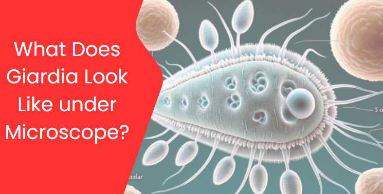
    Giardia is a microscopic parasite that causes a diarrheal illness known as giardiasis in humans. The organism exists in two forms: a motile, pear-shaped trophozoite and a non-motile, oval-shaped cyst. When examining Giardia under a microscope, you would typically observe the trophozoite and cyst stages. Here’s a brief description of each:
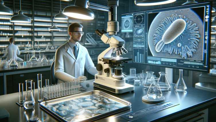
    1. Trophozoite Stage:
      • Shape: The trophozoite is pear-shaped or teardrop-shaped.
      • Size: It is relatively large for a single-celled organism, measuring about 10 to 20 micrometers in length.
      • Features: The trophozoite has a characteristic appearance with a pair of nuclei that are visible under the microscope. The organism is flagellated, meaning it has hair-like structures called flagella that it uses for movement.
    2. Cyst Stage:
      • Shape: The cyst is oval or round.
      • Size: It is smaller than the trophozoite, typically around 8 to 12 micrometers in diameter.
      • Features: The cyst is the dormant, resistant form of Giardia. It has a protective outer shell that allows it to survive outside the host in harsh conditions. Inside the cyst, you can find the infective structures that, when ingested, can cause infection.
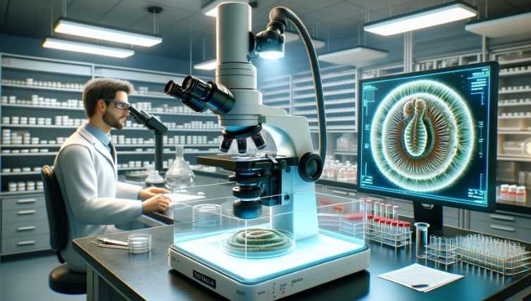
    When examining a sample under the microscope, special staining techniques are often used to enhance the visibility of Giardia. One commonly used staining method is the trichrome stain, which helps highlight the characteristic features of the trophozoite and cyst stages.

    Key Characteristics of Giardia under Microscope:

    1. Shape: Giardia trophozoites, the active and feeding form of the parasite, typically have a pear or teardrop shape. They are approximately 10 to 20 micrometers in length and 5 to 15 micrometers in width.
    2. Nuclei: Giardia trophozoites have two distinct nuclei, which are often visible under high magnification. The nuclei are positioned close to the center of the organism.
    3. Flagella: Giardia possesses flagella, whip-like appendages that extend from the body. These flagella are used for movement and contribute to the parasite’s distinctive appearance. There are four pairs of flagella: two anterior, two lateral, one caudal, and one ventral.
    4. Attachment Disk: The ventral side of Giardia trophozoites contains an adhesive structure called the ventral disk, which helps the parasite attach to the intestinal lining.
    5. Cysts: In addition to the trophozoite form, Giardia can also exist in a cyst form, which is a dormant and more resistant stage. Cysts are typically round and have a thick, protective wall. The cyst form is responsible for the transmission of Giardia between hosts.

    When diagnosing giardiasis, stool samples are often examined under a microscope to detect the presence of Giardia trophozoites or cysts. The appearance of Giardia under the microscope can vary slightly, but the characteristics mentioned above are typical for this parasitic organism. Keep in mind that the exact details may vary based on the staining methods used and the specific conditions of the microscope examination.

    Giardia: A Microscopic Perspective

    Giardia, a microscopic protozoan parasite, belongs to the genus Giardia and falls under the family Giardiidae. This classification places it among diplomonads, highlighting its unique biological features. Understanding the general characteristics of Giardia is crucial for effective microscopic observation and comprehensive knowledge of its behavior.

    Table: General Characteristics of Giardia

    CharacteristicDetails
    Classification and TaxonomyGenus: Giardia; Family: Giardiidae
    Habitat and PrevalenceIntestinal tracts of humans and animals; Global prevalence, especially in areas with poor sanitation and water treatment

    Classification and Taxonomy

    Giardia’s taxonomic classification places it within the genus Giardia, highlighting its distinct biological characteristics. The family Giardiidae further categorizes it among diplomonads, showcasing its evolutionary relationships within the microbial world.

    Habitat and Prevalence

    Giardia predominantly inhabits the intestinal tracts of humans and various animals. Its prevalence is notable on a global scale, with a higher incidence observed in regions characterized by inadequate sanitation and water treatment. This distribution emphasizes the relevance of understanding Giardia’s general characteristics for global health considerations.

    Lifecycle of Giardia

    Giardia’s lifecycle is a fascinating process involving two main stages: trophozoites and cysts. This intricate life cycle plays a pivotal role in the transmission and infection dynamics of Giardia.

    Table: Lifecycle of Giardia

    StageDescription
    Trophozoite StageActive, feeding stage; pear-shaped with flagella and adhesive discs; resides in the small intestine of the host
    Cyst StageInactive, survival stage; oval-shaped with a protective cyst wall; formed as a response to harsh environmental conditions

    Trophozoite and Cyst Stages

    1. Trophozoite Stage: This is the active, feeding stage of Giardia. Trophozoites are pear-shaped, measuring approximately 10-20 micrometers in length. They possess flagella and adhesive discs, aiding in attachment to the host’s intestinal wall.
    2. Cyst Stage: The cyst stage is an inactive, survival form of Giardia. Cysts are oval-shaped and exhibit a robust cyst wall, providing protection against harsh environmental conditions. Cysts are formed as a response to factors like dehydration, facilitating transmission between hosts.

    Transmission and Infection

    The transmission of Giardia primarily occurs through the ingestion of cysts, which are resistant to environmental challenges. Once ingested, cysts release trophozoites in the host’s small intestine, leading to infection. Understanding these stages is vital for developing effective strategies for prevention, diagnosis, and treatment of Giardia infections.

    Microscopic Techniques for Giardia Observation

    Microscopic observation of Giardia requires a careful and systematic approach to ensure accurate identification and analysis. This section explores the essential techniques involved in observing Giardia under a microscope, encompassing sample collection, preparation, staining methods, and the overall importance of employing proper microscopy techniques.

    A. Sample Collection and Preparation

    Table: Sample Collection and Preparation

    TechniqueDetails
    Sample CollectionFecal samples are commonly collected for Giardia observation, ensuring representation of the intestinal environment.
    Sample PreservationImmediate fixation or refrigeration helps prevent deterioration of the sample, preserving the integrity of Giardia cysts and trophozoites.
    Concentration TechniquesCentrifugation or sedimentation may be employed to concentrate parasites, enhancing their visibility during microscopy.

    Proper sample collection and preparation are foundational steps in the microscopic observation of Giardia. Fecal samples, often the primary source, should be collected meticulously to ensure representative specimens. Immediate fixation or refrigeration of samples is crucial to prevent degradation and maintain the viability of Giardia cysts and trophozoites. Additionally, concentration techniques such as centrifugation help enhance the concentration of parasites, facilitating more accurate observations under the microscope.

    B. Staining Methods for Enhanced Visibility

    Table: Staining Methods for Giardia Observation

    Staining MethodDescription
    Direct Wet MountInvolves placing a fresh sample directly on a microscope slide with a cover slip; provides a quick observation of motile trophozoites.
    Modified Iron-Hematoxylin StainingUtilizes a staining solution containing iron and hematoxylin to enhance contrast and visibility of Giardia cysts and trophozoites.
    Immunofluorescence StainingUtilizes specific antibodies labeled with fluorescent dyes to target Giardia antigens, allowing for highly specific and sensitive detection under fluorescence microscopy.
    1. Direct Wet Mount: This technique offers a rapid observation method by placing a fresh sample directly on a microscope slide with a cover slip. It allows for the visualization of motile trophozoites, providing quick insights into Giardia activity.
    2. Modified Iron-Hematoxylin Staining: In this method, a staining solution containing iron and hematoxylin is used to enhance the contrast and visibility of Giardia cysts and trophozoites. This staining technique improves the clarity of cellular structures for more detailed microscopic examination.
    3. Immunofluorescence Staining: Immunofluorescence staining employs specific antibodies labeled with fluorescent dyes. This highly targeted approach allows for the specific and sensitive detection of Giardia antigens under fluorescence microscopy. Immunofluorescence staining is particularly valuable for enhancing specificity in identifying Giardia.

    C. Importance of Proper Microscopy Techniques

    The success of Giardia observation hinges on employing proper microscopy techniques.

    Table: Importance of Proper Microscopy Techniques

    AspectDetails
    Accuracy in IdentificationProper techniques enhance accuracy in identifying Giardia cysts and trophozoites, reducing the risk of misdiagnosis.
    Timely DiagnosisEfficient microscopy techniques contribute to timely diagnosis, enabling prompt initiation of appropriate treatment for giardiasis.
    Research AdvancementsContinuous refinement of microscopy techniques supports ongoing research, leading to advancements in our understanding of Giardia and related diseases.

    Proper microscopy techniques are paramount for accurate identification and timely diagnosis of Giardia. The use of accurate methods ensures precision in differentiating Giardia from other microorganisms, reducing the likelihood of misdiagnosis. Additionally, these techniques contribute to ongoing research advancements, fostering a deeper understanding of Giardia and its impact on human health.

    Microscopic Techniques for Giardia Observation

    Microscopic observation of Giardia is a meticulous process that involves specific techniques for sample collection, preparation, and staining to enhance visibility. These techniques are crucial for accurate identification, aiding in the diagnosis and understanding of Giardia-related diseases.

    A. Sample Collection and Preparation

    Sample Collection and Preparation Table

    TechniqueDetails
    Fecal Sample CollectionCollect fecal samples meticulously to ensure a representative specimen.
    Sample PreservationImmediately fix or refrigerate samples to prevent degradation and maintain viability.
    Concentration TechniquesUtilize centrifugation or sedimentation to enhance the concentration of parasites.

    Proper sample collection is fundamental for successful Giardia observation. Fecal samples, commonly used for this purpose, should be collected carefully to ensure they represent the intestinal environment accurately. Immediate fixation or refrigeration of samples is essential to prevent degradation, preserving the integrity of Giardia cysts and trophozoites. Concentration techniques such as centrifugation enhance the visibility of parasites under the microscope.

    B. Staining Methods for Enhanced Visibility

    Staining Methods Table

    Staining MethodDescription
    Direct Wet MountPlace a fresh sample directly on a microscope slide with a cover slip for a quick observation of motile trophozoites.
    Modified Iron-Hematoxylin StainingUse a staining solution containing iron and hematoxylin to enhance contrast and visibility of Giardia cysts and trophozoites.
    Immunofluorescence StainingUtilize specific antibodies labeled with fluorescent dyes to target Giardia antigens, allowing for highly specific and sensitive detection under fluorescence microscopy.
    1. Direct Wet Mount: This technique involves placing a fresh sample directly on a microscope slide with a cover slip. It offers a rapid observation method, allowing for the visualization of motile trophozoites and providing quick insights into Giardia activity.
    2. Modified Iron-Hematoxylin Staining: This method employs a staining solution containing iron and hematoxylin to enhance the contrast and visibility of Giardia cysts and trophozoites. The staining improves the clarity of cellular structures for more detailed microscopic examination.
    3. Immunofluorescence Staining: This technique uses specific antibodies labeled with fluorescent dyes. It allows for the specific and sensitive detection of Giardia antigens under fluorescence microscopy, enhancing specificity in identifying Giardia.

    C. Importance of Proper Microscopy Techniques

    Importance of Proper Microscopy Techniques Table

    AspectDetails
    Accuracy in IdentificationProper techniques enhance accuracy in identifying Giardia cysts and trophozoites, reducing the risk of misdiagnosis.
    Timely DiagnosisEfficient microscopy techniques contribute to timely diagnosis, enabling prompt initiation of appropriate treatment for giardiasis.
    Research AdvancementsContinuous refinement of microscopy techniques supports ongoing research, leading to advancements in our understanding of Giardia and related diseases.

    Proper microscopy techniques play a pivotal role in the accurate identification of Giardia. These techniques contribute to reducing the risk of misdiagnosis by enhancing accuracy in differentiating Giardia from other microorganisms. Timely diagnosis is facilitated through efficient microscopy techniques, enabling the prompt initiation of appropriate treatment for giardiasis.

    What Does Giardia Look Like?

    A. Detailed Description of Giardia Morphology

    Understanding the detailed morphology of Giardia is essential for accurate identification under a microscope. Giardia exists in two primary forms: trophozoites and cysts.

    1. Trophozoite Appearance

    a. Size and Shape

    Trophozoites, the active and feeding stage of Giardia, typically measure between 10-20 micrometers in length. Their pear-shaped bodies are easily distinguishable, and this size range allows for efficient movement within the host’s small intestine.

    b. Flagella and Adhesive Discs

    Giardia trophozoites exhibit characteristic flagella—hair-like structures that protrude from the body. These flagella play a crucial role in the motility of the parasite. Additionally, adhesive discs located at the anterior end of the trophozoite aid in attachment to the host’s intestinal wall, facilitating colonization.

    2. Cyst Characteristics

    a. Wall Structure

    Giardia cysts represent the dormant, survival stage of the parasite. They possess a resilient cyst wall that provides protection against environmental challenges. This cyst wall is essential for the transmission of Giardia between hosts.

    b. Size and Shape

    Cysts are typically smaller than trophozoites and exhibit an oval shape. Their smaller size contributes to the ease of transmission and dissemination in various environments.

    B. High-Resolution Microscopy Images

    To provide a visual representation of Giardia morphology, high-resolution microscopy images are invaluable. These images offer a closer look at the intricate details of trophozoites and cysts, allowing for a more comprehensive understanding of their structural features.

    High-Resolution Microscopy Images Table

    StageImage Description
    TrophozoitePear-shaped trophozoite with visible flagella and discs.
    CystOval-shaped cyst with a distinct and protective wall.

    C. Comparison with Other Microscopic Organisms

    Giardia exhibits unique features that distinguish it from other microscopic organisms commonly encountered in various environments. A comparative analysis highlights these distinctions.

    Comparison Table

    CharacteristicGiardiaOther Microorganisms
    MotilityFlagella-driven motilityVaried modes of locomotion
    AttachmentAdhesive discs for host attachmentAttachment mechanisms vary widely
    Life CycleAlternation between trophozoite and cyst stagesDiverse life cycles among different organisms
    Size10-20 micrometers (trophozoites)Size ranges widely across microorganisms

    Giardia’s flagella-driven motility, adhesive discs for host attachment, and unique life cycle set it apart from other microscopic organisms. Size variations, attachment mechanisms, and diverse life cycles among different organisms highlight the diversity within the microscopic world.

    Identifying Giardia-Associated Diseases

    A. Giardiasis and Its Symptoms

    Giardiasis, the disease caused by the protozoan parasite Giardia, manifests with a range of symptoms affecting the gastrointestinal system. Recognizing these symptoms is crucial for prompt diagnosis and effective treatment.

    Giardiasis Symptoms Table

    SymptomDescription
    DiarrheaFrequent, loose, and often foul-smelling bowel movements
    Abdominal CrampsIntermittent or continuous discomfort in the abdomen
    NauseaFeeling of queasiness or an urge to vomit
    DehydrationReduced fluid levels in the body due to persistent diarrhea
    Weight LossUnintentional weight loss resulting from malabsorption

    B. Link Between Giardia Morphology and Disease Severity

    The morphology of Giardia plays a significant role in determining the severity of associated diseases. Variations in the appearance of trophozoites under microscopic observation may correlate with the intensity of infection and clinical symptoms.

    Understanding Giardia morphology allows healthcare professionals to assess the potential impact on the patient’s health. For instance, an increased number of trophozoites or specific morphological characteristics may indicate a more severe infection, guiding clinicians in tailoring appropriate treatment strategies.

    C. Importance of Early Detection Through Microscopy

    Early detection of Giardia through microscopy is paramount for several reasons. Microscopic observation allows for the identification of Giardia cysts and trophozoites in clinical samples, confirming the presence of the parasite in the patient’s gastrointestinal tract.

    Importance of Early Detection Table

    AspectDetails
    Prompt TreatmentEarly detection enables timely initiation of specific anti-Giardia medications.
    Prevention of TransmissionIdentifying Giardia early helps implement preventive measures to limit further spread.
    Reduction of Disease SeverityEarly intervention may mitigate the severity of giardiasis, preventing complications.
    Public Health SurveillanceSwift identification supports public health efforts in monitoring and controlling outbreaks.

    Swift identification of Giardia through microscopy facilitates the prompt initiation of specific anti-Giardia medications, reducing the duration and severity of symptoms. Additionally, early detection aids in implementing preventive measures to limit further transmission, protecting both individual patients and the broader community. By reducing disease severity, early intervention can prevent complications associated with giardiasis, contributing to improved patient outcomes.

    3 Tips for Efficient Giardia Observation

    A. Proper Microscope Usage

    Efficient Giardia observation begins with mastering microscope usage. Regular calibration and maintenance ensure optimal performance. Adjusting lighting and focus settings enhances clarity, aiding in the identification of Giardia cysts and trophozoites.

    B. Sample Handling and Preparation Tips

    Meticulous sample handling is crucial. Ensure accurate representation by collecting fecal samples carefully. Immediate fixation or refrigeration prevents sample degradation, preserving Giardia integrity. Utilize concentration techniques like centrifugation for enhanced visibility during microscopy.

    C. Common Challenges and Troubleshooting

    Be prepared to tackle common challenges encountered during Giardia observation. Issues such as debris interference or insufficient staining require troubleshooting. Regularly check equipment and adjust techniques to overcome challenges, ensuring accurate and reliable results in Giardia identification.

    Facts and Statistics

    A. Key Facts about Giardia

    1. Ubiquitous Parasite: Giardia is a ubiquitous protozoan parasite that infects the small intestine of humans and animals, causing giardiasis.
    2. Waterborne Transmission: The primary mode of transmission is through contaminated water sources, emphasizing the importance of water hygiene.
    3. Resilient Cysts: Giardia exists in two stages, with cysts being the dormant, environmentally resistant form, allowing for survival outside a host.

    B. Statistics on Global Prevalence and Incidence

    1. Worldwide Distribution: Giardia has a global presence, affecting both developed and developing countries, with varying degrees of prevalence.
    2. High Incidence in Developing Regions: Developing regions often experience higher incidences due to inadequate sanitation and limited access to clean water.
    3. Common Waterborne Outbreaks: Waterborne outbreaks contribute significantly to the prevalence of giardiasis, emphasizing the importance of clean water initiatives.

    C. Impact on Vulnerable Populations

    1. Children and Immunocompromised Individuals: Giardia infections can have severe consequences for vulnerable populations, particularly children and those with compromised immune systems.
    2. Malnutrition and Growth Impairment: Chronic giardiasis in children may lead to malnutrition and growth impairment, affecting long-term health.
    3. Increased Susceptibility in Immunocompromised Individuals: Immunocompromised individuals, such as those with HIV/AIDS, face a higher risk of prolonged and severe Giardia infections, posing additional health challenges.

    Understanding these facts and statistics is crucial for public health initiatives, emphasizing the need for global efforts in sanitation, clean water provision, and targeted interventions to protect vulnerable populations.

    Final Words

    In exploring Giardia under a microscope, we unveiled its distinct trophozoite and cyst stages, each with unique morphological features. High-resolution microscopy images provided a visual journey into the microscopic world of Giardia, showcasing its pear-shaped trophozoites and oval-shaped cysts.

    Microscopic studies are pivotal in deciphering Giardia-related diseases. Accurate identification through microscopy guides effective treatment, early detection prevents transmission, and ongoing research enhances our understanding. The microscope remains a powerful tool in unraveling the mysteries of Giardia, contributing significantly to public health efforts worldwide.

    Resources and References

    A. Recommended Textbooks and Journals

    1. Textbooks:
      • “Parasitic Diseases” by Despommier et al.
      • “Medical Microbiology” by Murray et al.
    2. Journals:
      • “Journal of Parasitology”
      • “Clinical Microbiology Reviews”

    B. Online Resources and Databases

    1. Websites:
    2. Databases:

    C. Citations for Scientific Studies and Research Papers

    1. Scientific Studies:
      • Thompson RCA, Hopkins RM, Homan WL. “Nomenclature and genetic groupings of Giardia infecting mammals.” Parasitology Today, 2000.
    2. Research Papers:
      • Adam RD. “Biology of Giardia lamblia.” Clinical Microbiology Reviews, 2001.

    These resources provide a comprehensive foundation for studying Giardia, from authoritative textbooks and journals to online databases and key scientific studies.

  • Are Electron Microscope Images Coloured? Unveiling the Truth!

    Are Electron Microscope Images Coloured? Unveiling the Truth!


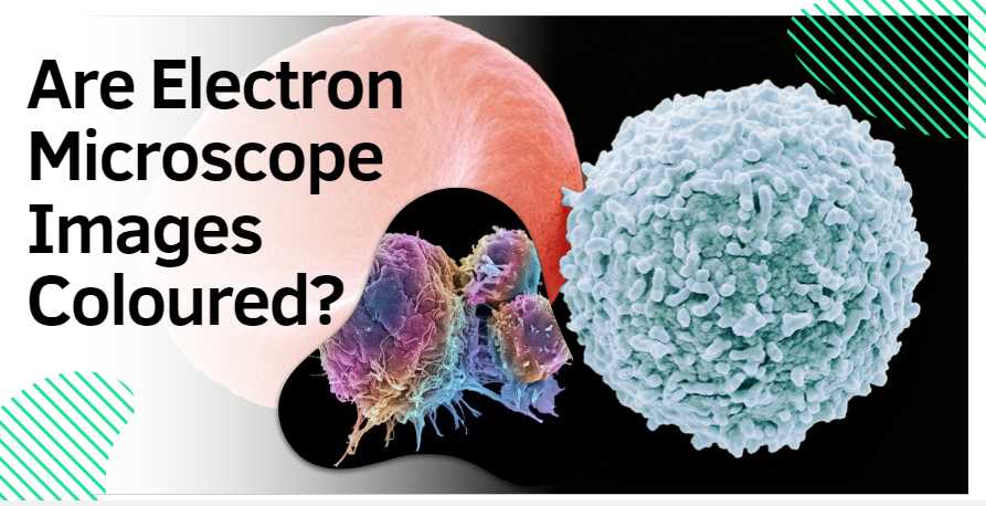
    No, electron microscope images are not inherently colored. By default, these images are captured in black and white, reflecting the monochromatic nature of the electron signals used in imaging. The grayscale representation offers high contrast and detailed views of microscopic structures. However, scientists often apply color to these images through pseudocoloring techniques for specific purposes. Pseudocoloring helps highlight distinct features or materials within the specimen, aiding in the interpretation and communication of scientific findings. The choice of colors is strategic and does not signify inherent colors present in the specimens. While artistic renderings and false coloration techniques exist, the core of electron microscope imaging lies in its ability to provide precise, high-resolution, and uncolored representations of the microscopic world.

    The Basics of Electron Microscopy

    Delving into the microscopic universe, electron microscopy serves as a transformative tool, revealing intricacies beyond the reach of traditional optical microscopes. Understanding the fundamentals of electron microscopy is essential to appreciate its capabilities and applications.

    Types of Electron Microscopes

    Electron microscopes come in diverse types, each tailored to specific scientific needs. The three primary types are Transmission Electron Microscope (TEM), Scanning Electron Microscope (SEM), and Scanning Transmission Electron Microscope (STEM). Each type offers unique advantages, allowing scientists to explore different aspects of specimens in unprecedented detail.

    Types of Electron Microscopes

    Electron Microscope TypeCharacteristics
    Transmission Electron Microscope (TEM)Penetrates thin specimens for detailed internal structures
    Scanning Electron Microscope (SEM)Provides 3D surface images with excellent depth of field
    Scanning Transmission Electron Microscope (STEM)Combines features of TEM and SEM for versatility

    How Electron Microscopes Work

    1. Electron Beam Generation:
      • Electron microscopes use electron guns to generate focused beams of electrons.
      • Magnetic lenses focus these beams onto the specimen.
    2. Interaction with Specimens:
      • When the electron beam strikes the specimen, interactions with atoms generate signals.
    3. Detection of Signals:
      • Detectors capture signals like secondary electrons, backscattered electrons, and transmitted electrons.
    4. Image Formation:
      • Captured signals are translated into high-resolution images, providing detailed views of specimen structures.

    Characteristics of Electron Microscope Images

    Characteristics of Electron Microscope Images

    AspectDescription
    High ResolutionExceptional detail and clarity
    Monochromatic NatureNative images are black and white
    Depth of FieldSEM provides excellent depth perception
    Internal StructuresTEM reveals internal structures in fine detail

    Electron microscope images stand out for their high resolution, monochromatic default state, and the ability to unveil internal structures with precision.

    Advantages and Limitations

    • Advantages:
      • High resolution for detailed imaging.
      • Capability to explore internal structures.
      • SEM excels in revealing fine surface details.
    • Limitations:
      • Meticulous sample preparation is essential.
      • Vacuum conditions are required.
      • Artefacts may be introduced during sample processing.

    Innovations in Electron Microscopy

    Continual innovations enhance electron microscopy’s capabilities:

    • Cryo-Electron Microscopy (Cryo-EM):
      • Allows imaging of biological specimens in their native state.
      • Minimizes damage from sample preparation.
    • Correlative Light and Electron Microscopy (CLEM):
      • Integrates data from light and electron microscopy for comprehensive analysis.
      • Bridges information across macro and micro scales.

    Understanding these basics sets the stage for exploring the colorful and captivating world of electron microscope images, debunking misconceptions and appreciating the intersection of science and art at the microscopic level.

    Black and White: The Native State of Electron Microscope Images

    Contrary to popular belief, the native state of electron microscope images is monochromatic, devoid of the vibrant hues that often characterize other imaging techniques. Electron microscopy relies on the interaction of electron beams with specimens and the subsequent capture of signals emitted during this interaction. The result is a raw, high-resolution image that is inherently black and white.

    Characteristics of Native Electron Microscope Images

    AspectDescription
    Monochromatic NatureLack of inherent color in native images
    Electron Signal InteractionDetectors capturing electron signals
    Raw RepresentationUnaltered depiction of specimen structures

    The monochromatic nature of these images arises from the fundamental principles of electron microscopy. Electron beams, focused onto the specimen, interact with the atoms present. Detectors capture the signals generated by this interaction, forming an image that faithfully represents the structural details without the introduction of artificial colors.

    These native black and white images offer a stark beauty, emphasizing the intricate details of the microscopic world. The absence of color allows scientists to focus on the structural information, discerning the fine nuances within cells, materials, or any specimen under examination.

    Appreciating the native state of electron microscope images is crucial for accurate interpretation and communication of scientific findings. While the absence of color might seem surprising to those new to the field, it underscores the precision and objectivity inherent in electron microscopy, where the focus is on revealing the true nature of microscopic structures. As we delve deeper into the world of electron microscopy, it becomes evident that the beauty lies not in the colors that adorn the images but in the rich tapestry of details that monochromatic images unveil.

    Pseudocoloring in Electron Microscopy

    In the realm of electron microscopy, the quest to unveil intricate details often extends beyond the limitations of black and white images. Pseudocoloring emerges as a powerful technique, enabling scientists to enhance visibility, highlight specific features, and convey additional information in electron microscope images.

    Pseudocoloring involves the deliberate assignment of colors to different parts of an image, creating a visually enriched representation without altering the inherent grayscale nature of electron microscope images. This technique is particularly valuable when researchers seek to emphasize specific elements within a specimen.

    The application of pseudocoloring is not arbitrary; instead, it adheres to scientific principles. By assigning distinct colors to different structures or materials, scientists can create a more intuitive visual interpretation of the microscopic world. For instance, in a biological sample, cell membranes may be pseudocolored in one hue, while organelles or other structures are represented in contrasting colors.

    Pseudocoloring Techniques

    TechniquePurpose
    Heatmap PseudocoloringEmphasizes variations in temperature or intensity
    Rainbow PseudocoloringEnhances contrast by assigning colors across the spectrum
    Grayscale with Color HighlightsRetains black and white base while accentuating specific details

    Heatmap pseudocoloring is often employed to visualize variations in temperature or intensity within a specimen, providing a gradient of colors that intuitively represent different levels. Rainbow pseudocoloring, on the other hand, enhances contrast by assigning colors across the visible spectrum, making subtle differences more apparent.

    Pseudocoloring is not limited to scientific utility; it also serves an essential communicative role. By introducing colors, researchers can create images that are not only scientifically informative but also visually engaging. This aids in effective communication of complex findings to diverse audiences, from fellow scientists to students and the general public.

    While pseudocoloring undeniably enhances the interpretability of electron microscope images, researchers must use it judiciously. The colors assigned should accurately reflect the scientific context, ensuring that the visual representation aligns with the underlying data. In navigating the microscopic landscape with pseudocoloring, scientists embark on a journey where art and science converge, bringing forth a richer understanding of the hidden world magnified by electron microscopy.

    False Coloration and Artistic Rendering in Electron Microscopy

    Beyond the realm of pseudocoloring, electron microscopy offers another layer of visual complexity through techniques such as false coloration and artistic rendering. These approaches elevate scientific images beyond mere representations, transforming them into captivating works that marry the precision of science with the aesthetics of art.

    False Coloration:

    False coloration involves the intentional application of colors to an image, departing from the natural grayscale representation. Unlike pseudocoloring, where colors are assigned based on scientific principles, false coloration allows for more subjective choices. This technique is commonly employed to emphasize specific details or highlight different elements within a specimen.

    False coloration often serves to enhance contrast and clarity, making subtle structures more discernible to the human eye. For example, in biological samples, false coloration may be used to distinguish between different cell types or to highlight areas of interest such as cellular organelles.

    Artistic Rendering:

    Artistic rendering takes electron microscope images to a different realm, where scientific visualization meets creative expression. While the primary goal of scientific imaging is to accurately represent the specimen, artistic rendering allows for a departure from strict realism. This technique involves applying artistic elements such as shading, texture, and even unconventional colors to create visually striking representations.

    Examples of Artistic Rendering:

    1. Neuronal Connections:
      • False coloration is used to distinguish between neural pathways, creating a visually appealing representation of the intricate connections within the brain.
    2. Cellular Landscape:
      • Artistic rendering transforms a typical cellular landscape into a visually engaging masterpiece, where scientific accuracy converges with creative expression.

    Artistic rendering not only produces aesthetically pleasing images but also invites viewers to appreciate the beauty inherent in the microscopic world. It bridges the gap between science and art, making complex scientific concepts more accessible to a broader audience.

    Both false coloration and artistic rendering contribute to the narrative of scientific discovery. They go beyond the mere documentation of structures and delve into the realm of visual storytelling. These techniques play a crucial role in communicating the wonder of the microscopic universe, fostering a deeper appreciation for the beauty concealed within the seemingly mundane world magnified by electron microscopy.

    Factors Influencing Coloration in Electron Microscopy

    The introduction of color to electron microscope images is a deliberate process influenced by various factors. Understanding these factors is crucial for accurately interpreting the visual information conveyed through the vibrant hues that adorn the microscopic world.

    Specimen Composition:

    The materials constituting the specimen play a pivotal role in influencing coloration. Different substances interact with electrons in distinctive ways, leading to variations in the colors observed. For instance, biological specimens may exhibit different colors based on the composition of cellular structures or the presence of specific molecules.

    Imaging Techniques:

    The choice of imaging techniques in electron microscopy can significantly impact color representation. Different techniques, such as transmission electron microscopy (TEM) and scanning electron microscopy (SEM), may produce varied color schemes. The interaction of electrons with specimens and the subsequent detection methods contribute to the diversity of colors observed in the final images.

    Post-processing:

    The coloration of electron microscope images is not solely determined during the imaging process but can be further modified during post-processing. Adjustments in contrast, brightness, and color balance can influence the final appearance of the image. Researchers often fine-tune these parameters to enhance the visual clarity and emphasize specific features within the specimen.

    Factors Influencing Coloration

    FactorInfluence on Color Representation
    Specimen CompositionDifferent materials may exhibit distinct colors
    Imaging TechniquesVaried techniques may result in different color schemes
    Post-processingAdjustment of contrast and brightness impacts color

    Navigating the intricacies of coloration in electron microscopy requires a nuanced understanding of these factors. The interplay between specimen composition, imaging techniques, and post-processing intricacies contributes to the rich palette of colors that brings the microscopic world to life. As scientists continue to refine their methods, the resulting images become not only scientific tools but also visual masterpieces that capture the essence of the microscopic realm in vibrant detail.

    Common Misconceptions about Color in Electron Microscopy

    Misconceptions often surround the representation of color in electron microscope images, stemming from assumptions and unfamiliarity with the underlying principles. Let’s address and debunk some of these common misconceptions:

    Debunked Misconceptions

    MisconceptionClarification
    Inherent Color in Electron ImagesElectron microscope images are naturally black and white
    Unrestricted PseudocoloringPseudocoloring is deliberate and guided by scientific principles
    1. Inherent Color in Electron Images:
      • Misconception: Assuming that electron microscope images inherently possess color.
      • Clarification: Native electron microscope images are monochromatic, capturing the grayscale nuances of specimen structures. Color is introduced through deliberate techniques.
    2. Unrestricted Pseudocoloring:
      • Misconception: Believing that pseudocoloring involves arbitrary assignment of colors.
      • Clarification: Pseudocoloring follows scientific principles, with colors assigned purposefully to enhance visibility and convey specific information without distorting the underlying data.

    Understanding these misconceptions is essential for appreciating the accuracy and intentionality involved in the coloration of electron microscope images. By dispelling these myths, we foster a more informed interpretation of the mesmerizing visuals that emerge from the microscopic world.

    6 Tips for Interpreting Electron Microscope Images

    Interpreting electron microscope images requires a nuanced approach to fully grasp the intricate details captured at the microscopic level. Here are six tips to enhance your understanding:

    1. Recognize Pseudocoloring:
      • Be aware that colors introduced to electron microscope images through pseudocoloring are intentional and serve a specific purpose. Understanding this technique aids in accurate interpretation.
    2. Understand the Native State of Images:
      • Acknowledge that native electron microscope images are monochromatic. This awareness helps in distinguishing between inherent features and those highlighted through coloration techniques.
    3. Context Matters:
      • Consider the scientific context in which the images are presented. Colors may be used to emphasize specific structures or materials, providing vital clues to the researcher’s intended focus.
    4. Be Mindful of Artistic Rendering:
      • Understand the distinction between scientifically accurate representation and artistic rendering. While the former is based on data, the latter introduces creative elements for visual appeal.
    5. Consider Specimen Composition:
      • Different materials within a specimen may naturally exhibit distinct colors. Take into account the inherent properties of the materials being imaged for a more accurate interpretation.
    6. Stay Informed About Imaging Techniques:
      • Different electron microscopy techniques may produce varied color schemes. Familiarize yourself with the imaging method used to capture the images, as this influences the visual outcome.

    3 Major Challenges in Electron Microscopy Imaging

    While electron microscopy has revolutionized our ability to visualize the microscopic world, it is not without its challenges. Three major hurdles persist in the field:

    1. Color Representation:
      • The debate over standardized colorization in scientific imaging remains ongoing. Establishing consistent color representation is challenging, as different labs may employ varied methods, impacting the reproducibility and comparability of results.
    2. Technological Advancements:
      • Despite continuous innovations, electron microscopy faces technological limitations. Improving spatial resolution, reducing specimen damage during imaging, and enhancing imaging speed are ongoing challenges that researchers strive to overcome.
    3. Standardization in Electron Microscopy:
      • The field lacks standardized protocols for colorization and imaging procedures. The absence of uniform standards hinders collaboration and makes it challenging to establish a universal language for interpreting electron microscope images. Efforts to standardize practices are underway but remain a complex endeavor.

    Do Electron Microscope Images Have Inherent Color?

    Electron microscope images are inherently monochromatic. The default representation is black and white, devoid of inherent color. This monochromatic nature arises from the fundamental principles of electron microscopy, where electron beams interact with specimens to create high-resolution images captured in grayscale.

    How are Colors Introduced to Electron Microscope Images?

    Colors are introduced through deliberate processes such as pseudocoloring and false coloration. Pseudocoloring involves assigning colors to specific features, enhancing visibility without altering the native grayscale. False coloration, on the other hand, allows for more subjective color choices, often used to highlight structures or create visually engaging representations.

    Is Pseudocoloring Arbitrary, or Does it Follow Scientific Principles?

    Pseudocoloring is not arbitrary; it follows scientific principles. Colors are assigned purposefully to enhance the interpretation of specific features within a specimen. This intentional approach ensures that the coloration aligns with the scientific context and aids accurate interpretation.

    Can Electron Microscope Images Be in True Color?

    True color, as perceived by the human eye, is not present in electron microscope images. These images are formed by capturing electron signals, resulting in a monochromatic representation. The introduction of color is a deliberate and strategic process aimed at enhancing scientific interpretation.

    What is the Difference Between Pseudocoloring and False Coloration?

    Pseudocoloring and false coloration both involve introducing color to electron microscope images, but they differ in their approach. Pseudocoloring follows scientific principles, assigning colors deliberately, while false coloration allows for more subjective and artistic color choices to enhance contrast and visual appeal.

    Are There Different Techniques for Pseudocoloring?

    Various techniques are employed for pseudocoloring in electron microscopy. Heatmap pseudocoloring emphasizes variations in temperature or intensity. Rainbow pseudocoloring enhances contrast with a spectrum of colors, and grayscale with color highlights retains a black and white base while accentuating specific details. These techniques cater to different imaging needs and objectives.

    Why Use Coloration in Electron Microscopy?

    Coloration in electron microscopy serves multiple purposes. It enhances visibility, making specific features more discernible. It allows for the conveyance of additional information, aiding in the interpretation of complex structures. Moreover, coloration adds a visual dimension to microscopic images, making them more accessible and engaging for a broader audience.

    What is the Role of Artistic Rendering in Electron Microscopy?

    Artistic rendering in electron microscopy involves creative embellishments to scientific images. While scientific accuracy remains crucial, artistic rendering adds aesthetic elements, such as shading and texture, contributing to visually striking representations. This approach bridges the gap between science and art, making microscopic imagery more captivating.

    Are Electron Microscope Images Ever in True Color?

    True color, as perceived by the human eye, is not inherent in electron microscope images. The introduction of color is a deliberate process, whether through pseudocoloring, false coloration, or other techniques. This deliberate approach ensures that the colors used in the images serve a scientific purpose, aligning with the researcher’s objectives.

    What Challenges Exist in Representing Color in Electron Microscopy?

    Challenges in representing color in electron microscopy include debates on standardized colorization, technological limitations, and the lack of universal standards. Achieving consistency in color representation remains a complex task, requiring ongoing efforts to enhance imaging technology and establish standardized practices for accurate and reproducible results.

    Final Words

    As we conclude our journey into the world of electron microscope images, the truth becomes evident—electron microscope images are not inherently colored. Instead, deliberate processes such as pseudocoloring and artistic rendering bring these microscopic wonders to life. Armed with newfound knowledge, we can appreciate the marriage of science and art that defines the captivating images revealing the hidden beauty of the microscopic realm.

    Resources and References

    For those eager to delve deeper into the realm of electron microscopy, the following resources provide a wealth of knowledge and insight:

    1. “Principles of Electron Optics: Applied Geometrical Optics” by Peter W. Hawkes
      • This comprehensive book delves into the principles of electron optics, providing a solid foundation for understanding the intricacies of electron microscopy.
    2. “Introduction to Electron Microscopy” by John J. Bozzola and Lonnie D. Russell
      • An excellent introductory text covering the fundamentals of electron microscopy, from basic principles to advanced techniques.
    3. “Cryo-EM: A Unique Tool for the Visualization of Macromolecular Complexity” by Sriram Subramaniam
      • This seminal article, published in Nature, explores the application of cryo-electron microscopy in visualizing macromolecular structures.
    4. “Current Trends in SPM Instrumentation for Nanoscale Imaging and Spectroscopy” by Andrea Schwartz and Enrico Gnecco
      • A valuable resource on scanning probe microscopy, offering insights into nanoscale imaging and spectroscopy techniques.

    These references, coupled with the firsthand experiences shared in this article, offer a comprehensive understanding of the intricacies surrounding the coloration of electron microscope images. Whether you are a novice or an experienced researcher, these resources provide valuable insights into the evolving field of electron microscopy.

  • What Does Skeletal Muscle Look Like under Microscope: A Microscopic Marvel

    What Does Skeletal Muscle Look Like under Microscope: A Microscopic Marvel

    Observing skeletal muscle under a microscope unveils a mesmerizing world of intricate structures and dynamic interactions. The detailed examination of muscle tissue provides insights into its composition, organization, and functional elements. Here’s a closer look at what skeletal muscle looks like under a microscope:

    1. Muscle Fibers:

      • At the cellular level, skeletal muscle is composed of long, cylindrical cells known as muscle fibers. These fibers exhibit a striated appearance under the microscope due to the orderly arrangement of contractile units called sarcomeres.
    2. Sarcomeres:

      • The sarcomere is the fundamental unit of muscle contraction and relaxation. When viewed under high magnification, sarcomeres reveal alternating dark and light bands. The dark bands, called A bands, consist of overlapping myosin and actin filaments, while the light bands, called I bands, contain actin filaments.
    3. Connective Tissue:

      • Delicate layers of connective tissue, such as endomysium, perimysium, and epimysium, surround and support the muscle fibers. Under the microscope, these structures appear as thin, fibrous layers intertwining with the muscle fibers.
    4. Neuromuscular Junctions:

      • Microscopic examination also unveils neuromuscular junctions, where nerve endings communicate with muscle fibers. This critical point of contact is essential for transmitting signals that initiate muscle contraction.
    5. Fiber Types:

      • Different fiber types, such as slow-twitch (Type I) and fast-twitch (Type II) fibers, exhibit distinctive microscopic features. Slow-twitch fibers appear darker due to higher myoglobin content, while fast-twitch fibers appear lighter with fewer mitochondria.
    6. Mitochondria and Organelles:

      • Electron microscopy reveals the subcellular details, including the abundance of mitochondria. These organelles, often referred to as the powerhouse of the cell, are responsible for energy production and play a crucial role in muscle function.
    7. Blood Vessels:

      • Microscopic examination allows for the visualization of blood vessels within the muscle tissue. The network of capillaries ensures oxygen and nutrient supply to the muscle fibers, supporting their metabolic needs.
    8. Adaptations to Exercise:

      • Through microscopic analysis, researchers can observe adaptations in muscle microstructure in response to exercise. These adaptations include changes in fiber size, increased capillarity, and alterations in the distribution of mitochondria.

    In essence, exploring skeletal muscle under a microscope is a journey into the marvels of cellular architecture. The nuanced details revealed through microscopic examination contribute to our understanding of muscle physiology, pathology, and the adaptive responses that shape this remarkable tissue.

    Basics of Skeletal Muscle

    As I journeyed into the microscopic world of skeletal muscle, the foundational understanding of these intricate structures became the cornerstone of my exploration.

    Composition of Skeletal Muscle Tissue

    At the core of skeletal muscles lies a remarkable composition of muscle fibers, connective tissue, blood vessels, and nerves. This symphony of elements orchestrates the complex dance of voluntary movements.

    Muscle Fibers

    Muscle fibers, elongated cells responsible for contraction, are the building blocks of skeletal muscle. These fibers house the machinery for muscle function, including myofibrils, the contractile units within each cell.

    Connective Tissue

    Weaving through the muscle fibers are layers of connective tissue that provide structural support. The endomysium, a delicate layer, envelops individual muscle fibers. Perimysium surrounds bundles of muscle fibers, forming fascicles. Lastly, the robust epimysium encases the entire muscle, ensuring cohesion and protection.

    Voluntary Muscle Control

    One of the captivating aspects revealed under the microscope is the voluntary nature of skeletal muscles. Unlike smooth muscles, which function involuntarily, skeletal muscles respond to conscious commands. This voluntary control is made possible by the intricate network of nerves intertwining with muscle fibers.

    Microscopic Features

    Sarcomeres

    The microscopic marvels known as sarcomeres define the functional units of skeletal muscles. Comprising thin actin and thick myosin filaments, these repeating structures dictate muscle contraction. The alternating dark and light bands visible under the microscope are a testament to the precise organization of sarcomeres.

    Neuromuscular Junctions

    Zooming in further, the microscopic theater unveils neuromuscular junctions. These specialized points of contact between nerve endings and muscle fibers play a pivotal role in transmitting signals for muscle contraction. Witnessing this microscopic dialogue adds a layer of appreciation for the intricate communication within our bodies.

    Fiber Types and Characteristics

    Fiber Type Contraction Speed Fatigue Resistance Key Function
    Slow-twitch (Type I) Slow High Endurance and Sustained Activity
    Fast-twitch (Type II) Fast Low Rapid, Powerful Contractions

    Understanding these fiber types, each with its unique characteristics, provides insights into the diverse roles that skeletal muscles play in our daily activities.

    In essence, the basics of skeletal muscle, when unraveled under the microscope, showcase the harmony of structural elements and the marvel of voluntary control. This foundational knowledge sets the stage for a deeper exploration of the microscopic intricacies within our muscles.

    Microscopic Techniques for Muscle Examination

    Diving into the microscopic realm of skeletal muscle, I found myself captivated by the array of techniques that unveil the intricate details hidden within muscle tissues. These techniques, ranging from traditional histology to advanced electron microscopy, are the lenses through which we gain insight into the microscopic landscape of our muscles.

    Traditional Histology

    Staining Methods

    Histology, the study of tissues, is the bedrock of microscopic examination. Staining methods enhance the visibility of cellular structures, making them stand out under the microscope. H&E (hematoxylin and eosin) staining, a standard in histology, reveals the general architecture of muscle tissues, distinguishing cell nuclei and cytoplasm.

    Immunohistochemistry

    For a more targeted exploration, immunohistochemistry comes into play. This technique employs antibodies to detect specific proteins within the muscle tissue. It allows for the identification of cellular components, such as myosin and actin filaments, enabling a more detailed analysis of the molecular makeup.

    Electron Microscopy

    Transmission Electron Microscopy (TEM)

    Taking the exploration to a finer scale, transmission electron microscopy (TEM) offers unparalleled resolution. This technique allows us to visualize structures at the subcellular level. Under TEM, the sarcomeres, mitochondria, and other organelles reveal their intricate details, providing a deeper understanding of the ultrastructure of skeletal muscle.

    Scanning Electron Microscopy (SEM)

    On the surface level, scanning electron microscopy (SEM) provides a three-dimensional view of the muscle tissue. SEM captures the external features of the tissue, offering insights into the surface morphology of muscle fibers. This technique is particularly valuable for studying the topography of the tissue and the arrangement of fibers.

    Fluorescence Microscopy

    Live Imaging

    Fluorescence microscopy, with its ability to visualize specific molecules labeled with fluorescent dyes, adds a dynamic dimension to the study of muscle tissues. Live imaging allows researchers to observe real-time cellular processes, providing valuable insights into the dynamic behavior of muscle cells during contraction and relaxation.

    Table: Comparison of Microscopic Techniques

    Technique Resolution Key Features
    Traditional Histology Moderate Provides an overview of tissue architecture with basic staining methods.
    Immunohistochemistry Variable Allows for the identification of specific proteins within the muscle tissue.
    Transmission Electron Microscopy High Offers ultrastructural details at the subcellular level.
    Scanning Electron Microscopy High Provides a three-dimensional view of the surface morphology of muscle tissue.
    Fluorescence Microscopy Variable Enables live imaging and visualization of specific molecules within the tissue.

    Practical Tips for Effective Microscopic Examination

    1. Careful Tissue Sectioning: Properly prepared tissue sections are crucial for accurate microscopic observation. Precision in sectioning ensures that the desired structures are visible.

    2. Optimal Staining Techniques: Choosing the right stain is paramount. Different stains highlight specific cellular components, providing clarity in the microscopic image.

    3. Calibration and Maintenance of Equipment: Regular calibration and maintenance of microscopes and imaging equipment are essential for obtaining reliable and consistent results.

    4. Integration of Multiple Techniques: Combining different microscopic techniques enhances the depth of analysis. For instance, integrating immunohistochemistry with electron microscopy provides both molecular and structural insights.

    Variations in Skeletal Muscle Fiber Types

    As my exploration of skeletal muscle continued under the microscope, the diversity within muscle fibers emerged as a captivating aspect of their microscopic landscape. Skeletal muscles, far from being homogenous, exhibit distinct fiber types that influence their function and performance.

    Slow-Twitch (Type I) Fibers

    Under the lens, slow-twitch fibers revealed their characteristic features. These fibers, also known as Type I, contract slowly but sustainably. Their microscopic appearance is marked by a higher density of mitochondria, the cellular powerhouses responsible for energy production. The abundance of mitochondria is a testament to the endurance capabilities of slow-twitch fibers, making them well-suited for activities requiring prolonged effort, such as long-distance running or cycling.

    Fast-Twitch (Type II) Fibers

    In contrast, the fast-twitch fibers, or Type II fibers, presented a different microscopic profile. These fibers contract rapidly but fatigue more quickly. Under the microscope, the distinguishing feature is a lower mitochondrial density compared to slow-twitch fibers. Instead, fast-twitch fibers boast a higher concentration of glycolytic enzymes, emphasizing their role in activities demanding bursts of power, like weightlifting or sprinting.

    Microscopic Variations

    Microscopically, slow-twitch fibers showcase a darker appearance due to the higher myoglobin content, a protein responsible for oxygen storage. This characteristic enhances their ability to utilize oxygen efficiently, contributing to their endurance.

    On the other hand, fast-twitch fibers appear lighter under the microscope due to lower myoglobin content. The reliance on anaerobic metabolism, without significant oxygen utilization, marks these fibers as powerhouses for short bursts of intense activity.

    Practical Implications

    Understanding these microscopic variations in muscle fiber types is not only academically intriguing but also holds practical implications. Athletes and fitness enthusiasts can tailor their training regimens based on the predominant fiber types in their muscles. For example, an endurance athlete may focus on activities that engage and optimize slow-twitch fibers, while a sprinter may prioritize training that targets fast-twitch fibers.

    Adaptations in Response to Training

    Remarkably, the microscopic landscape of muscle fibers is not fixed. Training-induced adaptations can alter the composition of muscle fibers. Endurance training, for instance, may prompt an increase in the number and efficiency of slow-twitch fibers.

    3 Factors Affecting Skeletal Muscle Microstructure

    The microscopic tapestry of skeletal muscle is not static; rather, it weaves a dynamic narrative influenced by various factors that leave their imprint on its microstructure. As I delved into the intricate details under the microscope, it became apparent that several elements shape the landscape of skeletal muscle at the cellular level.

    Exercise and Physical Activity

    Regular physical activity emerged as a prominent factor influencing skeletal muscle microstructure. Through the lens, I observed the effects of exercise on muscle fibers—adaptations such as increased mitochondrial density and altered fiber type composition. The microscopic changes reflected the muscle’s response to the demands placed upon it, showcasing the remarkable plasticity inherent in skeletal muscles.

    Aging and Microscopic Alterations

    Aging, another influential factor, cast its effects on the microscopic landscape of muscle tissue. Microscopic observations unveiled changes such as a decline in muscle mass, alterations in fiber type distribution, and disruptions in the integrity of connective tissue. These age-related modifications underscored the importance of considering the dynamic nature of skeletal muscle microstructure over the lifespan.

    Diseases Impacting Microstructure

    Certain diseases cast a shadow on the microscopic canvas of skeletal muscle. Conditions such as muscular dystrophy, myopathies, and neuromuscular disorders were evident in the alterations observed under the microscope. Microscopic analysis served as a diagnostic tool, unraveling the structural aberrations associated with these conditions and aiding in the understanding of their pathophysiology.

    Practical Tips for Microscopic Observation

    Navigating the microscopic landscape of skeletal muscle requires a keen eye, precision, and a thoughtful approach. As I honed my skills in microscopic observation, several practical tips emerged to ensure accurate and insightful analysis.

    1. Tissue Preparation and Sectioning Techniques:

      • The foundation of successful microscopic observation lies in meticulous tissue preparation. Optimal sectioning techniques ensure that the tissue slices are thin and free from artifacts, allowing for clear and accurate observations. Uniformly sliced sections enhance the visibility of key structures, facilitating a comprehensive analysis of muscle microstructure.
    2. Stain Selection for Enhanced Visibility:

      • The choice of stains significantly influences the clarity of microscopic images. Tailoring stain selection to specific cellular components enhances visibility. For instance, using hematoxylin and eosin (H&E) stains provides a general overview of tissue architecture, while immunohistochemistry allows for the targeted visualization of proteins. Thoughtful stain selection amplifies the microscopic details relevant to the research or diagnostic goals.
    3. Regular Calibration and Maintenance of Equipment:

      • Microscopes and imaging equipment are intricate tools that require regular calibration and maintenance. Ensuring that the equipment is in optimal condition guarantees the accuracy and consistency of microscopic observations. Regular calibration checks, lens cleaning, and alignment adjustments contribute to reliable results.
    4. Integration of Multiple Techniques:

      • The combination of various microscopic techniques enriches the depth of analysis. Integrating immunohistochemistry with traditional histology or electron microscopy provides a multi-dimensional perspective. This synergistic approach allows for a comprehensive understanding of both structural and molecular aspects, offering a more nuanced insight into skeletal muscle microstructure.
    5. Documentation and Record Keeping:

      • Accurate documentation is fundamental for reproducibility and future reference. Detailed notes on the experimental setup, staining protocols, and observed structures ensure that findings can be validated and built upon. Comprehensive record-keeping also aids in the identification of patterns or anomalies over time.

    Case Studies and Examples

    In the realm of skeletal muscle microscopy, real-world case studies and examples vividly illustrate the practical applications of microscopic analysis in diverse fields, from sports science to pathology.

    1. Athlete Performance Optimization:

      • Microscopic analysis has been instrumental in tailoring training regimens for elite athletes. By examining muscle biopsies, researchers can identify the predominant fiber types in an athlete’s muscles. This information guides personalized training programs, optimizing performance based on the specific demands of their sport.
    2. Neuromuscular Disorders Diagnosis:

      • Microscopic examination plays a crucial role in diagnosing neuromuscular disorders. For example, muscle biopsies from individuals with muscular dystrophy reveal characteristic structural abnormalities under the microscope. These findings aid in confirming diagnoses, understanding disease progression, and developing targeted therapeutic interventions.
    3. Rehabilitation Monitoring:

      • Post-injury or surgery, microscopic analysis of muscle tissue provides valuable insights into the effectiveness of rehabilitation. Observing changes in fiber type composition and connective tissue integrity helps assess the progress of recovery, guiding adjustments to rehabilitation protocols for optimal outcomes.
    4. Research on Age-Related Changes:

      • Studies examining age-related changes in muscle microstructure contribute to our understanding of sarcopenia, the age-related loss of muscle mass. Microscopic observations of muscle biopsies from different age groups reveal alterations in fiber size, distribution, and connective tissue, shedding light on the mechanisms underlying age-related muscle decline.

    These case studies underscore the practical significance of microscopic analysis in various domains. From enhancing athletic performance to aiding in medical diagnoses and rehabilitation, microscopic insights into skeletal muscle microstructure have far-reaching implications, showcasing the integral role of this technique in advancing both scientific knowledge and clinical practice.

    Current Advancements in Muscle Microscopy

    The dynamic field of muscle microscopy continues to evolve, driven by technological advancements that push the boundaries of resolution and functionality. One notable development is the integration of super-resolution microscopy techniques. These methods, such as structured illumination microscopy (SIM) and stimulated emission depletion microscopy (STED), break the traditional resolution limits, enabling researchers to visualize cellular structures with unprecedented clarity.

    Additionally, advancements in live-cell imaging bring a dynamic dimension to muscle microscopy. Techniques like confocal microscopy and two-photon microscopy allow researchers to observe real-time cellular processes, capturing the dynamic behavior of muscle cells during contraction and relaxation. These technological strides not only enhance our understanding of muscle microstructure but also open new avenues for studying physiological processes in vivo, promising exciting possibilities for both research and clinical applications.

    What Does Skeletal Muscle Look Like under a Microscope: A Microscopic Marvel

     Conclusion

    The intricate structure of skeletal muscle when observed under a microscope is nothing short of remarkable. These observations not only deepen our basic scientific understanding but are also crucial in medical contexts for diagnosing and treating muscle-related conditions. With advances in microscopic techniques and imaging technology, we continue to unlock more secrets held within our muscles, one fiber at a time.

    Resources and References

    For those eager to embark on their own microscopic adventure, the following resources provide a roadmap to further exploration:

    1. Alberts B, Johnson A, Lewis J, et al. (2002). “Molecular Biology of the Cell.” 4th edition. Garland Science.
    2. Junqueira LC, Carneiro J. (2003). “Basic Histology: Text & Atlas.” 11th edition. McGraw-Hill Education.
    3. Goldspink G. (2005). “Mechanical signals, IGF-I gene splicing, and muscle adaptation.” Physiology (Bethesda).

    These references offer a comprehensive foundation for delving deeper into the microscopic wonders of skeletal muscle.

  • What Does a Diamond Look Like Under a Microscope: A Sparkling Reveal

    What Does a Diamond Look Like Under a Microscope: A Sparkling Reveal

    Embarking on a microscopic journey into the heart of diamonds has been nothing short of an enchanting experience. As a gem enthusiast, the allure of these precious stones had always captivated me, but it wasn’t until I peered through the lens of a powerful microscope that the true magic unfolded. Join me as we unravel the hidden world of diamonds, discovering the intricate details that lie beneath their sparkling façade

    Inclusions and Blemishes

    One of the most fascinating features to look for in a diamond under magnification is the presence of inclusions and blemishes. These are imperfections that occur naturally and can take various forms. Inclusions are internal characteristics, while blemishes are found on the surface of the diamond. Both play a crucial role in the clarity grading of the diamond.

    Common Types Of Inclusions

    • Clouds are clusters of tiny pinpoints that can appear hazy under magnification.
    • Feathers: These are small cracks within the diamond that can sometimes reflect light in unique ways.
    • Crystal: A crystal inclusion is another mineral trapped inside the diamond, which can be visible as a small colored dot or a tiny crystal.

    Typical Surface Blemishes

    • Scratches: Fine lines on the surface, usually caused by wear or the cutting process.
    • Pits: Small holes that can occur naturally or as a result of the diamond being cut and polished.
    • Nicks: Small chips along the edges or facets of the diamond.
     
    What Does a Diamond Look Like under a Microscope: A Sparkling Reveal

    Credit: www.itshot.com

    Internal Graining and Growth Patterns

    Another aspect that can be seen under a microscope is the internal graining and growth patterns. These are lines or waves that reflect the diamond’s growth history and can impact its optical properties. Sometimes, these features are not visible to the naked eye, but under magnification, they can be quite prominent.

    Grades of Diamond Clarity

    Inclusions and blemishes affect a diamond’s clarity, which is one of the 4Cs (Cut, Color, Clarity, and Carat Weight) used to assess the quality of a diamond. The Gemological Institute of America (GIA) classifies diamond clarity into the following grades:

    Clarity Grade Description
    Flawless (FL) No inclusions or blemishes visible under 10x magnification.
    Internally Flawless (IF) No inclusions visible under 10x magnification.
    Very Very Slightly Included (VVS1 and VVS2) Inclusions so slight they are difficult for a skilled grader to see under 10x magnification.
    Very Slightly Included (VS1 and VS2) Inclusions are observed with effort under 10x magnification but can be considered minor.
    Slightly Included (SI1 and SI2) Inclusions are noticeable to a skilled grader under 10x magnification.
    Included (I1, I2, and I3) Inclusions are obvious under 10x magnification and may affect transparency and brilliance.

    Why Examine Diamonds Under a Microscope? 

    My journey into the microscopic world of diamonds began with a burning question: why bother examining these precious gems under a microscope when their brilliance is evident to the naked eye? The answer lies in the hidden stories and unique characteristics that only reveal themselves when magnified.

    Unveiling Impurities and Inclusions

    Under the microscope, diamonds expose a world of imperfections that are invisible to casual observation. These imperfections, often termed “inclusions,” are like nature’s fingerprints, providing a distinct identity to each diamond. Pinpoint inclusions, clouds, and feathers, when viewed closely, tell a tale of the diamond’s geological journey and the intense pressures it endured deep within the Earth. Examining these features is not about finding flaws but appreciating the natural history inscribed within the gem.

    Assessing Quality and Value

    While a diamond’s size and sparkle are key factors in its allure, true connoisseurs understand that microscopic features significantly influence its overall quality and value. Scrutinizing a diamond under magnification allows for a precise evaluation of clarity, one of the Four Cs (carat, cut, color, and clarity) that determine a diamond’s worth. The presence or absence of inclusions, their type, size, and location all contribute to the unique fingerprint of each stone.

    Scientific Research Applications

    Beyond the realm of gemology, the microscopic examination of diamonds serves vital scientific purposes. Researchers delve into the microscopic structure to gain insights into the Earth’s geology and the processes that lead to diamond formation. The study of diamonds under a microscope contributes to a broader understanding of the planet’s history, providing a window into the geological forces that shaped our world.

    Importance in the Jewelry Industry

    For jewelers and craftsmen, a microscope is not merely a tool but a window into perfection. Polishing marks, surface blemishes, and any minute irregularities are magnified, allowing artisans to refine their work and ensure that each facet reflects light flawlessly. The meticulous craftsmanship, observed at a microscopic level, elevates the artistry of jewelry-making and sets the standard for excellence in the industry.

    Microscopic Examination Table

    Microscopic Feature Significance
    Inclusions Provide a unique identity and reveal the diamond’s history
    Clarity Assessment Affects the diamond’s overall quality and monetary value
    Scientific Insights Contributes to geological research and understanding of Earth
    Craftsmanship Observation Ensures perfection in jewelry-making at a microscopic level

    In essence, examining diamonds under a microscope transcends the surface glamour, offering a deeper understanding of these extraordinary gems. It’s a journey into the unseen, where the beauty lies not just in what meets the eye but in the microscopic intricacies that make each diamond an unparalleled work of art.

    Microscopic Features of Diamonds

    The microscope, my trusted companion in this exploration, unveiled a world of wonders. Surface characteristics, normally invisible to the naked eye, were laid bare. Scratches and abrasions, the scars of a diamond’s encounters with the earth, became apparent. Polishing marks, like delicate brushstrokes, told a story of the meticulous craftsmanship involved in enhancing the gem’s brilliance.

    Delving deeper, the internal features emerged—tiny inclusions that resembled nature’s fingerprints. Each imperfection, a testament to the diamond’s natural origin, added character and uniqueness. The blemishes, far from diminishing the gem’s allure, became the signature strokes in a masterpiece, making each diamond a one-of-a-kind creation.

    Microscopic Tips for Diamond Examination

    As I delved deeper into the microscopic exploration of diamonds, I quickly learned that a magnified view reveals a world of intricacies that escape the naked eye. Here, I share invaluable tips gathered through my journey, offering insights into the nuances of using a microscope for an enriching diamond examination experience.

    1. Choosing the Right Microscope

    Selecting an appropriate microscope is the first crucial step in this journey. Opt for a microscope with sufficient magnification capabilities, typically ranging from 10x to 40x. Additionally, ensure that the microscope has adequate lighting options for a detailed examination. A well-lit view is essential for spotting surface features, inclusions, and assessing the overall quality of the diamond.

    2. Proper Lighting Techniques

    Lighting plays a pivotal role in revealing the microscopic features of a diamond. Experiment with different lighting angles to highlight surface characteristics and internal features. Side lighting can bring out scratches and abrasions, while top lighting emphasizes the brilliance of the gem. Use diffused light to minimize glare, allowing for a clearer view of the diamond’s details.

    3. Adjusting Magnification Levels

    The art of diamond examination lies in mastering magnification levels. Start with a lower magnification to appreciate the diamond’s overall appearance. As you become more accustomed to the microscope, gradually increase the magnification to explore finer details. Focus on specific areas of interest, such as inclusions or polishing marks, to uncover the nuances that define the diamond’s character.

    4. Recognizing Common Features and Abnormalities

    Familiarizing yourself with common features visible under the microscope is essential. This includes pinpoint inclusions, cloud-like formations, and feather-like fractures. Understanding these features helps in distinguishing between natural characteristics and potential irregularities that may impact the diamond’s clarity. Note any anomalies that deviate from the expected patterns, as these may influence the gem’s overall quality.

    5. Tips for Microscopic Examination

    • Clean the Diamond: Before placing the diamond under the microscope, ensure it is clean. Dirt or residue can obscure microscopic features, affecting the accuracy of your observations.

    • Stabilize the Diamond: Use a stable holder or a gem clamp to secure the diamond in place. This minimizes movement and allows for a steady examination.

    • Use Different Lighting Angles: Rotate the diamond under the microscope to view it from various angles. This helps in capturing different facets, revealing a comprehensive picture of the gem’s features.

    • Document Your Observations: Take notes or use a camera attached to the microscope to document your findings. This serves as a valuable reference for future comparisons or discussions with fellow gem enthusiasts.

    Microscopic Examination Table

    Microscopic Tip Description
    Choosing the Right Microscope Opt for a microscope with 10x to 40x magnification and adequate lighting options.
    Proper Lighting Techniques Experiment with side and top lighting to emphasize different features.
    Adjusting Magnification Levels Start with lower magnification, gradually increasing to explore finer details.
    Recognizing Common Features Familiarize yourself with pinpoint inclusions, clouds, and feather-like fractures.
    Clean the Diamond Ensure the diamond is free from dirt or residue before microscopic examination.
    Stabilize the Diamond Use a stable holder or gem clamp to prevent movement during examination.
    Use Different Lighting Angles Rotate the diamond to capture various facets and angles under the microscope.
    Document Your Observations Take notes or use a camera to record your findings for future reference.

    Armed with these tips, my own journey into the microscopic world of diamonds became not just a visual exploration but a methodical and insightful adventure. Each adjustment of the microscope, each change in lighting, revealed new facets of the diamonds, transforming the examination into a personal discovery of the gems’ hidden intricacies.

    Notable Facts and Data about Diamonds Under a Microscope

    In my quest for understanding, I stumbled upon fascinating facts and data that shed light on the microscopic world of diamonds. Statistical insights into diamond clarity grades provided a framework for assessing their quality. The frequency of inclusions in natural diamonds, like tiny time capsules, hinted at the geological processes that shaped these gems over millions of years.

    Frequency Chart: Common Inclusions in Diamonds
    Inclusion Type Frequency
    Pinpoint Inclusions High
    Cloud Inclusions Moderate
    Feather Inclusions Variable

    Moreover, the impact of microscopic features on diamond pricing became evident. Diamonds, like art, derive value not just from their size and brilliance but from the intricate details discernible only through a microscope. Exploring examples of famous diamonds and their microscopic characteristics added a historical dimension, connecting the present to the rich tapestry of diamond lore.

    Microscopic Characteristics Influencing Diamond Grading
    Characteristic Influence on Grading
    Inclusions Lower clarity grades
    Surface Blemishes Lower clarity grades
    Polishing Marks Evaluation of craftsmanship

    Comparative Analysis: Natural vs. Synthetic Diamonds

    As my journey progressed, I found myself drawn to the intriguing realm of synthetic diamonds. The microscope became a tool for differentiation, revealing subtle disparities between natural and synthetic counterparts. Through comparative analysis, it became evident that while synthetic diamonds mimicked nature’s brilliance, microscopic distinctions set them apart.

    Tables:

    Comparison Table: Natural vs. Synthetic Diamond Features
    Feature Natural Diamonds Synthetic Diamonds
    Crystal Lattice Distinctive natural crystal lattice Uniform, consistent synthetic lattice
    Inclusions Varied, nature-induced inclusions Rare, if any, artificial inclusions
    Formation Process Geological processes over millions of years Created in controlled laboratory environments

    Continuing the exploration, I delved into the manufacturing processes behind synthetic diamonds. The microscope, a discerning eye, exposed the nuances that betrayed their artificial origin. The significance of microscopic examination became paramount in distinguishing between the two, safeguarding the integrity of the diamond market.

    Final words

    I am left in awe of the intricate world concealed within diamonds. The microscopic features, once abstract concepts, have become tangible wonders, adding depth and character to these timeless gems. The journey has been more than a visual feast; it’s been a revelation of the stories etched within the heart of each diamond.

    As we step away from the microscope, let us carry a newfound appreciation for the complexity of diamonds. Whether you’re a gem enthusiast, a jeweler, or a curious soul, the microscopic exploration of diamonds is a journey that promises perpetual fascination. So, go ahead, peer through the lens, and uncover the secrets that diamonds are so eager to share.

    References and Additional Reading

    To further your understanding of diamonds and what they look like under a microscope, you may consider these resources:

    1. Gemological Institute of America (GIA) – Diamond Education
    2. American Gem Society (AGS) – Diamond Information
    3. European Gemological Center (EGC) – Diamond Clarity