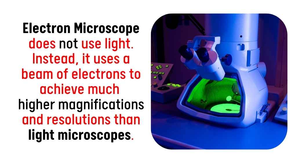
Electron microscope does not use light. Instead, it uses a beam of electrons to achieve much higher magnifications and resolutions than light microscopes.
| Image | Product | Detail | Price |
|---|---|---|---|
 | Carson MicroBrite Plus 60x-120x LED Lighted Pocket Microscope |
| See on Amazon |
 | Elikliv LCD Digital Coin Microscope |
| See on Amazon |
 | AmScope M150 Series Portable Compound Microscope |
| See on Amazon |
 | PalliPartners Compound Microscope for Adults & Kids |
| See on Amazon |
 | Skybasic 50X-1000X Magnification WiFi Portable Handheld Microscopes |
| See on Amazon |
| Type of Microscope | Method of Imaging | Magnification | Key Advantage |
|---|---|---|---|
| Electron Microscope | Electron beam | Up to 50,000,000x | Ultra-high resolution |
| Scanning Probe Microscope | Physical probe | Atomic resolution | Surface analysis at atomic scale |
| X-ray Microscope | X-rays | High resolution | Internal imaging without sectioning |
| Focused Ion Beam Microscope | Ion beam | Similar to electron microscopes | High resolution with sample preparation capabilities |

Types of Microscopes That Do Not Use Light
There are several microscopes that do not use light. These microscopes rely on different principles and techniques to magnify and analyze specimens. Let’s take a closer look at the types of microscopes that do not use light:
1. Electron Microscopes
Electron microscopes (EMs) use electron beams instead of light to create an image. The electrons have much shorter wavelengths than visible light, allowing these microscopes to achieve much higher resolution and magnification. There are two main types of electron microscopes: the scanning electron microscope (SEM) and the transmission electron microscope (TEM).
Scanning Electron Microscope (SEM)
A SEM works by scanning a focused beam of electrons across the surface of a specimen. The electrons interact with the atoms on the surface, producing secondary electrons that are detected to form an image. SEMs provide detailed, three-dimensional images of surfaces, and they can magnify objects up to around 1,000,000x.
Transmission Electron Microscope (TEM)
TEMs work by transmitting electrons through a thin sample. The electrons pass through the sample and are detected on the other side, creating a two-dimensional image. TEMs provide ultra-high resolution and are used to observe the internal structure of specimens, such as cells, viruses, and nanomaterials. They can achieve magnifications of up to 50,000,000x.
2. Scanning Probe Microscopes (SPMs)
Scanning probe microscopes (SPMs) use a physical probe to interact directly with the surface of the sample. They do not rely on light or electron beams. Instead, they detect forces between the probe and the sample at an atomic level, allowing them to map out surface structures. The two main types of scanning probe microscopes are the atomic force microscope (AFM) and the scanning tunneling microscope (STM).
Atomic Force Microscope (AFM)
An AFM uses a sharp tip mounted on a cantilever to scan the surface of a specimen. As the tip moves across the surface, it detects forces such as van der Waals forces, magnetic forces, and electrostatic forces. These measurements are used to construct highly detailed, three-dimensional images of the surface at the atomic scale.
Scanning Tunneling Microscope (STM)
An STM works by scanning a sharp tip over the surface of a sample and measuring the tunneling current between the tip and the sample. The current is highly sensitive to the distance between the tip and the sample, allowing the microscope to map the surface with atomic resolution. STMs are particularly useful for studying conductive materials at the atomic level.
3. X-ray Microscopes
X-ray microscopes use X-rays, a form of electromagnetic radiation, instead of visible light or electron beams. X-ray imaging works similarly to traditional medical X-rays, but at a much higher resolution. This technique is particularly useful for studying the internal structure of materials and biological specimens without the need for sectioning or staining.
X-ray microscopes operate by directing X-rays onto a sample and detecting the transmission or reflection of the X-rays. The variations in X-ray absorption across the sample create an image of its internal structure. This technique is valuable for imaging biological tissues, metals, and other materials with complex internal structures.
4. Focused Ion Beam Microscopes (FIB)
Focused ion beam microscopes use a beam of ions rather than light or electrons to create images. FIBs are often combined with scanning electron microscopes to provide both imaging and sample preparation capabilities. The ion beam can mill away layers of material to expose new features in a specimen, allowing for highly detailed imaging and analysis.
FIBs work by focusing a beam of ions, such as gallium ions, onto the surface of the sample. The ions interact with the sample, causing the release of secondary electrons, which are detected to form an image. FIBs can achieve resolution similar to that of electron microscopes, and they are particularly useful in materials science and semiconductor research.
Advantages of Microscopes That Do Not Use Light

Microscopes that do not rely on visible light offer significant advantages over traditional light-based microscopes. These alternative techniques allow researchers to achieve much higher levels of detail and access new kinds of data, especially in fields like materials science, biology, and nanotechnology. Below are some of the key benefits of using microscopes that do not use light:
1. Higher Resolution
One of the primary advantages of microscopes that do not use light is their ability to achieve much higher resolution. Electron microscopes, for instance, use electron beams with much shorter wavelengths than visible light, enabling them to resolve much smaller features at the nanometer or even atomic scale. This high resolution allows scientists to observe fine details of specimens, such as individual atoms in a material, the structures within cells, or the arrangement of molecules on surfaces. In comparison, optical microscopes are limited in their resolution due to the longer wavelength of visible light, typically achieving a resolution of around 200 nanometers, whereas electron microscopes can reach resolutions in the range of picometers (trillionths of a meter).
2. Ability to Examine Internal Structures
Many microscopes that do not use light, such as electron microscopes and X-ray microscopes, are capable of imaging the internal structures of samples without requiring the sample to be cut or altered. For example, transmission electron microscopes (TEM) can pass electrons through a thin specimen to generate images of its internal features, allowing researchers to observe structures at the cellular or sub-cellular level. X-ray microscopes also excel at revealing internal details, especially in materials like metals, plastics, or biological tissues, which would otherwise require invasive techniques such as slicing or staining. The ability to view these internal structures non-invasively is critical for studying delicate biological specimens, including viruses and bacteria, or understanding the internal properties of materials used in engineering and manufacturing.
3. No Need for Staining or Sectioning
Unlike light microscopes, which often require samples to be stained or sectioned to improve contrast and visibility, microscopes that do not use light can examine samples in their natural, unstained state. Staining techniques used in optical microscopy can sometimes distort or damage delicate structures, especially in biological samples. For instance, cell membranes, proteins, and other cellular structures may be altered during the staining process, leading to potential loss of critical information. Electron microscopes and scanning probe microscopes avoid this issue, as they operate on different principles, such as electron interactions or surface scanning, allowing them to preserve the sample’s natural state. This is particularly beneficial for biological specimens that need to be studied in their true form, without the risk of staining-related artifacts.
Disadvantages of Microscopes That Do Not Use Light
While microscopes that do not rely on light provide significant benefits, they also come with certain challenges and limitations that users must consider. These challenges can impact their usability and accessibility.
1. Complex Sample Preparation
Many of these advanced microscopes, particularly electron microscopes, require complex and precise sample preparation. In the case of electron microscopes, samples must be prepared by cutting them into thin slices, often just a few nanometers thick, to allow the electron beam to pass through. Additionally, biological samples often need to be coated with a thin layer of conductive material, such as gold or carbon, to ensure that they do not charge up under the electron beam, which could distort the image. Some samples also need to be placed in a vacuum environment, which adds further complexity to the process. This preparation can be time-consuming, requires specialized equipment, and demands a high level of skill and expertise, making it less straightforward than working with light microscopes.
2. High Cost and Maintenance
Electron microscopes, scanning probe microscopes, X-ray microscopes, and other advanced instruments that do not use light are typically expensive. The initial cost of purchasing such microscopes can be prohibitively high for many research labs and educational institutions. In addition to the purchase price, these microscopes also require ongoing maintenance, which can add to the total cost of ownership. Regular calibration, servicing, and replacement of parts can be costly, and the instruments often require specialized technicians to maintain them. Furthermore, operating these microscopes requires advanced training, and users must be well-versed in the intricacies of handling the equipment. This makes these types of microscopes less accessible to labs with limited funding or expertise.
3. Limited Sample Size
Many microscopes that do not use light, such as electron microscopes and scanning probe microscopes, are designed to work with very small samples. Electron microscopes, for example, typically require specimens to be extremely thin (often on the order of nanometers) in order to allow the electron beam to pass through or interact effectively. Scanning probe microscopes are limited by the size of the probe, which is very small and precise, and thus only suitable for imaging surfaces at the atomic scale. These size limitations can make such microscopes less practical for examining larger specimens, which may require slicing, preparation, or adjustments to fit into the microscope’s working area. Additionally, large specimens may require more complex setups or may not be suitable for analysis at all with certain types of non-light microscopes.
Why Do Electron Microscopes Not Use Light?
Electron microscopes do not use light because electrons have much shorter wavelengths than visible light. This allows electron microscopes to resolve much smaller details, offering magnifications up to several million times. Light, on the other hand, cannot achieve this level of resolution due to the limitations of its wavelength.
Can Scanning Probe Microscopes Replace Light Microscopes?
While scanning probe microscopes offer extremely high resolution, they are not typically used as replacements for light microscopes in most applications. Light microscopes are more versatile and can be used in many fields like biology and education for general purposes. Scanning probe microscopes are specialized tools used mainly for research and very high-resolution imaging of surfaces at the nanoscale.
Are There Any Disadvantages to Microscopes That Don’t Use Light?
Microscopes that do not use light, such as electron microscopes and scanning probe microscopes, generally come with high costs and require special sample preparation. Electron microscopes, for example, need samples to be in a vacuum and may also require coating with a conductive material. Scanning probe microscopes require the sample to be very flat, and the scanning process can be slow.
Final Words
In summary, several types of microscopes do not use light, including electron microscopes, scanning probe microscopes, X-ray microscopes, and focused ion beam microscopes. These microscopes offer advantages such as higher resolution and the ability to examine internal structures without the need for staining or sectioning. However, they also come with challenges such as complex sample preparation, high costs, and limited sample sizes. Each type of microscope has its unique applications, and their choice depends on the specific research needs and the type of specimen being examined.

I am an enthusiastic student of optics, so I may be biased when I say that optics is one of the most critical fields. It doesn’t matter what type of optics you are talking about – optics for astronomy, medicine, engineering, or pleasure – all types are essential.
Table of Contents
