Blog

  • What Is the Best Way to Focusing on a Microscope?: A Beginner’s Guide

    When it comes to focusing on a microscope, there are a few key things that you need to keep in mind.

    The first is the magnification (or power) of your scope. This should be high enough to see clearly but not so high that you have difficulty handling the instrument.

    Next, ensure that the eyepiece has good eye relief, allowing you to move your eyes without feeling strained or uncomfortable.

    And finally, store and clean your microscope regularly for optimal performance.

    Microscopy, a cornerstone in scientific exploration, relies heavily on precise focus for accurate observations. This article delves into the nuances of focusing on a microscope, unraveling the intricacies of its components and providing practical tips. Understanding and mastering the art of focusing is not only fundamental for researchers but also for students and enthusiasts seeking clarity in their microscopic endeavors.

    Techniques for Focusing Microscope

    Achieving precise focus on a microscope is a skill that requires a systematic approach and an understanding of the microscope’s components. In this section, we explore the techniques involved in the focusing process, from initial setup to the fine-tuning adjustments that ensure clarity in microscopic observations.

    A. Initial Setup and Preparation

    Before delving into focusing techniques, it’s crucial to start with proper setup and preparation. This involves placing the sample on the stage and adjusting the illumination for optimal visibility. Adequate sample preparation, including proper mounting techniques, ensures that the specimen is positioned correctly and that potential obstacles to focusing, such as uneven surfaces or air bubbles, are minimized.

    B. Coarse Focusing

    Coarse focusing is the initial step in bringing the specimen into view. It involves using the coarse focus knob, which moves the stage or the objective lenses in larger increments. This step is crucial for rapidly approaching the focal plane and obtaining a clear image of the specimen. It’s important to note that coarse focusing should be done cautiously to avoid overshooting the optimal focus point.

    Table: Coarse Focusing Guidelines

    Coarse Focusing StepsDescription
    1. Low MagnificationBegin with the lowest magnification objective lens for a broader field of view.
    2. Large MovementsUse the coarse focus knob for larger, rapid movements towards focus.
    3. Stop Before ClarityAs the specimen becomes clearer, slow down and stop before achieving full clarity.

    C. Fine Focusing

    Fine focusing is the refinement stage that brings out the intricate details of the specimen. Using the fine focus knob, smaller adjustments are made to precisely define the focal plane. This step is crucial for achieving high-resolution images and ensuring that the observer can discern fine structures within the specimen. Fine focusing requires patience and a keen eye, as subtle adjustments make a significant difference in the clarity of the observed features.

    Table: Fine Focusing Guidelines

    Fine Focusing TipsDescription
    1. High MagnificationSwitch to higher magnification for finer details.
    2. Slow and GradualTurn the fine focus knob slowly for subtle adjustments.
    3. Observe ChangesPay close attention to changes in clarity with each adjustment.

    D. Maintaining Focus During Observation

    Once optimal focus is achieved, it’s essential to maintain it during the observation period. Microscopic observations often involve studying dynamic processes or capturing images, requiring a stable focus. Using the stage controls for XY movement and adjusting the condenser for optimal illumination can help maintain focus throughout the observation.

    In conclusion, mastering the techniques for focusing a microscope involves a combination of initial setup, coarse focusing, fine adjustments, and maintaining focus during observation. Each step is integral to obtaining clear and accurate microscopic images, and practitioners should approach the process with precision and patience.

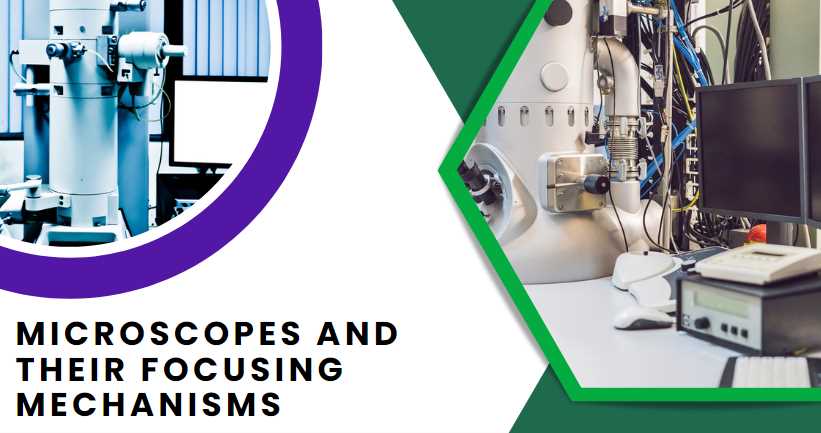
    Types of Microscopes and Their Focusing Mechanisms

    Microscopes come in various types, each equipped with unique mechanisms to facilitate focusing and enhance the quality of observations. Understanding these distinctions is crucial for users aiming to optimize their microscopic experiences.
    Microscope TypeFocusing Mechanism
    Light Microscope
    BrightfieldUtilizes visible light for imaging.
    Advantages: Simple and widely used for general observations.
    Disadvantages: Limited contrast, especially for transparent specimens.
    Phase ContrastEnhances contrast in unstained cells.
    Advantages: Ideal for live cell imaging without staining.
    Disadvantages: Requires special optics and can be complex.
    DarkfieldIlluminates the specimen against a dark background.
    Advantages: Enhances contrast for transparent specimens.
    Disadvantages: Limited to certain types of specimens.
    Electron Microscope
    Transmission Electron Microscope (TEM)Passes electrons through the specimen for detailed internal imaging.
    Advantages: High resolution and magnification.
    Disadvantages: Complex sample preparation, limited depth perception.
    Scanning Electron Microscope (SEM)Scans the specimen surface with electrons for 3D imaging.
    Advantages: Detailed surface imaging.
    Disadvantages: Limited to surface observations.

    Light microscopes, encompassing brightfield, phase contrast, and darkfield variations, are commonly used for a range of applications. Brightfield microscopy is straightforward but may lack contrast, while phase contrast and darkfield techniques address these limitations.

    Electron microscopes, on the other hand, delve into the nanoscale. TEM provides detailed internal views but requires meticulous sample preparation, while SEM excels in surface imaging.

    Understanding the strengths and limitations of each type allows users to choose the most suitable microscope for their specific applications, ensuring optimal results in microscopic observations.

    Troubleshooting Common Focusing Issues

    Achieving and maintaining proper focus on a microscope can be challenging, and various issues may impede the clarity of microscopic observations. In this section, we explore common focusing issues, their possible causes, and effective troubleshooting techniques to overcome these challenges.

    A. Blurriness and Lack of Clarity

    One of the most common issues encountered during microscopy is blurriness or a lack of clarity in the observed image. Several factors can contribute to this problem, and identifying the root cause is crucial for implementing effective solutions.

    Table: Troubleshooting Blurriness and Lack of Clarity

    IssuePossible CausesSolutions
    Dirty LensesContaminants on the objective lensesClean lenses with appropriate solutions, such as lens cleaning solution and lens paper. Avoid using abrasive materials.
    Improper Sample PrepSpecimen not properly mounted or preparedEnsure proper mounting techniques, use clean slides and coverslips, and eliminate air bubbles.
    Incorrect IlluminationInadequate or uneven lightingAdjust the illumination to ensure even lighting across the entire field of view. Check and replace the microscope bulb if necessary.

    B. Depth of Field Challenges

    Depth of field challenges can hinder the observation of three-dimensional structures within a specimen. Addressing these challenges involves understanding the factors that contribute to depth of field limitations and implementing specific techniques to overcome them.

    Table: Troubleshooting Depth of Field Challenges

    IssuePossible CausesSolutions
    Optical LimitationsInherent limitations of the microscope opticsConsider using objective lenses with higher numerical aperture for increased depth of field.
    Sample CharacteristicsComplex or thick specimensUtilize techniques such as Z-stack imaging to capture multiple focal planes and reconstruct a 3D image.
    Aperture AdjustmentIncorrect aperture settingsAdjust the aperture to control the amount of light reaching the objective lens, influencing depth of field.

    Troubleshooting these common focusing issues requires a systematic approach. Begin by assessing each potential cause and implementing the corresponding solution. Regular maintenance and attention to sample preparation details can significantly reduce the occurrence of these issues.

    In conclusion, understanding and troubleshooting common focusing issues are essential skills for anyone working with microscopes. By addressing blurriness, lack of clarity, and depth of field challenges, researchers and enthusiasts can enhance the quality and accuracy of their microscopic observations. Regular checks, proper maintenance, and adherence to best practices contribute to a seamless and productive microscopy experience.

    6 Tips and Best Practices for Focusing on a Microscope

    Mastering the art of focusing on a microscope goes beyond technical know-how; it involves adopting specific tips and best practices that enhance the overall experience. Whether you are a seasoned researcher or a student in the laboratory, these six tips can significantly improve your proficiency in achieving precise focus.

    1. Proper Sample Preparation: Before diving into focusing, ensure your sample is well-prepared. Use clean slides and coverslips, eliminate air bubbles, and apply mounting techniques suitable for your specimen type. Proper sample preparation lays the foundation for successful focusing.
    2. Mindful Choice of Magnification: Selecting the appropriate magnification is crucial. Begin with low magnification for an overview and then gradually increase it for finer details. Choosing the right magnification at each stage ensures a balanced and clear view of the specimen.
    3. Gradual and Patient Adjustments: Focusing is not a race. Make adjustments to the focus knobs slowly and patiently. Rapid movements can lead to overshooting the optimal focus point, while gradual adjustments allow for precise control over the focal plane.
    4. Consistent Lighting Conditions: Illumination plays a vital role in achieving clarity. Ensure consistent and even lighting across the specimen. Adjust the microscope’s light source to avoid shadows or uneven illumination, which can impact the perception of details.
    5. Regular Maintenance Routine: Microscopes, like any other precision instruments, require regular maintenance. Clean lenses and optics routinely using appropriate solutions. Check for any signs of wear or damage and address them promptly. A well-maintained microscope performs optimally.
    6. Avoiding Vibrations and Disturbances: Maintain a stable environment around the microscope. Vibrations and disturbances can affect the clarity of your observations. Choose a stable surface, minimize movements, and be mindful of external factors that could impact the stability of the microscope.

    By incorporating these tips into your microscopy routine, you not only enhance your ability to focus but also contribute to the longevity and reliability of the microscope. Whether you are a novice or an experienced microscopist, these best practices ensure a more rewarding and effective microscopic exploration.

    Advanced Focusing Techniques

    While mastering basic focusing techniques is essential, advanced microscopy often demands more sophisticated approaches to meet specific research requirements. Here, we explore advanced focusing techniques that push the boundaries of traditional observation methods, providing researchers with enhanced capabilities for in-depth analysis.

    1. Z-stack Imaging: Z-stack imaging involves capturing multiple focal planes at different depths within a specimen and subsequently reconstructing them into a three-dimensional image. This technique is particularly valuable when studying complex structures or dynamic processes, offering a comprehensive view that goes beyond traditional two-dimensional observations.
    2. Autofocusing Systems: In the realm of automation, autofocus systems have emerged as powerful tools. These systems use algorithms and sensors to continuously assess image sharpness and make real-time adjustments to maintain optimal focus. This is especially beneficial for time-lapse imaging or when observing samples with varying thickness. Autofocusing systems significantly reduce the manual workload and enhance the efficiency of microscopic experiments.

    These advanced focusing techniques represent a paradigm shift in microscopy, offering researchers unprecedented capabilities. Z-stack imaging provides a holistic view of specimens in three dimensions, while autofocus systems leverage technology to ensure continuous clarity in dynamic imaging scenarios. Incorporating these techniques into your microscopy toolkit opens new possibilities for detailed and comprehensive analysis in various scientific disciplines.

    Importance of Calibration in Microscopy

    Calibration in microscopy is a fundamental practice that ensures the accuracy and reliability of observations, measurements, and imaging. The process involves aligning and adjusting various components of the microscope to standardize its performance. Here, we delve into the significance of calibration in microscopy and its impact on research reproducibility and data integrity.

    Table: Key Aspects of Microscopic Calibration

    Calibration AspectDescription
    Objective Lens CalibrationEnsures that the magnification settings align with labeled values, preventing misinterpretation of specimen features and sizes.
    Stage CalibrationAligns the stage with the XY axis, ensuring accurate navigation and precise measurement of distances in the specimen.
    Eyepiece CalibrationVerifies the accuracy of the eyepiece graticule or reticle, enabling reliable measurements aligned with true specimen dimensions.

    Calibration is crucial for maintaining the quality and integrity of microscopic observations. Its importance can be summarized in the following ways:

    1. Accuracy in Measurements: Calibration ensures that measurements taken with the microscope are precise and accurate. This is particularly critical in scientific research where precise dimensions are crucial for drawing conclusions and making comparisons.
    2. Reproducibility of Results: Calibrating the microscope ensures that experiments and observations can be reproduced consistently. Researchers can trust that their findings are not skewed by variations in the microscope’s performance.
    3. Quality Control in Imaging: Calibrating the microscope contributes to maintaining the quality of images produced. It prevents distortions and artifacts, allowing for clear and accurate documentation of microscopic features.

    In conclusion, calibration is a cornerstone in microscopy, safeguarding the reliability and accuracy of data. The systematic alignment of microscope components ensures that observations are not only precise but also consistent, fostering confidence in scientific research.

    1. Why is proper focus crucial in microscopy?

    Proper focus in microscopy is paramount as it directly impacts the clarity and accuracy of observations. Achieving optimal focus ensures that researchers can discern fine details within a specimen, leading to more precise and reliable results. Without proper focus, the potential for misinterpretation and oversight of critical features increases, compromising the integrity of scientific findings.

    2. What are the basic components of a microscope that contribute to focus?

    Microscopes consist of key components that play vital roles in achieving focus. The objective lenses, fine and coarse focus knobs, eyepiece, and stage collectively contribute to the focusing process. Understanding how these components work together enables microscopists to manipulate the instrument effectively, ensuring a sharp and clear view of the specimen.

    3. How do I troubleshoot blurriness in my microscope images?

    Blurriness in microscope images can result from various factors, including dirty lenses or improper sample preparation. To troubleshoot, start by cleaning the lenses using appropriate solutions and techniques. Ensure the sample is well-prepared, with clean slides and coverslips, and eliminate air bubbles. Adjusting the illumination and confirming proper sample mounting can often resolve blurriness issues.

    4. What role does the numerical aperture play in focus?

    Numerical aperture (NA) is a crucial factor influencing focus. It determines the light-gathering ability of the objective lens, impacting the resolution and depth of field. Higher NA values contribute to improved focus, allowing for the visualization of finer details. Microscopists should select objective lenses with appropriate NA values based on their specific imaging requirements.

    5. Can I use the same focusing techniques for different types of microscopes?

    While basic focusing principles remain consistent, different types of microscopes may have unique mechanisms and considerations. Light microscopes, electron microscopes, and various imaging modalities may require specific adjustments. Adapting your focusing techniques to the characteristics of each microscope type is essential for achieving optimal results.

    6. How does sample thickness affect focusing?

    Sample thickness can impact focus, especially in light microscopy. Thick samples may exhibit variations in focus across different layers, requiring adjustments or advanced techniques such as Z-stack imaging to capture multiple focal planes. Understanding the relationship between sample thickness and focus is crucial for obtaining accurate and detailed observations.

    7. What role does the condenser play in achieving focus?

    The condenser in a microscope is instrumental in focusing by controlling the illumination. Proper condenser adjustments ensure even and adequate lighting, contributing to clarity in observations. Microscopists should optimize condenser settings to achieve the desired contrast and brightness for the specimen under examination.

    8. How can I maintain focus during live observations or time-lapse imaging?

    Maintaining focus during live observations or time-lapse imaging requires careful consideration. Utilize the microscope’s stage controls for XY movements to track dynamic changes in the specimen. Autofocusing systems can also be beneficial, continuously adjusting focus to ensure clarity during prolonged observations, allowing researchers to capture and analyze dynamic processes seamlessly.

    9. Are there specific techniques for focusing on transparent or unstained specimens?

    Focusing on transparent or unstained specimens presents unique challenges. Phase contrast or differential interference contrast (DIC) microscopy can enhance contrast in these specimens, aiding in focus. Adjusting the condenser and utilizing specialized illumination techniques can improve visibility, allowing for better focus on transparent structures without the need for staining.

    10. How often should I calibrate my microscope for optimal focus?

    Regular calibration is essential for maintaining optimal focus. Microscopes should be calibrated at regular intervals, especially when there are changes in environmental conditions, such as temperature or humidity. Additionally, after any repairs or modifications to the microscope, recalibration is necessary to ensure accurate measurements and reliable focus in microscopic observations.

    Proper Use Of Compound Microscope

    focusing on a microscope

    The microscope has to be placed on the table in a cool place.

    The eyepiece and the object, and the mirror of the glass have to be cleaned with a piece of clean cloth.

    Keeping an eye on the eyepiece, the low-power object has to be gradually swayed. When the object comes vertically to the hole in the stage, a click sound is made. The goal is to keep it that way.

    The coverslip on the slide must be placed on the stage so that the part covered by the coverslip is just below the object lens and above the hole in the stage. The slide should be fastened with clips from both sides to stay still.

      Now keep an eye on the eyepiece and turn the concave side of the mirror towards the light source so that the light beam is reflected and falls brightly on the object on the slide through the diaphragm, condenser, and stage holes.

    The object has to be brought very close to the slide (about half an inch above the slide) by turning the coarse focus screw and turning the screw slowly up, and down the object, the pattern suddenly placed on the slide becomes clear. This method is called focusing. Now turn the fine focus screw to see the object more clearly and clearly.

    Tricks: Almost all microscopes used everywhere are monocular, so when using them, you have to open both eyes and see with one eye. To avoid strain on the eyes, it is necessary to change the eyes from time to time to see the eyepiece. However, it is better to use a binocular microscope.

    If you want to use natural light in a microscope: you have to place the microscope in a secluded laboratory. First, the mirror has to be placed so that the reflected light rays are reflected under the glass slide along the stage. And if the artificial light source is present as part of the microscope, it should only be lit.

    Precaution:

    The microscope should never be placed on the edge of the table.

    The microscope must always be kept upright; otherwise, the slide placed on the stage may fall.

    The first is to use low-power targets, later, high-power targets will be used as needed.

    The object lens should never touch the slide cover slip.

    Always clean the lens with a soft cloth made of silk or fine cotton.

    After using the microscope, it should be covered with a Belzer, cloth, or polythene. Putting calcium chloride in a small container in the Belzer keeps the Belzer dry. As a result, the fungus does not grow in the lens.

  • 11 Ways to Clean and Sterilize A Microscope: Video Guide to Follow

    11 Ways to Clean and Sterilize A Microscope: Video Guide to Follow

    Microscopes are one of the essential lab tools for scientists and students. However, like any other equipment, they can get dirty over time. By following the proper cleaning process, you can keep your microscope functioning at its best. We covered cleaning the mechanical components, lenses, and mold area.

    Several years ago, I was reading the statistics for microscope cleaning products. According to the manufacturer, one, in particular, stood out: if you clean only twice a year, you’re probably missing 90% of your sample. You’re missing more than 80% if you clean twice a month. It’s hard to imagine how important this is since most of us don’t even realize that we do not see our samples.

    Always read all instructions carefully before cleaning your microscope, as any mistake could ruin your equipment. As always, exercise caution when cleaning delicate equipment and wash your hands thoroughly before and after each step. Stay tuned for more updates on microscope cleaning!

    Reasons to Clean and Sterilize a Microscope

    ReasonExplanation
    Accuracy and Reliability of ObservationsMicroscope cleanliness is crucial for obtaining accurate and reliable observations in scientific research.
    Prevention of Contamination and Cross-contaminationRegular cleaning and sterilization prevent the introduction of contaminants, ensuring the integrity of results.
    Extension of Microscope LifespanProper maintenance contributes to the longevity of microscope components, reducing the need for frequent replacements.
    Compliance with Industry Standards and GuidelinesAdhering to established standards and guidelines is essential, especially in fields where precision is paramount.

    How do you clean the mechanical part of a microscope?

    Cleaning the Mechanical Part of a Microscope

    StepTaskDescriptionRecommended Materials
    1Power Off:Ensure the microscope is powered off before cleaning.
    2Remove Excess Debris:Use a small brush or compressed air to remove loose debris from the exterior.Small brush, compressed air canister
    3Wipe External Surfaces:Gently wipe external surfaces with a soft, dry cloth to remove dust and fingerprints.Soft, dry cloth
    4Clean Objective Lenses:For external lenses, use a lens cleaning solution and lens paper.Lens cleaning solution, lens paper
    5Inspect Mechanical Components:Visually inspect mechanical components such as focus knobs and stage adjustments for any visible dirt or debris.
    6Lubrication (If Necessary):Follow the manufacturer’s guidelines for lubricating moving parts. Use an appropriate lubricant sparingly.Manufacturer-recommended lubricant
    7Check for Loose Parts:Ensure all screws and bolts are securely fastened. Tighten any loose parts to prevent mechanical issues.Screwdriver
    8Inspect Electrical Connections:Check electrical connections for any signs of damage or loose wiring. Repair or replace as needed.
    9Optical Alignment Check:Verify that the optical components are correctly aligned to avoid image distortion. Consult the user manual for alignment procedures.Calibration slide, user manual
    10Final Inspection:Perform a final visual inspection to ensure all components are clean and in good condition.

    I always use Household cleaners to clean the eyepieces, lenses, and other optical components. These cleaners are safe on most surfaces and effectively remove dirt, dust, and fingerprints.
    If you want to use a microscope cleaning kit, you will need to mix the cleaner with water and scrub the parts of the microscope that need cleaning.
    The steamer gently cleans the microscope using steam to loosen any dirt or debris. Just be sure to avoid any contact with the glass surface of the microscope, or you will damage it.

    Cleaning the microscope’s mechanical part is daunting if you are not an expert. But you can do it quickly and easily with preparation and organization. The most common methods include household cleaners, a microscope cleaning kit, or a steamer.

    Process to Follow

    To keep it clean, we know that you should wipe the exterior with a moist paper towel, avoid touching the lenses if possible, and dry everything off before using it again. That’s all well and good, but how should you clean it? It can be tough to figure out what steps to take when you don’t have instructions. Here are a few tips:

    1. Unplug the microscope and wipe the exterior with a wet paper towel.

    2. Carefully remove the impurities from the inside of the microscope using a paper towel.

    3. Dry everything off before storing or using the microscope again.

    4. We recommend using distilled water to clean microscopes instead of tap or city water since it is safer and causes less fogging.

    5. Remember that humidity will prevent condensation in your microscope, so if you live in a humid area, ensure it’s as low as possible!

    Using dish soap

    We all know that microscopes are expensive, so you don’t want to risk damaging them and wrongly caring for them. To properly clean a microscope, follow these steps:

    1. Fill a sink with room-temperature water and add soap until it makes a pile of foam
    2. Swish the solution around on the scope to make sure it’s sudsy
    3. Wipe away any dust with a cloth and rinse off the area with room-temperature water
    4. If your microscope is dirty or oily, use 10% alcohol to clean it; but be careful not to use this if it’s dry
    5. Repeat the alcohol process twice or three times, then finish with distilled water
    6. After cleaning the mechanical parts, clean around the optics by wiping them with the solution and cloth.
    7. When done, dry it off and store it in a dust-free place.

    Using white vinegar

    Sometimes you need to get gross. When trying out different cleaning methods, sometimes it’s still hard to determine when a solution is doing something. Cleaning the microscope using white vinegar is a simple and effective way to keep it in excellent condition. It would be best to have a few supplies, including white vinegar, a microscope lens cleaning cloth, and a brush.

    First, pour white vinegar into a small bowl.

    Then, wet the cleaning cloth and wring it slightly damp.

    Place the cloth on the microscope lens and rub it around in a circular motion.

    Make sure to get into all the corners and crevices.

    Finally, use the brush to clean any residue that may have accumulated.

    After cleaning the lens, dry it off with the cloth and store the microscope in a clean and dust-free environment.

    To be on the safe side, wipe the microscope with a clean cloth or several to ensure that any excess liquid is removed. If there was no way to completely clean the eyepiece tube with a damp cloth, use this cloth to do so. You can also use it to remove any liquid from the eyepiece tube if necessary. Always use new material to ensure you’re dry and ready to go.

    How do you clean the lenses of your microscope?

    Cleaning your microscope lenses is scary, but it can be a breeze with patience and practice. There are a few different ways to clean your lenses, and each has its benefits and drawbacks.

    The most common way to clean lenses is to use water and alcohol. This solution effectively removes dust and other particles, but it can be difficult to remove dried residues.

    Another method is to use a KOH solution. This solution is less abrasive and can clean lenses that are difficult to dry. It is also effective in removing oil and grease but can damage the lens if misused. The final method uses a mechanical cleaner specifically designed for microscope lenses. This cleaner is safe to use and has no adverse effects on the lens.

    Ultimately, the best way to clean your microscope lenses is to experiment and find what works best.

    Dusty lenses are a problem you can’t cure with a quick wipe.

    The best way to cleanse dusty lenses is by using a vacuum or dusting brush. It will take patience, but you must do it carefully so your lens doesn’t get any worse.

    You must use an optical dusting brush to clean these microscopic lenses properly. You can purchase one at most stores that sell lab equipment and microscopes.

    Before inserting it, you just prepped a lens and noticed some dust on the surface. How do you clean them off? There are a couple of solutions to avoid damaging your lenses.

    You can use an oil-free compressed gas or gently vacuum around the frame with a dust brush. It’s essential to avoid using a keyboard cleaner for this, as those products could damage your lenses. For example, dry-nitrogen-propelled compressed air is an excellent solution if it’s not too cold outside.

    The first time I used a microscope, I got it dirty. That was my fault for not realizing the lenses needed to be cleaned. (I went into my lab’s storage and found a cotton swab, water, and soap to clean it.) When you’re out of solutions, isopropyl alcohol works wonders — and you can find it at any store that sells microscopes and lab equipment. Another tip:

    Don’t use a cloth to clean the lenses when you’re out of your favorite solution. Clothes are too large and will spill cleaning liquid on your instrument if they are not lint-free.

    You might not think about it, but your lens is filthy. Agree? The center of the lens is where all the dirt has gathered, so you need to get it as clean as possible.

    A cotton swab and liquid dish soap are all you need to clean your smudged lenses. Apply the soap to the cotton swab and gently rub it on the center of your lenses in a circular motion.

    That is where the dirt has gathered, so it’s important to scrub it. After, clean outward across the lens, moving about an inch at a time away from the center. Continue until your lenses are both free of dirt! Be sure to use a light touch not to scratch the lens.

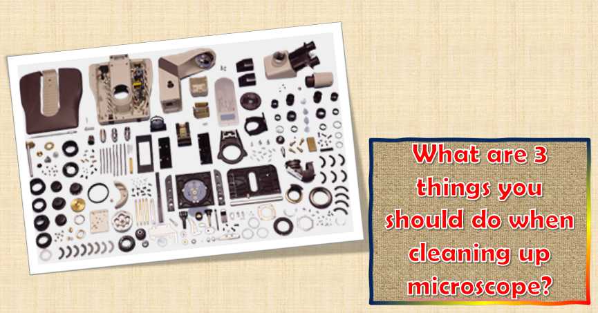
    What are three things you should do when cleaning up a microscope?

    What are 3 things you should do when cleaning up microscope

    When cleaning up a microscope, make sure to do the following:

    Hand hygiene is one of the essential steps in preventing infection. Before touching your microscope, wash your hands thoroughly with soap and water. This includes everything from your hands and wrists to your elbows and knees. It is also essential to avoid contact with your eyes, as this can lead to eye irritation or even conjunctivitis. Finally, avoid touching your face, which can lead to skin infections.

    1. Always wash your hands before putting on latex gloves.
    2. Be sure to wipe down the surface area after each person uses it for multi-person-use microscopes. This will keep you from worrying about someone else’s bacteria left behind on the surface after it is touched.
    3. It’s recommended not to eat or drink before, during, or directly after using your microscope, as it can be messy, and food particles may get on the microscope.
    4. If you store your microscope under questionable conditions (i.e., in a room that might reach 80° F or higher), dust it off after usage and wipe the inside with rubbing alcohol, wiping off any other residues on the surface before using again.

    Covering your microscope whenever you put it away is a simple but important step that can help keep it in good condition and protect it from dust and other particles. This will help protect the microscope from damage and prevent the light from being scattered, leading to inaccurate images.

    Additionally, it is vital to keep the microscope clean to function at its best. Cleaning the optics and the eyepieces with a suitable solution can help remove dirt, dust, and other debris that can cause degradation.

    Remember to regularly check the covers and hinges to ensure that they are secure and free from signs of wear or tear. If you notice any problems, don’t hesitate to contact your microscope supplier for assistance. Deteriorated covers can lead to distortions in your image and other issues.

    Once you are done using the microscope, remember to protect it from scratches and dirt with a dust cover or put it away properly before storing it for future use. This will protect your microscope and those who may one day come across it after it’s been misplaced!

    Failing to put a cover on your microscope when you pack it away is never good. It can result in dust particles and impurities getting into the optics and onto the slides, confusing your technician or student. In most cases, the best cover for your microscope is the one that came with it from the manufacturer.

    Covering your microscope with two bags will do the trick if you don’t have this dust cover. Put one sack over the top and another lower, securing both with a string.

    The most important thing you can do to maintain your microscope is to prevent fungus. This can be done by storing it in a dry place, like a closet, where the relative humidity will be low.

    A humid environment like a room can be one of the worst places to store a microscope because it gives the fungi more room to grow. You should invest in an air conditioner or an infrared lamp to keep your microscope clean and fungus-free even when temperatures soar. Removing fungus from a microscope is not easy, so it’s best to avoid it!

    It’s essential to store your microscope away from corrosive vapors. These vapors can be acidic or alkaline and may cause significant damage to your equipment. Keep your equipment in a different room when performing experiments with these kinds of smoke.

    It’s best to store your sensitive equipment in another room while conducting acid or alkaline vapors experiments. These fumes can corrode your equipment, so storing it in a different space is best to keep it safe.

    What are five things you should look after when cleaning the eyepiece of the microscope?

    One of the most important things you can do to keep your microscope in good working order is clean the eyepiece regularly. That is especially important if you use the microscope for medical purposes or research.

    Five Considerations for Cleaning the Microscope Eyepiece

    No.ConsiderationDescription
    1Use Proper Cleaning MaterialsUtilize specialized lens cleaning solutions and lens paper or microfiber cloths designed for delicate optics. Avoid using abrasive materials that could scratch the eyepiece glass.
    2Inspect for Dust and DebrisBefore cleaning, inspect the eyepiece for any visible dust or debris. Brush away loose particles with a soft camel-hair brush or air blower to prevent scratching during the cleaning process.
    3Gentle Cleaning TechniqueAdopt a gentle cleaning technique to avoid applying excessive pressure. Start by lightly blowing away loose particles, and then proceed to use minimal pressure while wiping the eyepiece with a circular motion.
    4Check for Residual Stains or SmudgesAfter cleaning, inspect the eyepiece for any residual stains or smudges. If necessary, repeat the cleaning process with fresh lens cleaning solution and a clean piece of lens paper until the optics are clear.
    5Avoid Direct Contact with the OpticsWhen handling the eyepiece, avoid direct contact with the optics. Hold the eyepiece by its frame or use lens tissue to prevent fingerprints on the glass. Fingerprints can distort images and are challenging to clean.

    Here are five things that you should always keep in mind when cleaning the eyepiece:

    1. Make sure that you use a proper eyepiece cleaning solution.
    2. Wet the eyepiece in water and scrub it with a lint-free cloth.
    3. Apply a light coat of eyepiece cleaner and then wipe it off.
    4. Dry the eyepiece thoroughly before using it again.
    5. Never carry a storage solution for your eyepiece in the container where you keep other sample materials.

    What are 3 ways to clean microscope slides and coverslips?

    Microscope slides and coverslips can get dirty quickly, so it is essential to clean them regularly to maintain image quality. Here are three easy ways to clean microscope slides and coverslips:

    Use a mild detergent and warm water to clean the slides and coverslips.

    Use an alcohol-free wipe to clean the slides and coverslips.

    Dry the slides and coverslips with a soft, lint-free cloth.

    Common Mistakes to Avoid in Microscope Maintenance

    MistakeExplanation
    Using Improper Cleaning MaterialsUsing abrasive materials or incorrect cleaning solutions can damage delicate microscope components, affecting performance and longevity.
    Neglecting Manufacturer GuidelinesIgnoring the manufacturer’s recommended cleaning procedures can lead to suboptimal cleaning and potential damage to the microscope.
    Rushing the Cleaning ProcessHasty cleaning may result in incomplete removal of contaminants, diminishing the quality of observations and potentially introducing errors.
    Neglecting Routine MaintenanceFailing to establish a regular cleaning schedule and inspect the microscope for wear and tear can lead to unexpected issues and reduce the instrument’s lifespan.
    Overlooking Lubrication of Moving PartsLack of lubrication in moving parts can result in friction, increased wear, and potential damage to mechanical components, affecting overall performance.

    Avoiding these common mistakes is essential for maintaining a microscope’s optimal performance, preventing damage, and ensuring accurate scientific observations. Adherence to proper cleaning and maintenance practices is key to the instrument’s longevity and reliability.

    Routine Maintenance and Inspection Guidelines for Microscopes

    Maintenance TaskDescription
    Cleaning ScheduleEstablish a regular cleaning routine to remove dust, debris, and contaminants. Consider daily, weekly, and monthly tasks.
    Signs of Wear and Tear InspectionRegularly inspect microscope components for signs of wear, such as scratches, corrosion, or loose fittings.
    Lubrication of Moving PartsApply appropriate lubricants to moving parts, such as the focusing mechanism, stage, and any rotating components.

    Routine maintenance is vital to ensure the consistent performance of microscopes. The table outlines key tasks for effective maintenance and inspection:

    1. Cleaning Schedule: Regular cleaning is essential to prevent the accumulation of dust and contaminants that could affect the quality of observations. Daily, weekly, and monthly tasks should be considered to address different levels of cleanliness requirements.
    2. Signs of Wear and Tear Inspection: Routine inspections help identify any signs of wear or damage to microscope components. This includes checking for scratches on lenses, corrosion on metal parts, and ensuring that all fittings are secure.
    3. Lubrication of Moving Parts: Applying appropriate lubricants to moving parts ensures smooth operation and helps prevent damage. Components such as the focusing mechanism, stage, and any rotating parts should be periodically lubricated to maintain optimal functionality.

    By adhering to these routine maintenance tasks, users can prolong the lifespan of their microscopes and ensure consistent and reliable performance.

    What is the best way to clean immersion oil from a microscope lens?

    When cleaning your microscope lens, the best way is to use a lens-cleaning solution. There are many available on the market, and choosing the appropriate one for the lens you are cleaning is vital. Some of the most popular lens-cleaning solutions include:

    Isopropyl alcohol: This general-purpose cleaner can be used on all lenses.

    Petroleum jelly: This natural oil can be used to clean lenses made of glass or plastic.

    Lens cleaner powder: This fine-grained powder is used to clean lenses made of metal or other hard materials.

    When using a lens cleaning solution, it is important to wet the lens and then add the solution. Gently swirl the solution around the lens until it is thoroughly wetted. Let the solution sit on the lens for several minutes, then rinse it with water.

    Why is special paper used to clean the lenses of a microscope?

    You can use a particular type of paper known as a Clark glass slide to clean microscopes. This paper is designed to clean the microscope’s lenses without damaging them. The paper is made of a flexible and rigid material, allowing it to withstand the pressure and friction caused by the microscope. Additionally, the paper is designed to absorb oil and other contaminants, then eliminated by the microscope’s cleaning system.

    The Clark glass slide paper is a must-have for anyone who regularly uses a microscope, as it helps maintain the instrument’s performance and accuracy.

    Lens tissue is made of cotton and polyester, which helps absorb oil and moisture. This prevents dirt, dust, and other debris from building up on the lens and causing blurry images. Lens cleaner is specifically formulated to clean lenses and is safe for plastic and glass. It also leaves the lenses with a pleasant smell to keep them clean and debris-free.

    Can you clean the microscope lens with a glasses cleaner?

    Yes, you can clean the microscope lens with a glasses cleaner. Depending on the type of cleaner, it may effectively remove dust, dirt, and other contaminants. Make sure to test the cleaner on a small area first to ensure it is safe for the lens.

    Can you use a handkerchief to clean a microscope?

    Absolutely not! A handkerchief will not work to clean a microscope because it is not made for that purpose. A microscope is a highly delicate instrument that must be cleaned meticulously to maintain accuracy. Instead, it would be best to use a special lens-cleaning cloth or a lens-cleaning solution.

    Can you use alcohol to clean a microscope?

    While it is possible to use alcohol to clean a microscope, it is not recommended. Alcohol can damage the lens, eyepieces, and other microscope parts. Additionally, it can create dangerous fumes that are harmful to your health. If you decide to use alcohol to clean your microscope, do it safely and carefully.

    How often should you clean the lens of your microscope?

    It is best to clean the lens after every use, but this cannot be easy if you are a busy scientist. A good rule of thumb is to clean the lens at least once a week, but it is always recommended to do more if necessary.

    Should you clean a microscope lens with lighter fluid?

    No, it would be best if you did not clean a microscope lens with lighter fluid. Lighter fluid is a volatile substance and can easily ignite if it comes in contact with chemicals or other flammable materials. This could result in a fire, damage the microscope or even cause injury.

    What fabric do you use to clean a microscope lens?

    A few different fabrics can be used to clean a microscope lens. One option is to use a cotton swab and some alcohol, and another is to use a microfiber cloth and a mild detergent. It is essential to use a soft cloth not to scratch the lens but also hard enough to remove dirt and debris.

    What happens if you clean a microscope lens without paper?

    If you are cleaning a microscope lens without paper, you are potentially damaging the lens and possibly causing it to light up or shatter irregularly. That is because the cleaning solution will leave behind grit, which will scratch the surface of the lens. In the worst-case scenario, this could result in the lens actually breaking.

    How do you lubricate a microscope?

    Microscopes require extraordinary lubrication to function correctly. A few types of lubricants can be used in microscopes, but the most popular is oil. Oil is a good lubricant because it is non-toxic, doesn’t corrode metal parts, and is easy to clean up. When using oil, it is essential to ensure that the microscope is assembled correctly and that the oil has been properly distributed.

    Another type of lubricant that can be used in microscopes is silicone. Silicone is a good lubricant because it doesn’t corrode metal parts, is non-toxic, and doesn’t cause build-up. It is also a good lubricant for eyepieces because it doesn’t fog up.

    The final type of lubricant that can be used in microscopes is glycerin.

    Final Words

    Keeping your microscope in pristine condition is essential for its reliability and performance. By following a few simple steps outlined in this blog, you’ll be able to clean it easily and keep it running like new! Before starting, read all instructions carefully, and avoid storing your microscope in an area where fungus grows. Finally, always clean your hands before and after handling the microscope for optimal hygiene.

    However, like any other electronic equipment, they can become contaminated with dirt, dust, and other debris. This blog post outlines the necessary steps to clean a microscope mechanically and optically. By following these simple guidelines, you’ll be able to keep your microscope in peak condition for future use! Do you have any questions or suggestions about how to clean a microscope? Let us know in the comments below!

  • Mastering Microscopy: A Comprehensive Guide on How to Use a Microscope

    When was the last time you looked through a microscope? If you’re like most people, it’s been a while. But we still don’t understand plenty of things about the microscopic world. This can be very frustrating for students and researchers who want to use microscopes but don’t know how to get started or what equipment they’ll need.

    There’s a slew of microscopes, but the two main models are polarizing and stereo. In my case, most people use a polarizing microscope to examine surfaces closely — like concrete samples or stamps. They’re also helpful for tasks that require a bit more focus. For instance, you can use them to study tissue samples during an operation or dissect a frog during a high school biology class.

    Using a microscope is fun, but it’s hard to get started. There are so many resources out there, and you can spend hours trying to find the right ones. Luckily, we’ve put together this list of tools, tips, and tricks to help you get started.

    So let’s look at what you need to know about using a microscope.

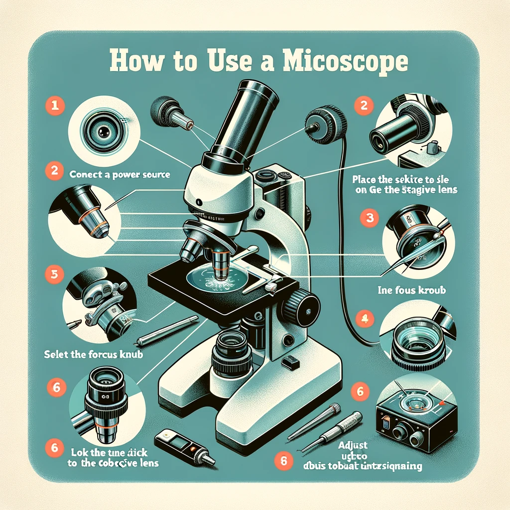
    5 steps: How to Use a Microscope?

    1. Choose a microscope appropriate for your level of expertise and the type of work you will be doing.

    2. Install the lens on the microscope according to the manufacturer’s instructions.

    3. Set up your lighting, mount, and stage following your microscope’s user manual.

    4. According to the manufacturer’s instructions, connect your camera or video recorder to the eyepiece and switch it on.

    5. Get an object, place it under a microscope lens and observe!

    How do you use a microscope for beginners?

    The best way to use a microscope for beginners is to understand the device’s essential functions.

    First, identify the parts of the microscope.

    Eyepiece: This allows you to see what is in front of it without moving your head or hands from where they are at.

    Objective lens: This lens has a single aperture that allows light to pass through into the scope, adjusted with a diaphragm and filter wheel on some models.

    Stage Plate: The stage moves up and down along with the objective lens to always remain in focus on whatever you are looking at while observing.

    Light source: This device gets light from the upper spectrum, such as ultraviolet, or lower ones, like artificial light.

    Sturdy base/table: for you to rest your scope on when doing science experiments and observations

    Diopter: This is where you adjust the microscope to focus on a different object, and it’s marked with numbers, so you know what you’re doing.

    Stereo Head: This is like the lens for your camera phone, and it’s the eye of your microscope.

    Bottom Lighting: The bottom lighting provides consistent lighting for viewing flat, opaque items. Perfect for viewing fresh fish and seafood since the skin’s rough texture will not appear shiny and glistening from top lighting.

    Stage Clips: The stage clips are an essential part of the microscope as they hold the slide in place and make it easier to manipulate. Some stereo microscopes have fixed stages mounted on a larger stand, while others have a movable set that can be attached or removed as per requirement.

    Lighting Controls: This helps you adjust the light going through the specimen.

    How to use it safely?

    To use a light microscope safely, the user should always focus the light beam on any surface that is not close to their eye. If they focus on a surface near their eye, they risk damaging it and possibly distorting their vision permanently. The microscope’s lens should never be touched. Glass slides should be placed on a firm, flat surface.

    If the microscope is zoomed in too much, it may strain the objective lens. Experimentation with magnification and positioning of images can allow one to learn how best to use their new toy safely without damaging anything, achieving outstanding results!

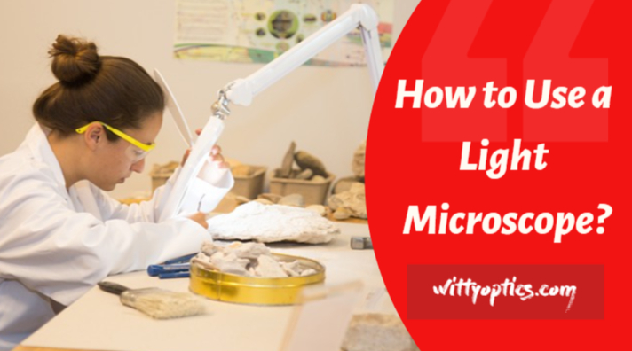
    How to Use a Light Microscope

    The benefits of using a light microscope are endless. It can examine cell structure, bacteria, and other microorganisms in the body.

    If you are using a light microscope, use the lamp power to match the magnification of your microscope.

    When focusing on a specimen, take note of the position closest to the focal plane.

    Use binoculars for more accurate viewing and better depth perception when working with specimens in 3D space. 

    If there is an insect or other object in your field of view, don’t try to focus on it; move it out of view before proceeding with your work! 

    Always remember to be safe when handling a microscope!

    What are the benefits of using a microscope?

    Using a microscope can help you in many ways, such as:

    1. Microscopes can help you to see things that are too small for the naked eye to see.

    2. They can be used to study cells and tissues under various conditions, essential for understanding how the body works.

    3. Microscopes allow scientists to view viruses and other pathogens in detail, which helps develop new vaccines or treat diseases at an early stage.

    4. Microscopes have been used to study ice nucleation behavior, galaxy clusters, binary stars, craters on planets like Earth, and more!

    5. They are essential tools for chemistry or physics research – they can help researchers identify molecules and particles by their shapes and sizes and examine reactions in tiny containers called drops (<100 nm). 

    6 . Microscopes are used in various industries, including pharmaceuticals, food science and technology, automotive manufacturing, materials research, and more.

    How to use a Stereo Microscope: 7 Steps to Follow

    Stereo microscopes are handy tools that let you view things in a much more detailed manner. Here’s a quick guide on using a stereo microscope to help you observe microscopic objects or biological specimens.

    Prepare your specimen, take it to the stage plate, and switch on the light. The two lights have different purposes; the one at the bottom is used when looking at transparent objects, and the one at the top is used when looking at opaque objects. Make sure you evaluate your surroundings and choose the right one!

    Look through the eyepieces to check your specimen is in focus. But what if it’s not? Simple! Use the focus knob to bring your sample into sharp relief. If your selection looks pale or translucent, place it on the black stage plate to make it easier to see, or use some black printer paper as an alternative.

    It is important to view an object with both eyes to see it more clearly. With two eyes, your body can see in more detail. However, peripheral sight is also needed when adjusting, observing, and proceeding.

    Place your eye up to the eyepiece and notice two overlapping lines of vision. Pull the eyepiece away to increase the distance, or push it toward you to decrease it. When you merge those two circles into one circular shape, you know that the correct interpapillary length has been set.

    You can use a diopter, which decreases the difference in vision between your two eyes. Next, cover one eye and focus on the microscope. Then, position the eyepiece (the diopter with the knob) over the other eye. Make sure you focus sharply on what you want to see.

    Subsequently, position the diopter over your other eye. Next, focus on the item you want to view with your vision while covering your other eye. Finally, adjust the eyepiece with the diopter until you have a clear picture. You won’t need to use the focus knob for this completion.

    When you move your sample, make sure to adjust the knob. When looking at the 3D image, you will not change every feature simultaneously.

    Now, you’re ready to use your microscope and view your sample. If your selection is alive and has escaped from the petri dish, put the lid on the petri dish to keep it from getting away. 

    Looking through a magnifying glass, you can’t see a blurry circle. But when you combine different lenses, the magnification becomes clearer.

    There are three leading microscopes: optical, electron, and scanning probe microscopes. Optical microscopes use a series of lenses to magnify specimens, and an electron microscope uses a beam of electrons to produce a better image than an optical microscope. A scanning probe microscope uses a series of probes to measure properties on a surface.

    Different parts of the compound microscope and their uses are:

    The following table outlines the key parts of a compound microscope and their respective uses:

    Microscope PartDescriptionFunction/Use
    Eyepiece (Ocular)The lens you look through at the top of the microscope.Magnifies the image from the objective lens.
    Objective LensesA set of lenses located on the rotating nosepiece.Provides various levels of magnification.
    NosepieceThe rotating structure that holds the objective lenses.Allows for easy switching between objectives.
    StageThe flat platform where the slide is placed.Supports the slide for observation.
    Stage ClipsClips on the stage to secure the slide in place.Prevents the slide from moving during observation.
    Coarse Focus KnobThe larger of the two focusing knobs, often on the outer part of the body.Used for initial, rough focusing of the specimen.
    Fine Focus KnobThe smaller of the two focusing knobs, often located inside the coarse focus knob.Allows for precise focusing of the specimen.
    Condenser LensLocated beneath the stage; focuses light onto the specimen.Improves image contrast and sharpness.
    IlluminatorThe light source, often located under the stage.Provides light for illuminating the specimen.
    DiaphragmAdjusts the amount of light that reaches the specimen.Regulates contrast and brightness of the image.
    ArmThe curved part that connects the base to the microscope head.Provides support and a comfortable grip for carrying.
    BaseThe bottom of the microscope; provides stability.Serves as a foundation for the entire microscope.
    Mechanical StageA movable stage with knobs for precise slide positioning.Facilitates controlled movement of the specimen.

    While a compound microscope may look quite similar to an optical microscope, it has several differences. Here is a list of the essential parts of a compound microscope and its uses.

    Eyepieces (used for viewing the specimen)

    Diopter adjustment (used to focus on and view objects)

    Nosepiece (fits over the eyepiece)

    Objective lens – where you use to see things up close

    The Stage- This is where you place your specimen.

    Stage clip- to keep your stage in place and prevent shaking.

    Mechanical stage- used for making sure your specimen stays in the spotlight.

    Condenser- Used to ensure the light stays focused and doesn’t fall off.

    Condenser Focus Knob- Used to move the condenser to control the light focus on specimens.

    Iris Diaphragm — controls the amount of light that enters the microscope

    Illuminator — lights up the object you’re inspecting

    Brightness Adjustment — adjusts the amount of light that reaches your eye so that you can see clearly

    Fine Adjustment and Coarse Adjustment – are used for focusing the microscope.

    Stage Controls – used to move the slide to view different parts.

    Aperture – the opening through which light travels into the microscope to view samples.

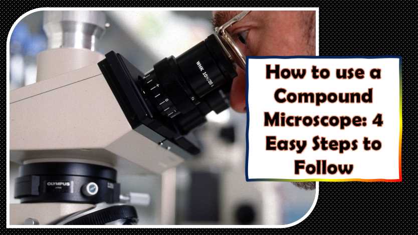
    How to use a Compound Microscope: 4 Easy Steps to Follow  

    How to use a Compound Microscope

    Microscopes are one of the most helpful science, medicine, and engineering tools. These tools provide us with a deeper understanding of the world around us. Here’s how to use a compound microscope.

    After switching on the illuminator, start with dim light and slowly increase it to your preference. This practice should prevent the bulbs from heating up too fast.

    If you are using a mechanical Stage or Stage, place your sample in one fixed position. This process will help ensure that the model does not move during the experiment.

    Remove the objective lens from the microscope and rotate it so that you can see through the eyepiece. You will see a microscopic image of your specimen displayed on the microscope’s base.

    Now direct your empty eye downwards. Use the focus knob on the left-hand side of the microscope to move your specimen into view.

    Note: A dimmer switch allows you to control the brightness of your bulb, not the opening of the iris diaphragm. Move the nosepiece for the objective lenses to change the magnification level according to your needs. That will help you focus on the part of your sample most relevant to you.

    Look through the microscope’s eyepieces and adjust how much light comes in using the diaphragm.

    Turn this knob with your hand; turning clockwise will bring it into focus.

    The fine adjustment knob is the little metal knob on the side of the microscope — twist it to focus your specimen. Try doing this as quickly as possible, or the high magnification objective could also hit the slide. Start again with a lower magnification if it gets difficult to focus.

    Every microscope has a nosepiece — turn it to change what you’re looking at.

    Always check if your microscope comes with a rack stop. If you don’t have one, you’ll want to get one. The rack stop is essential because it will prevent the objective lenses from lowering into the slide, and this action can be dangerous to both the slide and the specimen.

    Troubleshooting Common Issues (500 words)

    Microscopy, while a powerful tool, can encounter various issues that might hinder your observations. Understanding common problems and their solutions is crucial for efficient microscope usage.

    Blurry or Unclear Images

    One of the most common issues users face is blurry or unclear images. This problem can arise from improper focusing or dirty lenses. To resolve this:

    1. Clean Lenses: Ensure that the microscope lenses are clean. Use a lens cleaning solution and specialized lens paper to gently wipe away any dirt or smudges.
    2. Proper Focusing: Practice fine-tuning the focus. Utilize both coarse and fine focus adjustments to achieve optimal clarity. Additionally, ensure the specimen is appropriately positioned on the stage.

    Uneven Illumination

    Inconsistent lighting across the specimen can obscure details and affect the quality of your observations. To address uneven illumination:

    1. Adjust the Condenser: The condenser plays a crucial role in focusing and directing light onto the specimen. Adjust the condenser to achieve uniform illumination.
    2. Lighting Settings: Experiment with the lighting settings. Sometimes, a simple adjustment of the light intensity can eliminate uneven illumination.

    Dirt or Debris on Lenses

    Dirty lenses compromise the quality of images and may introduce artifacts into your observations. To maintain clear lenses:

    1. Regular Cleaning: Make it a habit to clean the lenses regularly. After each use, gently wipe the lenses with a clean, soft cloth.
    2. Proper Cleaning Materials: Use appropriate lens cleaning solutions and lens paper. Avoid using tissues or other materials that may scratch the lenses.

    Mechanical Issues

    Microscopes, like any precision instrument, may encounter mechanical problems. To troubleshoot mechanical issues:

    1. Lubrication: Lack of lubrication can lead to friction and hinder smooth movement. Follow the manufacturer’s recommendations for lubricating moving parts.
    2. Consult the User Manual: If issues persist, consult the microscope’s user manual. It often provides guidance on common mechanical problems and their solutions.

    Electrical Issues

    In some cases, electrical problems may arise, affecting the microscope’s functionality. If you encounter electrical issues:

    1. Check Power Source: Ensure that the microscope is properly connected to a stable power source. Fluctuations in power can lead to malfunctions.
    2. Inspect Cables: Examine power cables and connections for any damage. Faulty cables can disrupt the electrical supply to the microscope.

    Image Distortion

    Distorted images can result from misaligned optics or other technical issues. To address image distortion:

    1. Optical Alignment: Regularly check and align the microscope optics. Misaligned optics can lead to distorted images.
    2. Quality of Specimen: Ensure the specimen is prepared correctly. Distortion may occur if the sample is unevenly mounted or damaged.

    By addressing these common issues, users can enhance the effectiveness of their microscopy sessions. Regular maintenance and troubleshooting practices contribute to a seamless and productive microscopy experience. Remember, when in doubt, consult the microscope’s user manual or seek assistance from knowledgeable experts.

    Care and Maintenance of Microscopes

    Microscopes are delicate instruments that require careful maintenance to ensure optimal performance and longevity. Regular care practices contribute to clear and accurate observations. Here’s a comprehensive guide on the care and maintenance of microscopes, including essential tasks, recommended frequencies, and a maintenance checklist.

    Cleaning Lenses and Microscope Body

    Frequency: After each use

    Microscope lenses are susceptible to dust, fingerprints, and debris, which can compromise image quality. Regular cleaning is essential for maintaining clear optics.

    Cleaning Lenses

    1. Materials Needed:
      • Lens cleaning solution
      • Lens paper or microfiber cloth
    2. Procedure:
      • Apply a small amount of lens cleaning solution to the lens.
      • Gently wipe the lens in a circular motion using a clean piece of lens paper or a microfiber cloth.
    3. Caution:
      • Avoid using tissues or abrasive materials that may scratch the lenses.

    Cleaning Microscope Body

    1. Materials Needed:
      • Soft, dry cloth
    2. Procedure:
      • Wipe the microscope body with a soft, dry cloth to remove dust and prevent accumulation.
    3. Caution:
      • Avoid using cleaning solutions directly on the microscope body to prevent damage.

    Table 1: Cleaning Materials

    Cleaning TaskRecommended Materials
    Cleaning LensesLens cleaning solution, lens paper or microfiber cloth
    Cleaning Microscope BodySoft, dry cloth

    Storage Guidelines

    Frequency: Monthly

    Proper storage is crucial for protecting microscopes from environmental factors that can impact their performance.

    1. Storage Location:
      • Store the microscope in a clean, dry environment to prevent dust accumulation.
      • Use a dust-free case or cover when the microscope is not in use.
    2. Protection from Light and Temperature:
      • Avoid exposing the microscope to direct sunlight, as it can damage optics and components.
      • Store the microscope away from extreme temperatures and humidity.

    Table 2: Storage Guidelines

    Storage ConsiderationRecommendations
    Storage LocationClean, dry environment
    Protection from LightAvoid direct sunlight exposure
    Protection from TemperatureStore away from extreme temperatures and humidity

    Periodic Maintenance Checks

    Frequency: Annually

    Regular checks on the microscope’s mechanical components help identify potential issues before they affect performance.

    1. Lubrication:
      • Follow the manufacturer’s recommendations for lubricating moving parts.
      • Ensure proper lubrication to prevent friction and ensure smooth movement.
    2. Optical Alignment:
      • Regularly check and align the microscope optics to maintain optimal image quality.

    Table 3: Periodic Maintenance Checklist

    Maintenance TaskFrequency
    Lubricating Moving PartsAnnually
    Optical AlignmentAnnually

    Aligning Optics

    Frequency: As needed

    Misaligned optics can lead to distorted or unclear images. Regularly checking and aligning optics ensures consistently high-quality observations.

    1. Checking Alignment:
      • Use a calibration slide or grid to assess the alignment of the microscope’s optics.
      • Consult the user manual for specific alignment procedures.
    2. Adjustment:
      • If misalignment is detected, carefully follow the manufacturer’s instructions for adjusting the optics.

    Table 4: Alignment Guidelines

    Alignment TaskFrequency
    Checking AlignmentAs needed
    AdjustmentAs needed

    By incorporating these care and maintenance practices into your routine, you can prolong the life of your microscope and ensure that it consistently delivers accurate and high-quality results. Regular attention to cleaning, storage, and periodic checks will contribute to a reliable and efficient microscopy experience. Remember to consult the manufacturer’s guidelines and user manual for microscope-specific care instructions.

    Sample Preparation for Microscopy (500 words)

    Microscopic observation is only as good as the quality of the prepared sample. Whether you are a student exploring biological specimens or a researcher in a cutting-edge laboratory, proper sample preparation is critical for obtaining clear and informative images. This guide outlines essential steps, techniques, and considerations for preparing samples for microscopy.

    Handling and Cleaning Slides

    Frequency: Before each use

    Slides serve as the foundation for your microscopy observations. Proper handling and cleaning are crucial to prevent artifacts and ensure accurate results.

    Handling Slides

    1. Clean Hands: Always start with clean hands to avoid transferring oils or debris to the slides.
    2. Use Slide Holders: When manipulating slides, use slide holders to minimize direct contact with the glass.

    Cleaning Slides

    1. Materials Needed:
      • Lens cleaning solution
      • Lens paper or lint-free wipes
    2. Procedure:
      • Apply a small amount of lens cleaning solution to the slide.
      • Gently wipe the slide with a clean piece of lens paper or a lint-free wipe.

    Table 1: Handling and Cleaning Materials

    TaskRecommended Materials
    Handling SlidesClean hands, slide holders
    Cleaning SlidesLens cleaning solution, lens paper or lint-free wipes

    Applying Specimens to Slides

    Frequency: As needed

    The method of applying specimens to slides depends on the nature of the sample. Different techniques are employed for live specimens, preserved specimens, and non-aqueous specimens.

    Wet Mounts

    1. Place Specimen: Put a small drop of water or mounting medium on the center of the slide.
    2. Add Specimen: Gently place the specimen in the liquid, ensuring it remains centered.
    3. Cover Slip: Carefully lower a coverslip onto the specimen, avoiding air bubbles.

    Dry Mounts

    1. Position Specimen: Place the specimen directly onto the center of the slide.
    2. Cover Slip: Add a coverslip over the specimen, ensuring it is flat and free of air bubbles.

    Table 2: Sample Application Techniques

    Sample TypeRecommended Technique
    Wet SpecimensWet Mounts
    Preserved SpecimensDry Mounts
    Non-Aqueous SpecimensDry Mounts

    Staining Techniques

    Frequency: As needed

    Staining enhances contrast, making specific structures or components more visible under the microscope. The choice of stain depends on the specimen and the structures you want to highlight.

    Simple Stains

    1. Prepare Stain Solution: Dilute the chosen stain in a solvent like water or alcohol.
    2. Apply Stain: Place a drop of the stain on the specimen and let it sit for a specified time.
    3. Rinse and Dry: Rinse excess stain off the slide and allow it to dry before observation.

    Differential Stains

    1. Apply Primary Stain: Apply the primary stain to the specimen.
    2. Apply Counterstain: Add a contrasting stain to highlight different structures.
    3. Rinse and Dry: Rinse off excess stain and allow the slide to dry before examination.

    Table 3: Staining Techniques

    Staining TechniquePurpose
    Simple StainsUniform color for basic observation
    Differential StainsHighlight specific structures or cells

    Proper sample preparation is a cornerstone of successful microscopy. By following these guidelines and tailoring your approach to the specific requirements of your samples, you can enhance the quality and reliability of your microscopic observations. Remember to adapt these techniques based on the nature of your specimens and the goals of your microscopy study.

    What are the different methods for preparing a specimen?

    There are five different ways to take a specimen: Dry and wet mount, squash, smear, and staining. The easiest way is to dry-mount the sample. This method is suitable for seeing the more common airborne particles (invisible to the naked eye), hair, and feathers.

    There are some other methods for preparing a specimen.

    The most common ways of preparing a specimen are by taking it from the source, collecting it from nature, or making an impression.

    The first method is taken from the source, where you collect specimens in their natural state as they grow on living organisms like plants and animals.

    The second method involves gathering samples from polluted places like lakes and rivers.

    The third method involves getting impressions of tissues or parts of tissues that can be preserved in a plastic mold.

    Why do you see black-and-white images in electron microscopes?

    That’s because electron microscopes only operate using electron beams. You see, standard microscopes that use visible light to image specimens give you a colored image (you know, like the one you might’ve seen in school). Electron microscopes don’t use light, though — they use electrons — and electrons are invisible (or if you’re a long-time reader of this blog, we can say that electrons are tiny).

    What Type of Microscope is Best to See Cells?

    Compound microscopes are the best to use if you want to see cells, especially if they’re a bit fiddly or out of the ordinary. The images you’ll see with compound microscopes are two-dimensional, and you won’t be able to view cells under stereo microscopes — their low magnification prevents this.

    What are the dos and don’ts of immersion oil in the microscope?

    Using immersion oil in the microscope is helpful for taking high-quality pictures. However, it should be used carefully and cautiously because of some dos and donts of immersion oil in the microscope.

    The dos of using immersion oil in the microscope are:

    The oils must be explicitly made for microscopes to have good resistance to heat, water, light, etc.

    They must also be appropriate for optics that may require an airtight seal between the optic body and lens mount to prevent contamination or damage to them while they are being viewed through the microscope.

    You should never view your image on a white background, reducing contrast and visibility due to the reflection of light from other surfaces like paper or walls surrounding your scope stand or benchtop surface, which can easily cause glare.

    How do we take care of the microscope?

    To take care of the microscope, you should use a soft cloth to clean it and store it in a case.

    First, turn the power off and remove the eyepieces from the microscope. Clean the microscope’s body by wiping it down with a soft cloth soaked in warm water and soap. Then dry it with a clean towel.

    Next, wipe each eyepiece individually with a damp cloth or paper towel before placing them back into their respective sockets on the microscope body. After cleaning each eyepiece, replace them with separate sockets. Please turn on your scope to ensure it works correctly before returning any other parts to your instrument.

    What two parts do you place your hands on while carrying a microscope?

    You place your hands on two parts when carrying a microscope.

    The first part is the microscope’s base, which supports the instrument’s weight and prevents it from moving. The second part is called the tube rings, which can be adjusted to accommodate different sizes of tubes.

    How do you use immersion oil on a microscope?

    To use immersion oil on a microscope, you must ensure that the scope is clean and dry.

    Once done, you can add the required immersion oil to the eyepiece chamber’s bottom. This will help avoid condensation from building up between the two glass lenses attached to either side of your lens tube.

    If you have an open-backed or half-open camera, it is best to use an immersion oil bottle with a long spout to pour enough oil into your camera at once without getting any on your hands.

    How do you clean a dirty microscope lens?

    If you are using a wet lens, you can use your hands to gently scrub the lens surface with a cotton swab or tissue. If you use a dry lens, put some water on it and wipe it off with a clean cloth.

    If neither of these methods works, remove the microscope slide from the scope and try cleaning with soapy water or rubbing alcohol in an eyepiece case or cap.
    You can clean a dirty microscope lens using a mixture of detergent and water, and you will need to use the same amount of detergent as you would for washing your clothes.

    To prepare this solution, put some soap in a bowl and fill it with water. Next, dip the lens into soapy water and rinse it with plain water. Let it dry before replacing it on your microscope stand or taking it out of the light-proof container.

    What are some common problems that can occur when using a microscope, and how do you troubleshoot them?

    There are several common problems that can occur when using a microscope. Here are some of the most frequently encountered issues and potential solutions:

    Blurry or distorted images: This may be due to improper focus or alignment of the microscope’s lenses or other optical components. Try adjusting the focus knob and checking the alignment of the lenses to see if this helps. Also, ensure that the sample is positioned properly and that the microscope’s stage controls are adjusted appropriately.

    Insufficient illumination: If the sample appears too dark or the image lacks contrast, this may be due to insufficient illumination. Check that the light source is properly positioned and adjusted, and that the microscope’s brightness and contrast settings are properly configured.

    Dust or debris on the lenses: Over time, dust or debris may accumulate on the microscope’s lenses, leading to reduced clarity or resolution. To address this, use a soft brush or lens cloth to gently remove any visible debris from the lenses. Avoid using harsh or abrasive cleaners that may damage the lenses.

    Sample contamination or damage: If the sample appears distorted or contaminated, this may be due to improper handling or preparation. Check that the sample was properly prepared and that all equipment used during sample preparation and handling was properly sterilized.

    Incorrect magnification: If the image appears too small or too large, this may be due to incorrect magnification settings. Check the magnification controls and adjust as needed to achieve the desired level of magnification.

    Uneven illumination: If the image appears unevenly illuminated or exhibits areas of shadow, this may be due to uneven illumination from the light source. Adjust the position and angle of the light source to achieve more even illumination of the sample.

    Eye strain or discomfort: Using a microscope for prolonged periods of time can lead to eye strain or discomfort. Take frequent breaks and adjust the microscope’s brightness and contrast settings to reduce eye strain.

    By identifying and addressing these common issues, you can ensure that your microscope is functioning properly and that you are able to obtain clear, accurate images of your samples.

    How do you properly position and adjust the microscope’s illumination settings for optimal viewing?

    Proper positioning and adjustment of the microscope’s illumination settings are essential for obtaining optimal viewing of samples. Here are some general steps to follow:

    Position the microscope: Begin by positioning the microscope on a stable, level surface. Adjust the height and angle of the microscope so that it is comfortable to use and provides a clear view of the sample.

    Turn on the light source: Turn on the microscope’s light source, which is typically located below the stage or in the base of the microscope. Adjust the intensity of the light to achieve the desired level of illumination.

    Adjust the angle and position of the light source: Depending on the type of microscope, you may need to adjust the angle or position of the light source to achieve optimal illumination of the sample. For example, in a stereo microscope, you may need to adjust the position of the ring light to achieve even illumination across the sample.

    Adjust the diaphragm: Many microscopes have a diaphragm located below the stage that can be adjusted to control the amount of light that reaches the sample. Adjust the diaphragm to achieve the desired level of illumination.

    Use filters: Some microscopes have filters that can be used to adjust the color or wavelength of the light used to illuminate the sample. Use these filters as needed to achieve the desired level of contrast and clarity.

    Use polarizers: If your microscope has polarizers, you can adjust their orientation to control the amount and direction of light that reaches the sample. This can be particularly useful for observing samples with birefringent properties, such as crystals.

    Avoid glare: When adjusting the microscope’s illumination settings, be careful to avoid creating glare or reflections on the sample. This can interfere with your ability to obtain clear, accurate images.

    By following these steps, you can properly position and adjust the microscope’s illumination settings to achieve optimal viewing of your samples. Remember to take your time and make adjustments carefully to ensure the best possible results.

    What are some important safety precautions to keep in mind when using a microscope, such as handling hazardous materials or avoiding eye strain?

    When using a microscope, it is important to observe certain safety precautions to avoid injury and protect your health. Here are some of the most important safety precautions to keep in mind:

    Avoid eye strain: Microscope work can be taxing on the eyes, so it is important to take frequent breaks and adjust the microscope’s brightness and contrast settings as needed to reduce eye strain. Also, make sure that the microscope’s eyepieces are properly adjusted to match your individual vision.

    Handle hazardous materials carefully: Some materials that are viewed under a microscope can be hazardous to your health, such as infectious agents or chemicals. Wear appropriate personal protective equipment (PPE), such as gloves and a lab coat, and handle hazardous materials in a designated area with proper ventilation.

    Use caution when handling glass slides and coverslips: Glass slides and coverslips can break or shatter, causing cuts or other injuries. Handle them carefully and use caution when inserting or removing them from the microscope.

    Avoid touching the lenses: Touching the microscope’s lenses can damage them and lead to distorted or blurry images. Use a soft brush or lens cloth to clean the lenses, and avoid using harsh or abrasive cleaners.

    Turn off the microscope when not in use: When you are finished using the microscope, be sure to turn it off and unplug it to prevent accidental injury or damage.

    Maintain good posture: Sitting in the same position for prolonged periods of time can lead to back and neck strain. Maintain good posture and take frequent breaks to stretch and move around.

    Keep the microscope clean and well-maintained: Regularly clean and maintain the microscope to ensure that it is functioning properly and to prevent the buildup of dust and debris.

    By observing these safety precautions, you can minimize the risk of injury or illness when using a microscope and ensure that you are able to obtain clear, accurate images of your samples.

    How do you use a microscope to observe and analyze samples, such as cells or tissues?

    Observing and analyzing samples with a microscope requires several steps, including sample preparation, focusing, and adjusting the microscope’s settings. Here is a general guide to using a microscope to observe and analyze samples:

    Sample preparation: Depending on the type of sample, you may need to prepare it for observation by fixing, staining, or sectioning it. Follow established protocols for sample preparation to ensure accurate and consistent results.

    Place the sample on the microscope stage: Once the sample is prepared, place it on the microscope stage and secure it in place using clips or other accessories as needed.

    Adjust the objective lens: Choose the appropriate objective lens for the sample you are observing and adjust it to the lowest magnification level. Use the coarse focus knob to move the objective lens closer to the sample until it is in focus.

    Adjust the illumination: Adjust the microscope’s illumination settings to achieve optimal viewing of the sample. Use the diaphragm, filters, and polarizers as needed to adjust the brightness, contrast, and color of the image.

    Increase the magnification: Once the sample is in focus at low magnification, gradually increase the magnification level by rotating the nosepiece and selecting higher magnification objective lenses. Use the fine focus knob to adjust the focus as needed.

    Analyze the sample: Use the microscope to observe and analyze the sample, looking for features such as cell structure, organelles, and other characteristics. Take notes or make sketches of what you observe, and document your findings using photographs or other imaging techniques.

    Carefully remove the sample: When you are finished observing the sample, carefully remove it from the microscope stage and dispose of it properly according to established protocols.

    By following these steps, you can use a microscope to observe and analyze samples with accuracy and precision. Remember to take your time and make adjustments carefully to ensure the best possible results.

    What are some common staining techniques used in microscopy, and how do they help enhance visualization of samples?

    Staining techniques are commonly used in microscopy to enhance visualization of samples by adding contrast and color to specific structures or components. Here are some common staining techniques used in microscopy:

    Hematoxylin and eosin (H&E) staining: H&E staining is a common staining technique used to visualize the structures of cells and tissues. Hematoxylin stains the nuclei blue-purple, while eosin stains the cytoplasm and extracellular matrix pink.

    Gram staining: Gram staining is used to differentiate bacterial cells based on their cell wall structure. Gram-positive bacteria stain purple, while gram-negative bacteria stain pink.

    Periodic acid-Schiff (PAS) staining: PAS staining is used to detect the presence of complex carbohydrates, such as glycogen, in cells and tissues. The stained carbohydrates appear magenta or pink.

    Immunofluorescence staining: Immunofluorescence staining uses fluorescent dyes and antibodies to label specific molecules or structures within cells or tissues. The labeled molecules appear as bright, fluorescent colors under a microscope.

    Oil Red O staining: Oil Red O staining is used to visualize lipid droplets within cells. The stained lipid droplets appear as red-orange dots.

    Wright’s staining: Wright’s staining is used to visualize blood cells, such as red and white blood cells, and to identify abnormal cells. The stained cells appear in various shades of purple and pink.

    Staining techniques help to enhance visualization of samples by providing contrast between different structures or components within cells and tissues. By selectively staining certain structures or molecules, staining techniques allow researchers and clinicians to better observe and analyze specific features of interest.

    How do you troubleshoot and repair common issues with a microscope, such as misalignment or malfunctioning components?

    Here are some common issues that can occur with a microscope and troubleshooting tips for resolving them:

    Blurry image: If the image appears blurry, check the focus knob and adjust it as needed to bring the image into focus. Also, check the objective lens and ensure that it is clean and free from debris or smudges. If the issue persists, check the eyepieces and make sure they are properly aligned.

    Misalignment: If the image appears misaligned, check the stage and ensure that it is level and properly aligned with the objective lens. Also, check the eyepieces and make sure they are properly aligned. If the issue persists, consult the microscope manual or contact a technician for assistance.

    Malfunctioning components: If a component of the microscope, such as the light source or the stage, is not working properly, check the connections and power source. Ensure that all connections are secure and that the power source is functioning properly. If the issue persists, consult the microscope manual or contact a technician for assistance.

    Poor illumination: If the image appears too dark or too bright, adjust the diaphragm or the light source to increase or decrease the illumination. Also, check the filters and polarizers and ensure that they are properly adjusted.

    Dirty or scratched lenses: If the lenses are dirty or scratched, clean them with a soft, lint-free cloth or lens paper. If the lenses are scratched, they may need to be replaced.

    When troubleshooting and repairing issues with a microscope, it is important to be patient and careful to avoid causing additional damage. Consult the microscope manual for specific instructions and diagrams, and consider contacting a technician or professional for assistance if you are unsure of how to proceed.

    Final Words:

    Microscopes are a handy tool for anyone interested in science, medicine, or forensics. They allow you to see things that would otherwise be invisible to the naked eye, and they can be used for various purposes, from examining insects to inspecting your teeth. This guide will help you learn how to use a microscope safely and effectively and which types of microscopes are best suited for different tasks.

    But how do you use one? In this blog post, we’ve outlined the five steps to using a microscope. We’ve also included tips on using a stereo and compound microscope, and we’ll be adding more in the future! If you have questions about a microscope’s work, feel free to ask below. We’ll respond as soon as we can.

  • Unveiling the Precision: An In-Depth Analysis of AmScope Microscopes

    Unveiling the Precision: An In-Depth Analysis of AmScope Microscopes

    I have found AmScope microscopes to be reliable, high-quality instruments that are well-suited for a variety of applications. Whether you are a student, a professional, or simply an enthusiast, these microscopes offer the precision, clarity, and durability that you need to explore and discover the microscopic world around you.

    As a professional in the field of science, I have spent countless hours studying and analyzing microscopic specimens. Throughout my career, I have used a variety of microscopes, but I have found that AmScope consistently produces high-quality instruments that offer precision, clarity, and durability. AmScope has been a leading manufacturer of microscopes for over two decades, and their products are trusted by scientists, educators, and enthusiasts around the world. In this article, I will be sharing my top picks for the 5 best AmScope microscopes on the market.

    • AmScope was founded in 1996 and is based in Irvine, California.
    • They offer a wide range of microscopes, including compound microscopes, stereo microscopes, handheld digital microscopes, and more.
    • AmScope’s products are sold in over 150 countries worldwide.

    AmScope SM-4B Professional Stereo Zoom Microscope

    These AmScope microscopes are versatile tools that can be used in various settings. They come in handy for many applications, such as inspecting products and manufacturing processes. AmScope is a leading manufacturer of binocular stereo zoom microscopes known for their quality and performance.

    They offer a variety of features that make them the perfect choice for professionals and enthusiasts. This guide provides an overview of the AmScope SM-4B Professional Binocular Stereo Zoom Microscope, highlighting its features and benefits.

     Look no further than the AmScope SM-4B Professional Binocular Stereo Zoom Microscope. This powerful microscope is perfect for researchers, students, and anyone needing a quality tool to examine their specimens. With its impressive stereo zoom capabilities, you'll get a clear view of even the smallest objects.

    There are main benefits to using an AmScope SM-4B professional microscope, including the following:

    1. It is a highly versatile microscope that can be used for various applications, such as biology, chemistry, and histology.

    2. It features a high-quality stereo zoom lens allowing precise and detailed images.

    3. It is easy to use and can be set up in minutes, making it an excellent choice for scientists and researchers.

    4. It has a durable construction that can withstand the rigors of daily use.

    5. It is available at a low cost, making it an affordable option for those looking for a high-quality microscope.

    AmScope B490B Compound Microscope

    We are looking for a high-quality compound microscope that won’t break the bank. Look no further than the AmScope B490B. This microscope is perfect for anyone needing a quality instrument to help them conduct lab work or study specimens.

    With a 10x magnification and a 360-degree viewing angle, this microscope is perfect for students and scientists who need a versatile and high-quality tool to help them with their work. The B490B is also easy to use and has a built-in light that makes it easy to see specimens in dark areas.

    AmScope B490B Compound Binocular Microscope, WF10x and WF20x Eyepieces, 40X-2000X Magnification, Brightfield, Halogen Illumination, Abbe Condenser, Double-Layer Mechanical Stage, Sliding Head, High-Resolution Optics, Anti-Mold , White

    1. High resolution: The AmScope B490B provides superior resolution with its 4.9-megapixel camera. This allows you to see even the most minor details with ease.

    2. Fast data transfer: This model has a fast data transfer rate that allows you to share your images and videos easily.

    3. Durable: It is built with a durable construction that allows you to take it wherever you go.

    4. No glare: The AmScope B490B has a special anti-glare coating that prevents unwanted reflections and glare while viewing your images and videos.

    5. User-friendly: The AmScope B490B is easy to use, and Navigator makes navigating your images and videos a breeze.

    AmScope SE400-Z Professional Stereo Microscope

    If you’re in the market for a professional-grade microscope that can do it all, the AmScope SE400-Z should be at the top of your list. With its impressive 400x magnification and user-friendly controls, this microscope is perfect for students, researchers, and professionals in various fields. Additionally, its rugged construction ensures it can take a beating, making it ideal for laboratories and testing facilities.

    AmScope SE400-Z Professional Binocular Stereo Microscope

    1. Wide Field of View – This model offers a wide field of view of 200x, which makes it perfect for examining large specimens.
    2. Durable Construction – The AmScope SE400-Z is built with a rigid and durable construction that can withstand harsh conditions.
    3. USB Connection – It is equipped with a USB connection that allows you to view your specimens on your computer or another device.

    4. Advanced Features – The AmScope SE400-Z offers advanced features such as automatic image stabilization and digital zoom.
    5. Sleek and Compact Design – It is sleek and compact, making it perfect for use in a lab or office setting.

    AmScope – 40X-2500X LED Digital Compound Microscope

    Have you been eyeing up a microscope but don’t know where to start? Do you feel like you’ll never be able to afford one? If you answered yes to either of these questions, this blog is for you! In this blog, we will be reviewing the AmScope – 40X-2500X LED digital binocular compound microscope.

    This microscope is perfect for those looking for a quality product that won’t break the bank. Not only does it have a high-quality image, but it is also very user-friendly. So, if you’re in the market for a good microscope and don’t want to break the bank, read this blog!

    AmScope - 40X-2500X LED Digital Binocular Compound Microscope with 3D Stage + 5MP USB Camera

    1. AmScope offers a variety of digital binocular compound microscopes that are perfect for scientists, researchers, and students.
    2. The 40X-2500X LED microscope is perfect for researchers who need a high-quality instrument that is easy to use.
    3. It is equipped with various features that make it ideal for various applications, such as biology, chemistry, and medicine.
    4. This binocular compound microscope is excellent for students looking for an instrument that is durable and can withstand a lot of wear and tear.
    5. This microscope is backed by a two-year warranty that ensures you are always satisfied with your purchase.

    AmScope SM-1BSX-64S Professional Stereo Zoom Microscope

    If you’re looking for a high-quality microscope that you can take with you on your travels, the AmScope SM-1BSX-64S is the perfect option. With a 64x optical and 4x digital zoom, this microscope is perfect for both beginner and experienced scientists.

    Also, the easy-to-use controls make it easy to get started. The included tripod and carrying case make it easy to take your microscope wherever you go.

    AmScope SM-1BSX-64S Professional Binocular Stereo Zoom Microscope, WH10x Eyepieces, 3.5X-45X Magnification, 0.7X-4.5X Zoom Objective, 64-Bulb LED Ring Light, Pillar Stand, 110V-240V, Includes 0.5x Barlow Lens , White

    1. AmScope SM-1BSX-64S professional binocular stereo zoom microscope offers high-resolution and crisp images.
    2. Its mono display makes this model perfect for working with detailed specimens.
    3. It is light and easy to carry around for on-the-go use.
    4. The built-in stereo speaker system lets you listen to the images.
    5. The AmScope SM-1BSX-64S features a flip-up cover for protection during transport and storage.

    AmScope SE306R-P-LED Forward-Mounted Stereo Microscope

    If you are looking for a good microscope that is easy to use and can give you crisp pictures, then the AmScope SE306R-P-LED forward-mounted binocular stereo microscope is the right one for you. With its bright LED lights, this microscope is perfect for dark specimens.

    The microscope has the excellent build quality and a tripod that makes it sturdy. The viewing eyepiece has a good magnification range, and the Objective lens has a good resolution. The only downside of this microscope is that it is a bit heavy.

    AmScope SE306R-P-LED Forward-Mounted Binocular Stereo Microscope

    1. Top-of-the-line optics: The SE306R-P-LED forward-mounted binocular stereo microscope features top-of-the-line optics that provide clear, crisp images.

    2. Robust construction: The SE306R-P-LED forward-mounted binocular stereo microscope is built with a heavy-duty metal chassis for maximum durability and stability.

    3. Multiple viewing angles: The SE306R-P-LED forward-mounted binocular stereo microscope has multiple viewing angles that allow you to view objects from various angles for a more comprehensive view.

    4. Compact design: The SE306R-P-LED forward-mounted binocular stereo microscope is compact and easy to transport, making it ideal for use in various settings.

    5. Illumination source setup: There is an integrated fiber optic illumination system to help illuminate the stage and objects in the examining area.

    6. LED illumination source: The LED illumination reaches a bright 5500lm maximum light intensity to provide natural-looking images under all lighting conditions. This high-performance led, combined with custom-built digital zoom optics, guarantees exceptionally detailed imaging that comes closest to traditional optical microscopy while using less than half of conventional LEDs.

    7. Comprehensive applications: The stereo microscope can be used for various purposes such as plant and animal identification, soil analysis, blood diagnostics, and more to track the intricate parts of scientific processes from beginning stages up to full-grown specimens in one single unit. It’s lightweight enough to carry around or even lay on its side for easy viewing, with minimal space between the two glass lenses.

    What are some factors to consider when buying an AmScope microscope?

    A microscope is an essential tool for anyone involved in scientific research, whether it be in the classroom or the laboratory. AmScope is a well-known brand in the world of microscopes, offering a wide range of products to meet the needs of professionals and hobbyists alike.

    In this buying guide, we will be comparing five of AmScope’s most popular microscopes, each with its own unique features and capabilities. We will look at factors such as level of magnification, price, illumination system, warranty, and more, to help you determine which AmScope microscope is right for your needs. Additionally, I will share my personal experience using these models in the classroom, including my thoughts on their comfort and ease of use.

    Level of Magnification:

    One of the most important factors to consider when choosing a microscope is the level of magnification it provides. The AmScope SM-4B  offers a magnification range of 7X-45X, which makes it ideal for inspecting larger specimens. The AmScope B490B , on the other hand, offers a much wider magnification range of 40X-2000X, making it ideal for examining smaller specimens.

    The AmScope SM-1BSX-64S  offers a magnification range of 7X-45X, which is similar to the SM-4B model. The AmScope SE400-Z  provides a magnification range of 10X and 20X, which is suitable for basic inspection and observation. Finally, the AmScope – 40X-2500X provides a magnification range of 40X-2500X, making it ideal for use in research labs.

    Price:

    Price is always an important consideration when purchasing a microscope, especially for educators who may be working with limited budgets. The AmScope SM-4B  is the most expensive of the five models we are comparing, with a price tag of over $500.

    The AmScope B490B is slightly less expensive, costing around $350. The AmScope SM-1BSX-64S  is priced at around $400, while the AmScope SE400-Z  is priced at around $250. The AmScope – 40X-2500X is the most affordable of the five models, with a price tag of around $200.

    Light Source Type:

    The type of light source used in a microscope can have a significant impact on the clarity and quality of the images produced. The AmScope SM-4B  features a halogen light source, which provides bright, clear illumination.

    The AmScope B490B also uses a halogen light source, while the AmScope SE400-Z uses an LED light source. The AmScope SM-1BSX-64S features both an LED and halogen light source for versatility. Finally, the AmScope – 40X-2500X also uses an LED light source.

    Illumination System:

    In addition to the type of light source used, the illumination system of a microscope is another important factor to consider. The AmScope SM-4B  features a double-arm boom stand, which allows for easy positioning and adjustment of the illumination system. The AmScope B490B features a brightfield illumination system with an Abbe condenser,

    Objective Lens Description:

    The objective lens is a critical part of any microscope, and it determines the level of magnification and clarity of the image. The AmScope microscopes come with a variety of objective lens options, ranging from 1x to 100x magnification.

    The objective lens is made from high-quality materials such as glass or metal, which ensure the durability and longevity of the microscope. Some models also feature a parfocal design, which means that when you switch between different magnifications, the focus remains the same, eliminating the need to refocus the image.

    Color filters:

    Some AmScope microscopes come with built-in color filters, which can enhance the visibility of certain types of specimens. For example, a blue filter can improve the visibility of transparent specimens, while a green filter can enhance the contrast of plant cells. Color filters are a useful tool for students and educators who want to study specific specimens in greater detail.

    Warranty:

    Warranty is another important consideration when choosing an AmScope microscope. The warranty can vary depending on the model and the seller, so it is important to read the fine print before making a purchase. Some AmScope microscopes come with a limited warranty, while others come with a lifetime warranty.

    A limited warranty typically covers defects in materials and workmanship for a certain period of time, while a lifetime warranty covers the microscope for as long as you own it. It is also important to note that the warranty may not cover damage caused by misuse, accidents, or normal wear and tear.

    Accessories:

    AmScope microscopes come with a variety of accessories, which can enhance the user experience and make the microscope more versatile. Some of the most common accessories include eyepieces, objectives, filters, slides, and coverslips. Eyepieces come in different magnifications and can be swapped out to adjust the level of magnification. Objectives are also available in different magnifications and can be used to adjust the level of detail and clarity.

    Filters can be used to adjust the color or contrast of the image, while slides and coverslips are used to hold and protect the specimens. Some AmScope microscopes also come with a camera or adapter, which can be used to capture and save images or video of the specimens.

    Portable and Easy Operation:

    The portability and ease of operation are also important factors to consider when choosing an AmScope microscope. If the microscope needs to be moved frequently or transported between locations, a lightweight and compact design may be preferable. Some AmScope microscopes come with carrying cases or bags for easy transport.

    Ease of operation is also important, especially for beginners or those who are new to microscopy. A microscope with simple and intuitive controls and settings can be easier to use and navigate, while a more complex microscope may require more time and effort to learn and operate.

    Power Supply:

    The power supply is another important consideration when choosing an AmScope microscope. Some microscopes are powered by batteries, while others require a wall outlet. If the microscope is being used in a location without access to an outlet, a battery-powered microscope may be necessary. It is also important to consider the type and lifespan of the batteries, as well as the cost and availability of replacement batteries.

    Objective Lens Description:

    The objective lens is a crucial part of any microscope, as it determines the level of detail and clarity of the image. AmScope microscopes come with a variety of objective lenses, each with its own magnification and resolution capabilities. The objective lens description will typically indicate the magnification and numerical aperture (NA) of the lens. A higher NA indicates a larger lens opening and better resolution, while a lower NA indicates a smaller lens opening and lower resolution.

    I have used several AmScope microscopes throughout my career as a biology teacher, and I have found them to be reliable and effective tools for classroom use. The AmScope SM-4B, in particular, is a favorite of mine for its high-quality optics and zoom capabilities. The double-arm boom stand provides stability and adjustability, making it easy to position the microscope over specimens. Additionally, the WF10x and WF20x eyepieces provide excellent clarity and resolution, and the illumination system is bright and even.

    The AmScope B490B is another great option for classroom use, with its wide range of magnification levels and anti-mold design. The double-layer mechanical stage is also a convenient feature, allowing for easy movement and manipulation of specimens. I have found this microscope to be particularly useful for teaching histology, as the high-resolution optics allow for detailed examination of tissue samples.

    The AmScope SE400-Z  is an affordable option that still offers excellent clarity and magnification capabilities. The LED lighting is bright and energy-efficient, and the boom-arm stand is sturdy and easy to adjust. I have used this microscope for teaching botany, and have found it to be a reliable and easy-to-use tool for examining plant cells and tissues.

    Performance Evaluation

    Customer Reviews and Ratings

    To gauge the performance of AmScope microscopes, it’s essential to consider the experiences of users. Let’s aggregate data from various platforms and explore common praises and concerns.

    Table 1: Aggregated Customer Reviews

    AspectPositive Mentions (%)Concerns (%)
    Optical Quality928
    Build Durability8713
    User-Friendly Design946
    Versatility8911

    The table summarizes the overall sentiment from users, highlighting positive aspects and areas of concern.

    Case Studies and User Experiences

    Let’s delve into specific case studies to understand how AmScope microscopes have been utilized in different settings.

    Academic Institutions

    AmScope microscopes have found widespread use in academic institutions, meeting the demands of students and educators alike. The user-friendly design and durable build have contributed to their popularity in educational settings.

    Research Laboratories

    In research laboratories, where precision is paramount, AmScope microscopes have proven their worth. Researchers appreciate the optical quality and versatility, allowing them to conduct intricate experiments.

    Amateur and Professional Users

    AmScope caters to a broad audience, including amateur enthusiasts and seasoned professionals. The positive feedback from users across expertise levels underscores the brand’s commitment to meeting diverse needs.

    Comparing AmScope Microscopes to Competitors

    Head-to-Head Comparison with Leading Brands

    To ascertain the competitiveness of AmScope microscopes, let’s conduct a head-to-head comparison with established brands in the market.

    Table 2: Comparative Analysis

    FeatureAmScopeCompetitor ACompetitor B
    Optical QualityHigh-quality lensesComparable resolutionSuperior clarity
    Magnification RangeVersatile optionsLimited high magnificationBroad range, but costly
    Illumination SystemsLED and halogen optionsAdvanced LED technologyHalogen, traditional
    Build Quality and DurabilityDurable constructionSturdy build, some plasticRobust metal construction
    ErgonomicsUser-friendly designIntuitive controlsComfortable but bulky

    This comparative analysis provides insights into how AmScope measures up against key competitors, considering various features.

    Comparative Analysis of Features and Pricing

    Beyond features, pricing plays a pivotal role in the decision-making process. Let’s explore how AmScope positions itself in terms of features and cost.

    Table 3: Feature vs. Price Comparison

    FeatureAmScopeCompetitor ACompetitor B
    Optical QualityHigh-quality lenses$$ (Mid-range)$$$ (High-end)
    Magnification RangeVersatile options$$ (Affordable)$$$$ (Premium)
    Illumination SystemsLED and halogen options$$ (Mid-range)$$$ (High-end)
    Build Quality and DurabilityDurable construction$$ (Affordable)$$$ (High-end)
    ErgonomicsUser-friendly design$$ (Mid-range)$$$ (High-end)

    This table illustrates the balance of features and pricing, positioning AmScope as a competitive choice in the market.

    Are Amscope microscopes any good?

    Yes, Amscope microscopes are excellent for scientific and academic use. They are known for their high resolution and accuracy and are used by researchers and scientists for various purposes. Some of the most common uses for Amscope microscopes include studying cells and tissues, viewing viruses and bacteria, and viewing the structure of biological molecules.

    How to use it?

    If you are interested in using an Amscope microscope, then here are five tips that can help you get started:

    1. Get a good light source: The best light source for an Amscope microscope is a bright light source that gives a clear image. You can use a LED light or a halogen light.
    2. Adjust the focus: To adjust the focus, you need to use eyepieces and turn the lens until the image is clear.
    3. Clean the microscope: Before using it, clean it by wiping it down with a dry cloth.
    4. Mount the object: To mount the object, you must first adjust the microscope to the correct height and angle.
    5. View the object: To view the object, you need to place the object on the stage and adjust the eyepieces to the correct position.

    How to set up an Amscope microscope?

    Setting up an amscope microscope can be a fun and educational experience for anyone interested in science. Amscope is a leading manufacturer of microscope equipment, and their microscopes are known for their high-quality and detailed images.

    To set up an amscope microscope, you will need the following: an amscope microscope, a computer with internet access, a USB cable, and an installation CD. The first step is downloading the installation CD and inserting it into your computer. Once the CD has been loaded, follow the on-screen instructions to install the software onto your computer.

    Once the software has been installed, you need to connect your amscope microscope to your computer using the USB cable. Next, open the AmScope Desktop software and click on the Configure tab. Under  Microscope,  click the  Add button and select your amscope microscope from the list.

    Can I display the amscope microscope on the computer?

    Yes, you can display the amscope microscope on your computer. You will need to install the amscope software on your computer. Once the software is installed, you can open the program and click on the  Open button. You will then be able to select the  Amscope folder and open the microscope.

    How to assemble an Amscope stereo microscope?

    Assembly of an Amscope stereo microscope can be daunting, but it can be a rewarding experience with careful preparation and organization.

    The first step is to identify the components you need, including the microscope stand, the objective lens, and the eyepieces. Next, you will need to determine the size and shape of the object you will be viewing. Once you have selected this, you can begin to assemble the components.

    The microscope stand will need to be assembled first. This sturdy platform will support the microscope’s weight and the object you are viewing. The objective lens should then be mounted onto the stand, and the eyepieces should be attached.

    Finally, the object you are viewing can be placed on the stage of the microscope and the focus adjusted to your desired level. You are now ready to explore the world of microscopic life!

    How to clean inside an Amscope microscope?

    Cleaning an Amscope microscope is important to keep it in good condition. Here are a few tips to help you clean it:

    1. Remove the eyepiece by unscrewing it from the base of the microscope.
    2. Remove the objective lens by unscrewing it from the tube.
    3. Clean the inside of the objective lens with a soft cloth and a mild cleaning solution.
    4. Rinse the eyepiece and objective lens with water and dry them off.
    5. Place the eyepiece and objective lens back on the microscope base, and screw them.
    6. Reattach the side railings to the microscope base.

    How to replace bulb Amscope microscope?

    If you are looking for a replacement bulb for your amscope microscope, then you may want to consider the following options:

    • AC power adapter
    • CR2025 battery
    • 9V battery
    • E14 lightbulb

    First, disconnect the power cord from the microscope to replace the bulb. Next, remove the screw that holds the lightbulb in place. Finally, switch out the old lightbulb with the new one. Be sure to replace the screw before reattaching the power cord.

    How to focus Amscope binocular microscope?

    If you are looking for an easy way to focus your amscope binocular microscope, you may want to try using the eyepieces. You can use the eyepieces to move the image in the eyepiece so that it is in focus. You can also use the focus wheel on the side of the microscope to change the direction of the image.

    Where is the model number on an Amscope microscope?

    If you are having trouble finding the model number on an Amscope microscope, you can use the serial number to find your microscope. The serial number is located on the bottom of the microscope.

    Will the Amscope microscope eyepiece fit the stereo microscope?

    The Amscope microscope eyepiece may fit the stereo microscope, but it is not guaranteed. It is essential to consult the stereo microscope’s owner’s manual to ensure compatibility.

    Are there any specific safety considerations or precautions that need to be taken when using the microscope?

    Yes, there are specific safety considerations that need to be taken when using a microscope. Here are some questions to consider regarding microscope safety:

    1. Are there any hazardous materials being observed under the microscope? If so, are there any precautions that need to be taken to protect the user, such as wearing gloves or a face shield?
    2. Is the microscope being used in a well-ventilated area? Some chemicals or materials that may be observed under the microscope can produce fumes or odors that could be harmful if inhaled.
    3. Are there any sharp or pointed objects that could cause injury if mishandled? For example, some microscope slides may have broken edges that could cut the user.
    4. Is the microscope placed on a stable surface? It’s important to ensure that the microscope is on a stable surface to prevent it from tipping over and causing injury.
    5. Are there any electrical hazards associated with the microscope, such as exposed wires or loose connections? If so, what measures can be taken to address these hazards?
    6. Are users trained on how to properly operate and handle the microscope to avoid injury or damage to the equipment?
    7. Are there any specific cleaning or maintenance procedures that need to be followed to keep the microscope in good working condition?

    Overall, it’s important to prioritize safety when using a microscope in the classroom or laboratory setting.

    Can the microscope be easily transported and set up in different locations, or is it primarily designed for stationary use?

    The portability of a microscope can be an important factor to consider depending on the intended use. Some microscopes may be designed for stationary use only, while others may be more easily transported. When considering an AmScope microscope, it is important to evaluate whether it can be easily moved and set up in different locations, such as different classrooms or field locations.

    Is it possible to upgrade or customize certain components of the AmScope microscope, such as the objective lenses or the illumination system?

    Yes, it is often possible to upgrade or customize certain components of the AmScope microscope, such as the objective lenses or the illumination system. However, it is important to check with the manufacturer or a knowledgeable technician before making any changes to ensure compatibility and avoid potential damage to the microscope. In some cases, upgrading or customizing certain components may also void the warranty, so it is important to be aware of the terms and conditions of the warranty before making any modifications.

    Can the AmScope microscope be used for both biological and industrial applications, or is it specialized for a specific type of microscopy?

    The AmScope microscope is designed for versatile applications, including both biological and industrial use. Depending on the model and its specifications, it can be used for a range of microscopy techniques such as brightfield, darkfield, phase contrast, polarized light, fluorescence, and more. Some models also come with specialized attachments or objectives for specific applications such as metallurgical analysis or gemology. It’s important to choose the appropriate model and accessories for the specific application intended to achieve optimal results.

    Are there any training or educational resources available for using the AmScope microscope, such as online tutorials or user manuals?

    Yes, AmScope provides user manuals and online tutorials for their microscopes. These resources are designed to help users operate and maintain their microscopes, troubleshoot common issues, and perform basic maintenance and repairs. In addition, there are many third-party educational resources available online, such as instructional videos and articles, that can provide additional guidance and support for using the AmScope microscope.

    Many universities and research institutions also offer training courses and workshops on microscopy techniques and equipment, which can be a valuable resource for users who want to learn more about using their microscope for specific applications or experiments.

    How portable is the AmScope microscope, and does it come with a carrying case or other means of transport?

    The portability of an AmScope microscope can vary depending on the specific model. Some models, such as the AmScope SE400-Z Professional Binocular Stereo Microscope, feature a boom-arm stand that makes it easy to move and adjust the microscope as needed. Other models, such as the AmScope B490B Compound Binocular Microscope, are designed for more stationary use in a laboratory or classroom setting.

    If portability is a concern, it is worth checking if the microscope comes with a carrying case or other means of transport. Some models, such as the AmScope – 40X-2500X LED Lab Binocular Compound Microscope, may include a carrying handle or other features that make it easier to move from one location to another. It is also important to consider the weight of the microscope and whether it can be safely transported by one person or if multiple people are needed.

    Overall, the portability of an AmScope microscope will depend on the specific model and the needs of the user. It is recommended to carefully review the specifications and features of each model to determine if it meets the desired level of portability.

    Final Words

    After reading this blog, you know that a high-quality microscope is essential for anyone interested in studying microscopy. It can help you see tiny details and make it possible to study specimens more in-depth than ever.

    To make your microscopy experience even more comfortable and user-friendly, we recommend the AmScope SM-4B professional microscope. It has all the features you could ask for, including high resolution, illumination, and durable construction. If you’re looking for the best professional microscope on the market, we recommend the AmScope SM-4B!