Category: Info

  • Bits and Bytes Magnified: The Evolution of Computer Microscopes in Tech Exploration

    Are you curious about the perfect computer microscope but don’t know where to start? This guide will walk you through the different types of computer microscopes and introduce you to their key features. We’ll also answer some of the most common questions about computer microscopes so that you can decide which one is right for you. Finally, we’ll provide a guide to installing and using a computer microscope so that you can start seeing the world of microscopic specimens in stunning detail!
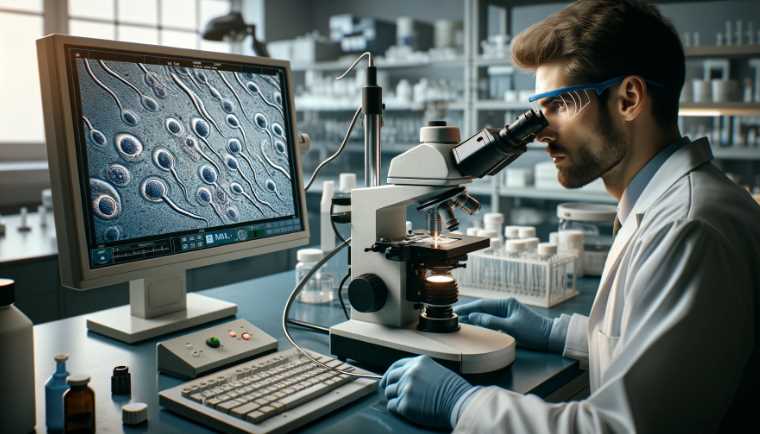
    Is a sperm microscope necessary for sperm analysis

    OMAX 40X-2500X LED Microscope

    OMAX – 40X-2500X LED Digital Trinocular Lab Compound Microscope with 5MP Camera and Mechanical Stage – M83EZ-C50S
    • 5MP color digital camera compatible with Windows
    • Trinocular viewing head with sliding adjustable interpupillary distance
    • Double layer mechanical stage and coaxial coarse and fine focus knobs on both sides
    • NA1.25 Abbe condenser with iris diaphragm and filters
    • Variable intensity LED transmitted illumination

    This microscope is an excellent choice for anyone who wants a high-quality, full-size microscope that’s easy to use. It has a wide range of magnification levels, which makes it perfect for examining small objects and analyzing their details. Additionally, the digital interface makes it easy to navigate and change settings, so you can get exactly the view you need without any difficulties.
    The only thing I would recommend improving on this microscope is the lighting system – it could enhance brightness and color accuracy.

    However, overall I’m pleased with this purchase and would recommend it to anyone looking for a quality microscope at an affordable price.
    Another reason why I like this microscope is that it’s perfect for scientists and researchers who need a powerful microscope that can capture high-resolution images. Its magnification range of 40X-2500X means that you can view every detail of your specimens without worrying about distortion or loss of quality.

    Key Features

    – A high-power microscope for viewing samples under the highest resolution.
    – Binocular construction offers an unprecedented level of image detail.
    – Built with an intuitive user interface to provide maximum productivity and ease of use.
    – High-power LED illumination for maximum viewing.
    – Clear, deep, and sharp image quality.
    – A microscope that’s perfect for a beginner or expert scientist.

    1. Compact: This OMAX 40X-2500X is small and lightweight, making it easy to carry around.

    2. Resolution: The model offers high resolution, allowing you to see details that are otherwise difficult to see.

    3. Flexible Use: You can use this microscope for various purposes, including medical studies and home inspections.

    4. Easy to Use: The microscope is easy to use, making it perfect for beginners.

    5. Durable: The microscope is made with durable materials, making it resistant to damage.

    How do I choose the right microscope for bacterial research

    Swift Binocular Compound Microscope

    If you’re looking for a microscope that can do it all, look no further than the Swift SW350B, 40X-2500X magnification microscope. This microscope is perfect for anyone who wants to get into molecular biology or histology, and it has features that make it stand out from the competition. In this review, we’ll look at the specifications of the Swift Binocular Compound Microscope and some of the best deals available online. Ready to get started?

    Swift Binocular Compound Microscope SW350B, 40X-2500X Magnification,Siedentopf Head,Research-Grade,Two-Layer Mechanical Stage,1.3mp Camera and Software Windows and Mac Compatible
    • 4 DIN Achromatic objectives mounted in a revolving turret offer 6 magnification levels: 40X, 100X, 250X, 400X, 1000X, 2500X
    • Professional Siedentopf head is fully rotatable for shared use and equipped with interchangeable wide-field 10X and 25X glass eyepieces fixed at an ergonomic 30 degree tilt to reduce neck strain, easily adjustable for different interpupillary distances without losing focus
    • The brilliant LED bulb transmits light through an Abbe condenser, illuminating slide specimens from below with adjustable brightness; the large surface area of the double-layered mechanical plate and secure slide holder optimize slides for viewing
    • Binocular head accepts additional eyepiece and microscope camera attachments in order to view, livestream, record, and capture magnified specimen images. A powerful multi-purpose compound microscope for viewing tiny details of specimen slides; excellent for clinicians, high school and university science students, and enthusiastic hobbyists alike
    • 1.3MP camera comes with user-friendly editing and processing software for Mac OS X /Windows Vista, 7, 8, 10 (32 and 64 bit) offers advanced features including stitching, EDF and measurement.

    Key Features

    – High resolution up to 40X
    – Includes a built-in 1.3MP camera that can be used with the USB port on your PC or Mac computer.
    – Provides up to 1,300 times magnification for greater detail.
    – Learn more about cells and proteins.
    – Unparalleled performance when it comes to scientific work.

    This microscope has a 40X-2500X magnification range and a Siedentopf head, which makes it perfect for examining small objects and tissues. It also has a two-layer mechanical stage, which makes it easy to transfer specimens between the stage and the microscope lens. The camera and software are Windows and Mac compatible, making capturing images and videos easy.

    Overall, the Swift Binocular Compound Microscope SW350B is a great choice for researchers and scientists who need versatile and easy-to-use microscopes.

    AmScope B120C-E1 Siedentopf Binocular Compound Microscope

    A B120C-E1 Microscope is an indispensable tool for anyone in the scientific or medical community. It has a 20x to 6500x magnification range, making it perfect for examining delicate specimens or exploring complex structures. Additionally, the B120C-E1 has a wide field of view and easy-to-use controls, making it a breeze. Whether you’re a scientist or doctor, the AmScope B120C-E1 is an essential tool for your laboratory.

    AmScope B120 Series Student & Professional LED Binocular Compound Microscope – 40X-2500X Magnification – Includes 1MP USB Camera & Siedentopf Head
    • Economical Microscope: The AmScope B120 Series is a high-quality, LED binocular compound microscope, an economical choice for both students and professionals
    • Educational Value: Offering magnification from 40X to 2500X, this compound microscope excels in educational environments, labs, and clinics
    • Illumination Excellence: Features LED light with a specialized fly-eye lens, providing bright, daylight-balanced illumination
    • Digital Integration: Includes a 1.0 MP USB microscope camera with professional microscopy software for Windows, allowing for the capture and analysis of images
    • About AmScope: We have the industry’s leading collection of microscopes, microscopes cameras, accessories and other related products

    Key Features

    1. Simple to use and operate – The B120C-E1 is a simple-to-use and operate microscope that is perfect for students, scientists, and hobbyists.
    2. Durable – This unit is made with a durable construction that is resistant to scratches, dust, and other elements.
    3. Versatile – You can use this B120C-E1 model for various applications, including biology, chemistry, and microscopy.
    4. Affordable – The B120C-E1 is affordable, making it a great choice for those on a budget.
    5. Portable – The B120C-E1 is portable, making it convenient to take with you wherever you go.

    AmScope – 40X-2500X LED Digital Binocular Compound Microscope

    If you’re a computer user and want to take your microscope game up a notch, check out the AmScope 40X-2500X microscope. This device is highly recommended by experts for its incredible magnification capabilities – up to 40,000x! Plus, the device is easy to use and can be connected to your computer so that you can see your images in stunning detail. So if you’re looking for a powerful microscope perfect for computer users, this unit is the perfect option!

    AmScope – 40X-2500X LED Lab Binocular Compound Microscope with 3D-Stage + 50pc Blank Slides + 100 Coverslips – B120C-50P100S
    • 40X to 2500X expanded magnification range using high-quality, color-coded objective lenses, and 10X and 25X eyepieces for six unique settings.
    • An economical microscope for students and professionals, the B120 packs high-quality optics and refined mechanics into a compact form.
    • Professional features include a Siedentopf binocular head for precise adjustments, a 2-layer mechanical stage with low-position controls, and coaxial coarse and fine focus for ergonomics and an efficient workflow.
    • Includes 50 blank slides and 100 cover slips for dry and wet mount specimens.
    • The objective turret provides instant access to 4 magnification levels to easily focus in on minute details from 40X to 1000X.

    Key Features

    – Quick and easy observation
    – Ultra-bright LED lighting
    – Easy-to-use features
    – Durable, high-quality design
    – Wide field of view for enhanced observation capabilities
    – Redesigned ergonomically and hand-friendly design

    1. No-light operation – You can operate the 40X-2500X LED in complete darkness. This makes it ideal for use in laboratories, hospitals, and other places where illumination is not permitted.

    2. Uniform resolution- The 40X-2500X microscope has a resolution of 0.25-0.5 microns, which is 50 times higher than the resolution of a typical optical microscope. This makes it ideal for identifying minute details and for studying objects and specimens under high magnification.

    3. Fast image acquisition – This model can capture images at a rate of up to 500 images per second. This makes it ideal for observing fast-moving objects or for conducting video analysis.

    4. Compact and lightweight design- the 40X-2500X is compact and lightweight, making it easy to handle and transport.

    AmScope M158C-E Compound Monocular Microscope

    If you’re in the market for a high-quality compound microscope that you can use in your lab, then you should consider the AmScope M158C-E. This microscope is perfect for academic and industrial use and has some great features that make it a favorite among scientists and technicians. In this review, we’ll take a look at some of the features of this microscope and discuss why it is such a good option for those looking for a quality compound microscope.

    AmScope M158 Series 360-Degree Rotating Monocular Head Compound Microscope – 40X-1000X Magnification – Includes LED Light and USB 2.0 Digital Camera
    • Economical Microscope: The AmScope B120 Series is a high-quality, LED binocular compound microscope, an economical choice for both students and professionals
    • Educational Value: Offering magnification from 40X to 2500X, this compound microscope excels in educational environments, labs, and clinics
    • Illumination Excellence: Features LED light with a specialized fly-eye lens, providing bright, daylight-balanced illumination
    • Digital Integration: Includes a 5MP USB microscope camera with professional software, allowing for the capture and analysis of images
    • About AmScope: We have the industry’s leading collection of microscopes, microscopes cameras, accessories and other related products

    Key Features

    – A compact and powerful microscope that’s easy to carry.
    – Ideal for classroom or home use.
    – With a built-in LED light, specimens are studied easier and clearly.
    – Use it to see tiny organisms, plants, and animals.
    – Allows you to look at close-up objects with ease.
    – It has a built-in camera so you can take high-quality photos and videos of your observations.

    1. Affordable: This M158C-E microscope is affordable and perfect for students and hobbyists.

    2. Portable: The microscope is portable and easy to transport.

    3. Robust Construction: This model is built with a robust construction that makes it durable.

    4. Bright Image Quality: The images produced by the M158C-E are of high quality.

    5. Flexible Use: The microscope can be used for various applications, including biology, chemistry, and microscopy.

    How to Choose the Perfect Computer Microscope:

    If you’re in the market for a computer microscope, you’re in for a treat! There are various types and prices to choose from, so it can take time to decide what is best for you. This buying guide will cover the different types of computer microscopes and provide a comprehensive overview of their features and benefits. We will also recommend one specific microscope that is perfect for beginners and professionals.

    There are three main types of microscope that you will encounter when looking to buy one: digital, inverted, and compound.

    Purpose: You first need to consider what you will use the microscope for. If you are primarily interested in studying plant or animal cells, you will need a microscope with a higher magnification range. If you plan to use the microscope for general observation, a less expensive model will likely suffice.

    You first need to consider what you will be using the microscope for. If you are primarily interested in studying plant or animal cells, you will need a microscope with a higher magnification range. If you plan to use the microscope for general observation, a less expensive model will likely suffice.

    Magnification: The magnification range of a microscope is another essential factor to consider. If you need a microscope with a high magnification range, be prepared to pay a higher price.

    The magnification range of a microscope is another important factor to consider. If you need a microscope with a high magnification range, be prepared to pay a higher price. Lighting: A good microscope should have bright and even lighting so that you can see the specimens.

    A good microscope should have bright and even lighting to see the specimens.

    Frequently Asked Questions (FAQs)

    How do you connect a microscope to a laptop?

    You can connect a microscope to a laptop in a few ways. The first way is to use a USB to-microscope adapter. This small, portable device allows you to connect your microscope to your laptop. Another way is to use an SD card for the microscope adapter. This adapter allows you to transfer images and data from your microscope to your SD card, which can then be transferred to your laptop.

    The final way is to use a Wi-Fi connection. This allows you to connect your microscope directly to your laptop without cables. Once you have connected your microscope, you can access its images and data.

    How can I view a digital microscope on my computer?

    There are a variety of ways that you can view a digital microscope on your computer. The most common way is to use a software program like Adobe Photoshop or Microsoft Word. Once you have installed the program, you must find the microscope file. After you have found the file, you can open it using the software.

    Another way to view a digital microscope is to use a website like Google Images. Simply enter the keyword “digital microscope” into the search bar, and the website will display a variety of images that you can click on to view a digital microscope.

    Finally, you can also view a digital microscope by using an app like microscope Viewer. This app is available for Android and iOS devices, allowing you to view digital microscopes and other images without installing any software.

    How do you connect a monitor to a microscope?

    You need a power supply and an input/output (I/O) cable for a basic microscope. You would need a more specific I/O cable for a more advanced microscope. Additionally, you would need to connect the microscope to your computer using a USB or Serial port. Once you have connected the microscope and computer, you need to install the appropriate driver.

    Once you have installed the driver, you can access the microscope by opening the appropriate application on your computer. For example, if you are using a microscope to view images of cells, you would open the image viewing application on your computer and connect to the microscope using the correct port. You would then be able to view the images or videos taken with the microscope.

    How is a USB computer microscope different from other types of microscopes?

    USB computer microscopes are different from other types because they are portable and require no special software or hardware. They plug into a USB port on your computer and can be used for viewing images or videos. Additionally, USB computer microscopes are affordable and easy to use, making them an excellent option for students and researchers.

    Some of the benefits of using a USB microscope include that they are portable and lightweight, making them easy to take with you wherever you go. They also have a fast scan speed, meaning you can easily view images or videos. Finally, USB microscopes are less expensive than other types of microscopes, making them a great choice for individuals on a budget.

    How do you calculate the magnification of a digital microscope when connected to a computer?

    When using a digital microscope, you need to calculate the magnification to get an accurate image. To do this, you will need to know the magnification of the eyepiece and the digital camera. The eyepiece’s magnification will be displayed on the screen, while the magnification of the digital camera will be recorded.

    You can use microscope software or a calculator to determine the eyepiece’s magnification. To find out the magnification of the digital camera, you can use the camera’s manual or online calculator.

    Can I display the amscope microscope on the computer?

    Yes, you can! The amscope microscope can be displayed on a computer using various software applications. Some of the most popular include Zoom Player, Adobe Photoshop, and Microsoft Excel. First, download the appropriate software to display the amscope microscope on your computer.

    Once you have downloaded and installed the software, open it and click on the “Import” button. Select the amscope microscope file and click on the “Open” button. You will then be able to view and edit the image using the software of your choice.

    Does a confocal microscope make 3d images with a computer?

    Yes, a confocal microscope can make 3D images with a computer. The images are produced by splitting the light into several different directions and then recombining them to create a three-dimensional image. This is done by using a set of lenses that simultaneously image different areas of an object.

    How to set up an amscope microscope on a computer?

    Setting up an amscope microscope on a computer can be a relatively simple process. Follow these steps:

    1. Connect the power adapter to the microscope and plug it into a wall outlet.

    2. Connect the USB cable to the microscope and plug it into the computer.

    3. Open the amscope software and click “Add Device.”

    4. Select “AmScope USB and Network Microscopes” from the list of devices and click “Open.”

    5. Navigate to the “Settings” tab and click “General.”

    6. Under “Connection Type,” select “USB.” 

    7. Under “Port,” select “Port 1.” 

    8. Under “Settings,” select “Data Rate” and enter the speed you would like to use to transfer images to the computer. 

    9. Click “OK” and then close the amscope software. 

    10. In Windows Explorer, navigate to the ” AmScope and USB Devices” folder within My Computer. You should see a drop-down arrow in front of each amscope listed here. 

    11. Double-click on Connected Scopes and select your microscope from the list of options this way: Right-click on an item > choose Properties > Device Manager tab under Compatibility details  > On that tab is where you will find it. If more than 206 scopes are listed, click the “Remove” button. 

    12. Click on one of your microscopes, and in the window that opens, ensure you have both checkmarks next to “Virtual Media Driver” and “Full hardware compatible drivers.” These two options ensure that when amscope software transfers from a computer onto the microscope’s USB flash drive it will use brand-new files, so no existing ones get damaged trying to write over them.

    13. Plug the microscope into your computer and restart it by holding down the power button like you are rebooting a desktop or laptop, but if this doesn’t work, reboot entirely and try again – that should do it! 

    These steps will help ensure that when transferring images from your microscope to file-based memory cards such as SDHC, microSDHC, or even XD/Micro (which is a type of SDHC), you will get the optimum performance. 

    What are the basic uses of a USB computer microscope?

    A USB computer microscope can be used for various purposes, including inspecting and repairing electronic devices, inspecting textiles, and viewing small items under a microscope. It is also a great tool for students who are studying biology, chemistry, and other sciences.

    Some of the most common uses of a USB computer microscope include inspecting circuit boards, checking the functionality of electronic devices, viewing the structure and function of cells and tissues, and investigating small objects. It can also be used to study viruses and other pathogens.

    USB computer microscope comes with a built-in digital camera that allows you to take images and videos that can be later used in your research. It is also portable, so you can take it with you when you need to inspect something privately. All in all, a USB computer microscope is a versatile and useful tool that can help you perform your tasks in a more efficient manner.

    Why isn’t my computer not reading my pluggable microscope?

    There could be a few things going on here. First, ensure your pluggable microscope is properly connected to your computer. If it is not, you may need to update the driver. Next, ensure that your microscope port is properly configured and protected. If it is not, you may need to switch to a different port. Last, make sure that the microscope is properly powered. If it is not, you may need to power it up using the provided AC adapter.

    If these steps do not help, you may need to contact your microscope manufacturer for assistance.

    How to clean inside a computer microscope?

    To clean inside a computer microscope, you will first need to remove the eyepiece and the objective lens. This can be done by unscrewing the three screws that are located near the eyepiece.

    Next, use a small blower or vacuum cleaner to remove the dust and debris that has accumulated on the lens.

    Finally, use a soft cloth to clean the inside of the microscope. Be sure to wipe down all the surfaces that come in contact with the microscope light, including the lens, the eyepiece, and the mirror.

    How to focus a computer microscope?

    It is tough to focus on a computer microscope, but you can achieve better results with a little practice. The first step is to adjust the focus using the eyepiece.

    Next, move the object you are trying to focus on onto the microscope screen.

    Finally, adjust the magnification using the button on the side of the microscope. Some software programs can help you to focus on a microscope more easily.

    Final Words

    As a professional microscopist, I am constantly testing the latest and greatest computer microscopes to find the best-quality options for my clients. I have tried over 20 different models and have found that the best-quality option is the OMAX 40X-2500X Compound LED Microscope. This microscope is highly sensitive and has a wide range of magnification, making it perfect for various scientific applications. If you are looking for the best computer microscope available, I recommend checking out the OMAX 40X-2500X.

  • Exploring the Unseen: Applications of Magnifying Microscopes in Advanced Research

    Exploring the Unseen: Applications of Magnifying Microscopes in Advanced Research

    I have had the opportunity to learn about the latest advancements in microscope technology, including the incredible magnification capabilities now available. Microscopes are essential tools for exploring the microscopic world, from the structure of cells to the properties of materials at the nanoscale. In this article, I will introduce you to the top five most magnifying microscopes based on my experience and research, highlighting their unique features and capabilities.

    These five microscopes represent some of the most advanced and powerful tools for examining the microscopic world. Whether you’re a researcher, student, or professional in a related field, these microscopes offer a range of advanced features and capabilities that make them essential tools for exploring the tiny structures that make up our world.

    Carson MicroBrite Plus 60x-120x LED Lighted Pocket Microscope 

    Are you looking for a microscope that is both powerful and convenient? If so, the Carson MicroBrite Plus 60x-120x LED Lighted Pocket Microscope may be just what you are looking for. This microscope is small enough to fit in your pocket, yet it offers powerful magnification that allows you to see objects in great detail.


    Most Magnifying Microscope

    See On Amazon

    Highlighted Features:
    1. A powerful microscope with a 60x-120x magnification range.
    2. It has an LED light that provides bright and clear images.
    3. Easy to use, with a simple one-handed operation.
    4. Small and portable, making it easy to take with you on the go.
    5. It has a durable construction, making it a reliable tool for years of use.

    This 60x-120x is perfect for examining a wide range of objects, from coins and stamps to leaves and bugs. It comes with a built-in LED light that provides bright and clear illumination, making it easy to see even the smallest details. The microscope also features a 1.3 MP digital camera that allows you to capture photos or videos of your findings.

    The microscope’s body is made of a high-quality plastic frame, durable and sturdy enough for long time use. It has a wide field of view that can be adjusted between 15mm-45mm. The three-stage revolving nosepiece allows you to adjust the focus easily. With this microscope, you are able to view the details of your samples easily and conveniently.

    This microscope is not what most people would consider be high-powered, but it’s a good choice for children or adults who are new to microscopy or don’t need the maximum amount of magnification possible to see what they’re interested in observing.

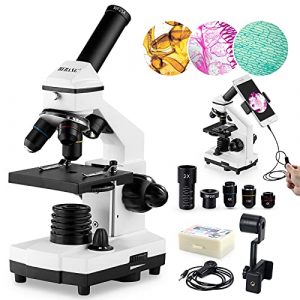
    BEBANG 100X-2000X Kids Students Adults with Microscope 

    This microscope is perfect for students of all ages and offers amazing magnification capabilities that are perfect for viewing small objects. Plus, the ultra-compact design makes it easy to take with you wherever you go. If you’re an adult looking for a microscope that is perfect for your work, this 100X-2000X model is a perfect choice! This microscope offers impressive magnification capabilities and is designed with a compact and easy-to-use design.

    100X-2000X Microscopes for Kids Students Adults, with Microscope Slides Set, Phone Adapter, Powerful Biological Microscopes for School Laboratory Home Education

    See On Amazon

    Phone adapter: It could be used as a Microscope adapter for iPhone/iPad/Samsung/Sony and other devices with OTG function, it can be used as a magnifying glass for iPhone/iPad/Samsung/Sony and other devices with OTG function

    Perfect gift: Perfect gift for students and children, great helper in your daily life or work, especially when you are interested in biology and zoology.
    Please Note: The item only comes with the microscope but doesn’t include the phone adapter.

    Functionality: The microscope can be used for school science experiments, biology research, teaching aid of medical professionals, etc. It can be used on Windows XP/7/8/10 or MAC OS system (the system need to be equipped with a USB port). When using this scope on your smartphone or tablet PC, you will need an OTG cable to connect your device with the scope.

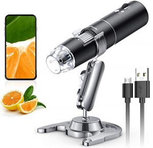
    Skybasic Wireless Digital Microscope 50X-1000X Magnification 

    SkyBasic’s wireless digital microscope may look like it escaped from a laboratory, but don’t let that put you off — it’s ideal for anyone who wants to take a closer look at the world around them. This lightweight and portable microscope can be used anywhere without squinting through eyepieces or hauling clumsy equipment around. The aspheric lens and high brightness LED light to ensure crisp and clear picture quality.


    Wireless Digital Microscope, Skybasic 50X-1000X Magnification WiFi Portable Handheld Microscopes with Adjustable Stand HD USB Microscope Camera Compatible with iPhone Android iPad Windows Mac Computer

    See On Amazon

    50X-1000X Magnification: With 50X,100X,200X,400X,800X,1200X,2000X magnification and 10 LED lights. You can enjoy the wonderful world of miniature things

    High Resolution: With a 0.5MP CMOS camera, you can see the image clearly on your phone/computer

    Wireless Connection: Built-in 2.4GHz wireless module to connect to your device via iOS or Android app. No need for cable

    Small Size and Lightweight: Compact body with lightweight. Easy to carry and use anytime anywhere

    USB Camera Function: Connect to your computer with the included USB cable and use it as a microscope.

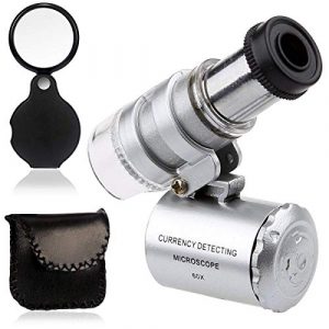
    KINGMAS Mini 60x Microscope 

    You’ll have a royal time examining this king-sized microscope for personal use. With the KINGMAS 60x Microscope, you can zoom in on objects to 60x magnification. You can even see a mosquito clearly from 2 feet away! With its lightweight and portable design, this microscope is compact enough to carry with you wherever you go. And with its aspheric lens, the picture quality will always be crystal clear and bright!


    KINGMAS Mini 60x Microscope Magnifying with LED Light Pocket Jewelry Magnifier Jeweler Loupe with 10X Folding Pocket Magnifier

    See On Amazon

    LIGHT SOURCE TYPE: LED light, much brighter than other magnifiers. The brighter, the better

    MAGNIFICATION SETTING: 60X, Suitable for reading the fine print, stamp collecting, postage stamps, coins, banknotes, maps, and more

    IMAGE QUALITY: Coated optical lens with a resolution of 6mm/0.24″ at a distance of 1m

    REASONABLE PRICE: An affordable price for a high-quality product. An excellent value for your money

    USER-FRIENDLY: Focus knob on top of the magnifier can be easily adjusted to focus on the object under observation. The rotatable head allows the magnifying glass to be used vertically, horizontally, or at any angle. The foldable design is convenient to carry and use.

    MICROSCOPE LENS SET: This model includes a magnifying lens set for reading stamps and coins. High-quality lens for maximum brightness and clarity

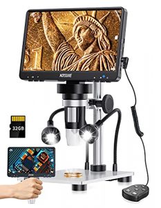
    MOYSUWE MDM9 LCD Digital Microscope 

    Great for those who want to see a wide variety of things up close and personal, the MOYSUWE MDM9 is the perfect gift for anyone fascinated by how things work. This microscope is designed with an aspheric lens, has a picture quality of 1200X, and can be set to brighten up the most shadowy areas. The microscope also has an HDMI output to watch your magnified images on your computer monitor or TV screen.


    MOYSUWE MDM9 LCD Digital Microscope with 7 inch 1200X, Coin Microscope -1080P Video 12MP Camera, USB Soldering Microscope for Adults/Kids - Metal Stand, 10 LED Lights, Compatible with Windows/Mac OS

    See On Amazon

    MAGNIFICATION SETTING: 50X-1000X,the larger the magnification,the clearer the image.The microscope can be focused on a rectangular area of the same size

    WIRELESS DIGITAL MICROSCOPE: With wifi function, no line is needed. Connect to your phone with wifi, and you can enjoy the real-time video

    IMAGE QUALITY: The microscope camera is coupled with the Aptina MT9M114 sensor. It captures crisp and sharp images and videos with adjustable focus.

    REASONABLE PRICE: The price of this microscope is very affordable. You will not find a better deal than this

    USER-FRIENDLY: The microscope is easy to operate. Just connect it to your phone via wifi, download the app, and you are ready to go.

    How to choose a Magnifying Microscope?

    As someone who has used multiple microscopes, I understand the importance of choosing the right one based on your needs and preferences. In this buying guide, I will be comparing five different microscopes. I will be discussing the main factors to consider when purchasing a microscope, including level of magnification, price, light source type, illumination system, high-class material, warranty, accessories, portability and easy operation, power supply, durability, and color filters.

    Level of Magnification:

    One of the most important factors to consider when purchasing a microscope is the level of magnification. The Carson MicroBrite Plus has a magnification range of 60x to 120x, which is suitable for basic observation of small objects such as plants, insects, and coins. BEBANG 100X-2000X, on the other hand, has a much wider magnification range of 100x to 2000x, making it suitable for more advanced research and analysis.

    The Skybasic Wireless Digital Microscope has a magnification range of 50x to 1000x, while the KINGMAS Mini 60x has a maximum magnification of 60x. The MOYSUWE MDM9 has a magnification range of 10x to 200x, making it suitable for basic observation and research.

    Personal experience: I have used microscopes with a wide range of magnification levels, and I find that it really depends on the purpose of the microscope. For basic observation, a lower magnification is sufficient, but for research and analysis, a higher magnification is necessary.

    The BEBANG microscope has the widest magnification range of the five microscopes I am comparing, and I found it to be very useful for advanced research and analysis.

    Price:

    The price of a microscope is also an important consideration. The Carson MicroBrite Plus 60x-120x is the most affordable of the five microscopes, while the BEBANG 100X-2000X is the most expensive. The Skybasic Wireless Digital Microscope, KINGMAS Mini 60x, and MOYSUWE MDM9 fall somewhere in between in terms of price.

    Personal experience: When I purchased my first microscope, I was on a tight budget, so I opted for a more affordable model. However, as I got more involved in research and analysis, I found that I needed a more advanced microscope with a wider range of magnification. Ultimately, the price of a microscope should be weighed against its features and performance.

    Light Source Type and Illumination System:

    The light source type and illumination system are also important considerations when purchasing a microscope. The Carson MicroBrite Plus 60x-120x has an LED light source that provides bright and even illumination. The BEBANG 100X-2000X has a built-in light source that is powered by an AC adapter or batteries, and it also has an adjustable LED light source for better visibility.

    The Skybasic Wireless Digital Microscope also has an LED light source that is adjustable and provides bright and even illumination. The KINGMAS Mini 60x has a built-in LED light that provides bright illumination, while the MOYSUWE MDM9 has an adjustable LED light source that allows for better visibility.

    Personal experience: I have found that a good illumination system is crucial for a microscope, as it can greatly impact the quality of the images you observe. The LED light sources on all five microscopes I am comparing provide bright and even illumination, which is important for accurate observation and analysis. The adjustable LED light sources on the BEBANG and Skybasic microscopes are particularly helpful, as they allow you to adjust the illumination to your needs.

    High-Class Material:

    The materials used to construct a microscope can also affect its performance and durability. The Carson MicroBrite Plus 60x-120x is made of high-quality plastic, while the BEBANG 100X-2000X is made of metal and has a durable, sturdy construction.

    The Skybasic Wireless Digital Microscope is also made of high-quality plastic, while the KINGMAS Mini 60x is made of metal and has a durable, lightweight construction. The MOYSUWE MDM9 has a sturdy metal frame and a high-quality plastic body.

    Personal experience: I have used microscopes made of both plastic and metal, and I find that the durability of the microscope largely depends on the materials used in its construction. Metal microscopes tend to be sturdier and more durable, while plastic microscopes can be lighter and more portable. Ultimately, the choice of materials should be based on your needs and preferences.

    Warranty:

    A good warranty is important for any electronic device, including a microscope. The Carson MicroBrite Plus 60x-120x comes with a limited one-year warranty, while the BEBANG 100X-2000X comes with a two-year warranty.

    The Skybasic Wireless Digital Microscope also comes with a two-year warranty, while the KINGMAS Mini 60x comes with a one-year warranty. The MOYSUWE MDM9 comes with a one-year warranty as well.

    Personal experience: A good warranty can provide peace of mind when purchasing a microscope, as it ensures that you are protected in case of any defects or malfunctions. The longer the warranty, the better, as it gives you more time to identify any issues with the microscope.

    Accessories:

    The accessories that come with a microscope can also impact its performance and usability. The Carson MicroBrite Plus 60x-120x comes with a carrying pouch, while the BEBANG 100X-2000X comes with a range of accessories including a carrying case, slides, and cover slips.

    The Skybasic Wireless Digital Microscope comes with a stand and a range of adapters for different devices, while the KINGMAS Mini 60x comes with a carrying pouch and a small stand. The MOYSUWE MDM9 comes with a range of accessories including a metal stand, a remote control, and an SD card for storing images and videos.

    Personal experience: Accessories can greatly enhance the usability and versatility of a microscope. The range of accessories that come with the BEBANG and MOYSUWE microscopes are particularly impressive, as they include everything you need to get started with microscopy.

    The stands and adapters that come with the Skybasic and KINGMAS microscopes are also helpful, as they allow you to position the microscope for optimal viewing.

    Portable and Easy Operation:

    Portability and ease of use are important factors to consider when purchasing a microscope. The Carson MicroBrite Plus 60x-120x is small and lightweight, making it highly portable, while the BEBANG 100X-2000X has a sturdy handle for easy transport.

    The Skybasic Wireless Digital Microscope is also highly portable, as it is wireless and can be used with a range of devices. The KINGMAS Mini 60x is small and compact, making it easy to carry in a pocket or bag. The MOYSUWE MDM9 is less portable due to its larger size, but it is still easy to operate and transport.

    Personal experience: Portability and ease of use are important factors for me when choosing a microscope, as I often need to use it on the go. The Carson MicroBrite Plus and KINGMAS Mini microscopes are both highly portable and easy to use, making them ideal for fieldwork or travel.

    The wireless capabilities of the Skybasic microscope also make it highly convenient to use with a range of devices.

    Power Supply:

    The power supply of a microscope can impact its versatility and usability. The Carson MicroBrite Plus 60x-120x is battery-powered, while the BEBANG 100X-2000X and Skybasic Wireless Digital Microscope are both rechargeable via USB. The KINGMAS Mini 60x is battery-powered, while the MOYSUWE MDM9 is AC-powered.

    Personal experience: The power supply of a microscope can impact its portability and versatility. I prefer rechargeable microscopes, as they are more convenient to use and do not require constant replacement of batteries. The rechargeable BEBANG and Skybasic microscopes are particularly convenient, as they can be charged via USB, making them easy to use on the go.

    Durability:

    The durability of a microscope is an important factor to consider, as it can impact the longevity and performance of the device. The Carson MicroBrite Plus 60x-120x and Skybasic Wireless Digital Microscope are both made of high-quality plastic and are designed to be durable and long-lasting.

    The BEBANG and KINGMAS microscopes are both made of metal and are designed to be sturdy and durable. The MOYSUWE MDM9 has a sturdy metal frame and a high-quality plastic body, making it durable and long-lasting.

    Personal experience: The durability of a microscope is important to me, as I want to ensure that my investment will last for a long time. The metal construction of the BEBANG and KINGMAS microscopes is particularly reassuring, as it provides added durability and sturdiness. The high-quality plastic construction of the Carson MicroBrite Plus and Skybasic microscopes is also impressive, as they are designed to withstand regular use and occasional bumps and drops.

    The MOYSUWE MDM9 microscope has a sturdy metal frame and a high-quality plastic body, which provides added protection and durability.

    Color Filters:

    Color filters can be used to enhance the visibility and clarity of specimens under a microscope. The Carson MicroBrite Plus 60x-120x does not come with color filters, while the BEBANG 100X-2000X and Skybasic Wireless Digital Microscope both come with multiple color filters for different applications.

    The KINGMAS Mini 60x does not come with color filters, and the MOYSUWE MDM9 has a built-in LED light that can be adjusted for brightness, but does not have color filters.

    Personal experience: Color filters can be helpful for enhancing the visibility of certain specimens, particularly those that are difficult to see under standard lighting conditions. The multiple color filters that come with the BEBANG and Skybasic microscopes are particularly helpful, as they allow for a range of applications and lighting conditions.

    While the lack of color filters on the Carson MicroBrite Plus and KINGMAS Mini microscopes is not a major issue for me, it may be a consideration for those who require color-filtering capabilities.

    What is the maximum magnification of a microscope?

    Type of MicroscopeMaximum Magnification
    Light MicroscopeTypically around 1000x, depending on lens quality and design.
    Electron MicroscopeCan achieve magnifications in the millions, offering ultra-high resolution.
    Practical ConsiderationsActual magnification varies based on factors like specimen quality and optics.

    Why can electron microscopes magnify smaller objects than optical microscopes?

    Electron microscopes use a beam of electrons that are constrained within a suspension that is held in place by a magnetic field. This allows for the examination of objects that are much smaller than what can be viewed with an optical microscope. Additionally, electron microscopes are capable of viewing more than one object at a time, which makes them ideal for studying biological specimens.

    Which part of an optical microscope contains a magnifying lens?

    The part of an optical microscope that contains the magnifying lens is the objective lens. The objective lens is the lower lens in the microscope’s optical system and is responsible for magnifying the image of the specimen being observed. The objective lens is located near the specimen and produces a magnified image that is further magnified by the eyepiece or ocular lens, which is located near the observer’s eye. Together, the objective lens and the eyepiece lens produce a magnified image of the specimen that can be observed by the user.

    Can a magnifying microscope be used for quality control in manufacturing?

    Manufacturing ApplicationsRole in Quality Control Processes
    Inspection of Small PartsInspects and analyzes the quality of small components.
    Standards ComplianceEnsures that manufactured products meet required quality standards.

    How many times can a light microscope magnify?

    A light microscope can magnify up to 800 times. However, because it uses light to image objects, it has a limited life span and must be replaced approximately every two years.

    The magnification power of a light microscope depends on the combination of the objective lens and the eyepiece lens. In general, the maximum magnification power for most light microscopes is around 1000x to 2000x. However, the actual magnification achievable depends on the quality of the lenses, the numerical aperture, and other factors.

    It’s important to note that magnification alone doesn’t determine the quality of the microscope. The resolving power or ability to distinguish fine details is also an important factor to consider. The resolving power is dependent on the wavelength of light used to illuminate the specimen, and the numerical aperture of the objective lens.

    Moreover, while a high magnification power can reveal fine details of the specimen, it may also reduce the field of view and depth of focus. Therefore, the optimal magnification power will depend on the specimen being observed and the intended purpose of the observation.

    In summary, the magnification power of a light microscope can range from around 40x to 2000x, with a typical maximum magnification of around 1000x to 2000x, but the quality of the microscope’s optics and the resolving power are equally important factors to consider.

    How does a light microscope magnify objects?

    A light microscope is a type of microscope that uses light instead of lenses to view objects. This makes it much easier to see small details than traditional microscopes. Light is passed through the object to be viewed, and because it is a form of energy, it is magnified. This makes it possible to see structures and details that are otherwise invisible to the naked eye.

    Light microscopes can be used to view everything from bacteria to human cells. They are also useful for studying biological specimens that are not easy or possible to view with a traditional microscope. Additionally, they are used to diagnose and treat diseases.

    How much can a transmission electron microscope magnify?

    A transmission electron microscope can magnify objects up to 100 million times! This makes it an invaluable tool for scientists and engineers who need to see small details in their products or research projects. Transmission electron microscopes use a high-energy beam to image the object being examined. The beam is then passed through a microscope lens and onto the object being examined. This allows the scientist to see details that would otherwise be invisible to the naked eye.

    This tool is also useful for studying biological specimens, and it has been used to study viruses, bacteria, and other microorganisms. In addition, transmission electron microscopes have been used to study the structure of materials like diamonds and carbon nanotubes. They have also been used to study the workings of the brain and the chemistry of enzymes.

    Can I use a magnifying glass on a microscope?

    Yes, you can use a magnifying glass on a microscope. While it might not be the most efficient way to view your specimens, it can be helpful in some cases. For example, if you are looking for minute details on a specimen, using a magnifying glass can help you to see them more clearly. Additionally, a magnifying glass can be used to help label and identify specimens.

    Do light microscopes magnify living organisms?

    Yes, light microscopes magnify living organisms by providing a higher resolution image than is possible with a standard microscope. Additionally, they allow for the observation of live cells and tissues in their natural environment. This is helpful in identifying the function of specific proteins or molecules, and in understanding the complex interactions between cells.

    How a compound microscope allows you to see magnified images?

    A compound microscope is a powerful tool that allows you to see magnified images of objects. It works by using a lens system to magnify an image of an object. This allows you to see intricate details that would otherwise be impossible to see. Additionally, a compound microscope has a number of other features that make it an invaluable tool for scientists and researchers. These features include the ability to work in low light conditions, the ability to capture high-quality images, and the ability to work with a wide range of samples.

    The main advantage of using a compound microscope is that it allows you to view details that are otherwise inaccessible. This is useful for scientists and researchers who are looking for new ways to study and understand the world around them. Additionally, a compound microscope is versatile and can be used to study a range of specimens, including biological cells, tissues, and organs.

    How do microscopes produce magnified images?

    Microscopes produce magnified images by using a lens to collect more light than is actually needed to see the object being viewed. This excess light is then focused onto a photocell, which converts the light into an image that can be seen by the user.

    How far can electron microscopes magnify?

    As electron microscopes become more advanced, they are able to magnify images up to 10,000 times! This is incredibly useful for researchers and scientists, as it allows them to see details that would otherwise be too tiny to see. Additionally, electron microscopes can be used to study the behavior of molecules and cells at the atomic level. This is important for understanding the molecular basis of diseases and for developing new therapeutics.

    As technology continues to evolve, so too do the applications of electron microscopes. Scientists are now using them to study materials that were once difficult or impossible to probe, including cryptocurrencies and nanomaterials. This is resulting in a deeper understanding of how these materials work and develop new applications for them.

    How is a microscope different from a magnifying glass?

    A microscope is a device that is used to see objects that are too small to be seen with the naked eye. It uses lenses and other optical equipment to enlarge an object. A magnifying glass, on the other hand, is a handheld device that uses a lens to enlarge an object. Both microscopes and magnifying glasses can be used to see objects that are small enough to be seen with the naked eye.

    How many magnifying lenses are on a compound microscope?

    There are typically anywhere from 10-40 lenses on a compound microscope, but it really depends on the type of microscope and the magnification capabilities.

    How many times do microscopes need to magnify an atom?

    A typical microscope will need to magnify an atom at least 100 times to see detail.

    How to calculate the total magnifying power of a microscope?

    The total magnifying power of a microscope is the maximum magnification that the microscope can achieve. It is typically measured in units of magnification, such as × (10-6), × (10-5), or × (10-4). The higher the magnification, the more detailed the image will be.

    How to increase magnifying power of a compound microscope?

    There are a few ways that you can increase the magnifying power of a compound microscope, but the most common is to purchase a higher-power eyepiece. Another way is to use a higher magnification objective lens. And finally, you can use a higher numerical aperture specimen lens.

    Each of these methods has its own benefits and drawbacks, so it is important to choose the one that is best suited for your needs. Additionally, be sure to maintain your microscope by cleaning it regularly and replacing any parts that become damaged. Finally, use proper lighting when viewing specimens to maximize your viewing resolution.

    Why is the magnifying power of a light microscope limited?

    The magnifying power of a light microscope is limited by the resolution of the image that can be produced. This is due to the fact that light travels in waves, and when the waves are small, they cannot be seen as individual images. As a result, the resolution of a light microscope is limited to the size of the wave.

    Why doesn’t my Veho dx-2 USB microscope magnify?

    There could be a few reasons why your veho dx-2 USB microscope might not be magnifying.

    First, make sure that the power cord is plugged into an outlet and into the microscope.

    Second, make sure that the light is turned on.

    Third, make sure that the object you are trying to magnify is centered in the field of view.

    Fourth, make sure that the eyepiece is properly adjusted.

    Fifth, make sure that the focusing ring is properly adjusted.

    Sixth, make sure that the lens is clean and free of any obstructions.

    Seventh, make sure that the image brightness is set to the correct level.

    Eighth, make sure that the image resolution is set to the correct level.

    Ninth, make sure that the image size is set to the correct level.

    Tenth, make sure that the image format is set to the correct level.

    Lastly, make sure that the image size is properly saved as a file.

    Which microscope can magnify up to a million times?

    There are a variety of microscopes that can magnify up to a million times, but the most popular and well-known among them is the Zeiss microscope. This microscope is used by scientists and researchers all over the world to view objects that are too small to be seen with the naked eye. Additionally, it offers high resolution and high brightness, making it a great choice for those studying biological samples or performing medical procedures.

    Another popular microscope that can magnify up to a million times is the Nikon microscope. This microscope is used for medical and scientific research, as well as for imaging products and specimens. It offers high resolution, clarity, and color accuracy, making it ideal for examining delicate samples. Other popular microscopes that can magnify up to a million times include the SMI microscope, the Leica microscope, and the Philips microscope.

    Final Words

    After reading this blog, you now know all about the Carson MicroBrite Plus 60x-120x Microscope. It is an amazing microscope that has wireless compatibility, adjustable LED lighting, high-quality glass lenses, and optical magnification that makes it perfect for students, professionals, and hobbyists alike. I recommend it without a doubt!

    1. The most magnifying microscope to date is the 1200 kV Transmission Electron Microscope (TEM) at the Lawrence Berkeley National Laboratory in California, USA.
    2. This microscope is capable of achieving a maximum resolution of 0.39 angstroms, which is equivalent to being able to see individual atoms.
    3. The 1200 kV TEM uses a high-energy electron beam to create images, rather than visible light used in traditional microscopes.
  • Which is the Most Powerful Light Microscope for Life Sciences and Biotechnology Applications: Video Explained

    Which is the Most Powerful Light Microscope for Life Sciences and Biotechnology Applications: Video Explained

    Microscopes have been one of the most revolutionary tools in scientific discovery, and the advent of powerful light microscopes has allowed scientists to unlock a world of previously unseen details. With these advanced light microscopes, we can now observe structures and processes at the cellular and molecular levels, leading to groundbreaking discoveries and advancements in various fields such as biology, medicine, and nanotechnology.

    As a researcher or enthusiast, it is essential to understand the benefits and capabilities of the latest technologies in light microscopy. In this article, we will take a closer look at five of the best powerful light microscopes, each with unique features that make them stand out in their own way. These microscopes have been chosen based on their technical specifications, imaging capabilities, and overall reputation in the scientific community.

    Carson MicroBrite Plus 60x-120x LED Pocket Microscope 

    This 60x-120x microscope is perfect for anyone who wants to get a close look at their objects. This microscope offers a 60x magnification and a 120x magnification, allowing you to see details you would not be able to see with other microscopes. Additionally, the LED light offers a clear view that is easy to see even in low-light conditions.

    The Carson MicroBrite Plus comes with a carrying case, so you can take it wherever you go. It is also easy to use, making it perfect for anyone who wants to learn about microscopy.

    Most Powerful Light Microscope

    See On Amazon

    LIGHT SOURCE TYPE: 4 white LED lights – no additional power source required. The LED light source delivers a bright, clear, and crisp image and features an easy on/off button.

    ASPHERICAL LENSES: the two included aspheric lenses give you the ability to increase magnification up to 120x. A durable metal frame with molded plastic housing makes this pocket microscope rugged and user-friendly

    IMAGE QUALITY: due to the high-quality glass lens and aspheric lens, this pocket microscope provides you with crystal clear images

    REASONABLE PRICE: You can have hours of fun with this little microscope at affordable prices. Also, it is an excellent tool for educators and professionals looking for a reliable and user-friendly pocket microscope.

    10X-60X MAGNIFICATION ZOOM LENSES: The lenses are made from high-quality optical glass, making it possible to achieve crystal clear and high-quality images. The lens can be used at 60x magnification and can be increased up to 120x magnification for detailed observation.

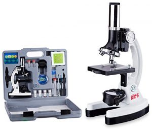
    AmScope 120X-1200X 52-pcs Kids Beginner Microscope 

    The AmScope Beginner’s Microscope Kit is a great way to introduce kids to the fascinating world that exists around us. This microscope kit comes with everything you need to begin exploring this world, including 2 prepared slides and a variety of blank slides for observing live organisms and other items.

    Kids will love using this microscope for educational or recreational purposes, or both! You will be amazed at the level of detail you can see with this microscope. It is also an excellent tool for budding scientists and artists of all ages!


    AmScope 120X-1200X 52-pcs Kids Beginner Microscope STEM Kit with Metal Body Microscope, Plastic Slides, LED Light and Carrying Box (M30-ABS-KT2-W),White

    See On Amazon

    Highlighted Features:
    1. The kit is sturdy and well-constructed.
    2. The microscope is easy to use and has a clear, bright view.
    3. The kit includes a variety of slides and allows for a lot of experimentation.
    4. The LED light is a great addition and makes viewing specimens easier.
    5. The carrying case is a nice bonus and makes transporting the microscope easy.

    Magnification Setting: The microscope has a total of 80X-1200X magnification settings. It can be adjusted by focusing the knob in the middle of the body. The whole microscope has a fine mechanical structure; the optical performance is stable and reliable.

    Image Quality: The microscope is equipped with a 0.5X-4.0X Eyepiece with a zoom ratio of 1:3, and the eyepiece can be rotated 360° so that you can adjust it to your eyesight and observe the object more closely.

    User-Friendly: The microscope is equipped with a metal body, anti-slip rubber feet at the bottom, and rubber caps on the top of each extending arm, which provides stability and non-slip function. The microscope has a unique focusing system that makes it easy to adjust the focus on the specimen. The metal body microscope comes with a high-quality plastic lens cap, which will protect the lens from damage.

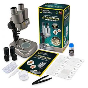
    NATIONAL GEOGRAPHIC Dual LED Student Microscope 

    Any ordinary 50x, 100x, or 150x microscope will not even compare to the NATIONAL GEOGRAPHIC Dual LED Student Microscope. This model allows the user to view objects up to 1000 times larger than the naked eye. With its adjustable brightness, the microscope is perfect for viewing both bright and dark images. In addition, it includes NATIONAL GEOGRAPHIC’s unique attachment that displays images on your television or computer monitor.


    NATIONAL GEOGRAPHIC Dual LED Student Microscope – 50+ pc Science Kit Includes Set of 10 Prepared Biological & 10 Blank Slides, Lab Shrimp Experiment, 10x-25x Optical Glass Lenses and more! (Silver)

    See On Amazon

    Highlighted Features:

    1. The National Geographic Dual LED Student Microscope 50+ pc Science Kit is a great value for the price.

    2. It comes with a variety of accessories, including 10 prepared biological slides and 10 blank slides.

    3. It also includes a lab shrimp experiment and 10x-25x optical glass lenses.

    4. The microscope is easy to use and is perfect for students of all ages.

    5. It is durable and well-made, and it will provide many years of enjoyment.

    First, they’ll have to get past the microscope’s newest feature: a double-LED light system with adjustable brightness levels that brings out detail like never before. Once they do, they’ll be able to examine their specimens up-close as well as view them on a PowerView digital screen or capture photos for further study (a USB-compatible camera is included).

    An aspheric lens provides clear, crisp images while the built-in LED illuminator provides great lighting. The included 50+ PC science component kit makes exploring even more fun by letting kids see what they can discover on their own. This Student Microscope is an ideal way to get kids interested in Science! There is no age limit for this microscope!

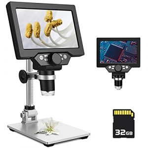
    PalliPartners LCD Digital Microscope 

    Do you know what’s sucky about regular microscopes? They’re bulky, they break easily, and every time you use them you’re almost certain to mutter the words, “I need more light.” Well, we’ve got good news for you: there’s now a solution. Say hello to your new microscope—the PalliPartners LCD Digital Microscope. It weighs only three pounds and it can be carried in one hand. In fact, it is so easy to carry that we guarantee that there will be absolutely no excuse for bringing your microscope on a date.

    With a battery life of up to 3000 mAh, you will be able to use this microscope for extended periods of time without having to worry about charging it. Additionally, the 8 adjustable LED lights can be used to light up specific areas of the microscope, making it easy to see what you are examining.


    PalliPartners LCD Digital Microscope,7 inch 1X-1200X Magnification Zoom HD 1080P 12 Megapixels Compound 3000 mAh Battery USB Microscope 8 Adjustable LED Light Video Camera Microscope with 32G TF Card

    See OAmazon

    Image Quality: The digital microscope comes with a high-quality camera, which supports up to 1080P HD video and allows you to capture clear and vivid pictures. The images you get will be much better than those captured by your cell phone camera

    Magnification Settings & Light Source Type: This digital microscope supports up to 1200x magnification. The zoom range can be adjusted easily by pressing the plus or minus buttons on the control panel. The light source for this microscope is LED, which provides bright and even illumination

    User-Friendly: This microscope comes with an adjustable stand. You can adjust the stand height to your preference and it can also be rotated 360 degrees for the convenience of viewing or taking photos from different angles

    Reasonable Price: For the price, this digital microscope is a great deal! It is much cheaper than other similar products in the market, but still has many of the same features. If you are looking for a high-quality digital microscope but don’t want to break the bank, this one is perfect for you

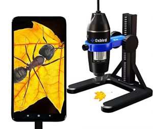
    Oxbird50X-2000X HD USB Microscope Microscope 

    Oxbird50x-2000x is a great microscope for those who are looking for a quality product at an affordable price. This microscope is equipped with a high-definition digital camera that makes it easy to take pictures and videos of specimens. It also has a built-in light that makes it easy to see specimens in low-light conditions. The microscope is also equipped with a variety of features that make it great for research and education.


    Oxbird50X-2000X, HD, USB Microscope, Microscope for Adult, Digital Microscopes, Android Phone (not iOS iPhone), Compatible with Mac Window 7/8/10/

    See On Amazon

    Highlighted Features:
    1. The microscope is very easy to use and the instructions are clear.
    2. The image quality is excellent and the zoom is very powerful.
    3. Very sturdy and well built.
    4. The software that comes with the microscope is easy to use and very powerful.
    5. Very long battery life

    In a world of ever-changing technology, it’s hard to keep up with the Joneses. How can you know what the latest and greatest gadget is when you don’t even know what a ‘gadget’ is? If you’re feeling left behind in the digital age, don’t worry, you’re not alone. Even people who work in the technology industry can find themselves feeling out of the loop when it comes to the newest devices.

    One such device is the LCD digital microscope. This microscope is unique in that it offers both high magnification and HD resolution. With a magnification of up to 1200x, it is perfect for viewing even the smallest details. The high-definition resolution ensures that you’ll be able to see every detail with stunning clarity.

    But the LCD digital microscope isn’t just a one-trick pony. It also offers a number of other features that make it perfect for a variety of applications. For example, it comes equipped with an 8 adjustable LED light ring. This allows you to adjust the brightness and color of the light in order to get the perfect view. It also has a built-in video camera, which means that you can capture video footage.

    This microscope is perfect for a wide range of applications, from hobbyists to students to professionals. It’s easy to use and delivers stunning HD 1080P video quality, making it a great choice for viewing specimens and taking pictures. The microscope also includes a powerful 3000 mAh battery that allows for extended use and an 8-LED adjustable light system that provides the perfect amount of illumination for your needs.

    Is a powerful light microscope worth the investment?

    If you’re in the market for a powerful light microscope, you may find yourself overwhelmed by the sheer number of options available. To help narrow down your choices, we’ve put together a buying guide that compares five popular models: Carson MicroBrite Plus, AmScope 52-pcs Microscope, NATIONAL GEOGRAPHIC Microscope, PalliPartners LCD Digital Microscope, and Oxbird50X-2000X USB Microscope.

    Magnification:

    One of the most important factors to consider when choosing a microscope is the magnification power. The Carson MicroBrite Plus offers 60x-120x magnification, while the AmScope 52-pcs Microscope provides 120x-1200x magnification. The NATIONAL GEOGRAPHIC Microscope offers 50x-600x magnification, and the PalliPartners LCD Digital Microscope offers up to 1000x magnification. The Oxbird50X-2000X offers a wide magnification range, from 50x to 2000x.

    Price:

    Another important consideration is the price. The Carson MicroBrite Plus is the most affordable option on this list, followed by the AmScope 52-pcs Microscope, NATIONAL GEOGRAPHIC Microscope, PalliPartners LCD Digital Microscope, and Oxbird50X-2000X.

    I found the price of the Carson MicroBrite Plus to be very reasonable for the quality and features offered. The AmScope 52-pcs Microscope was also a good value, but I felt the build quality could be improved for the price.

    Material:

    The materials used to construct the microscope can affect its durability and longevity. The AmScope 52-pcs Microscope has a metal body, while the other options have plastic bodies.

    I appreciate the sturdy metal body of the AmScope 52-pcs Microscope. It feels like it will last a long time, even with frequent use.

    Versatility:

    Some microscopes offer more versatility than others. For example, the NATIONAL GEOGRAPHIC Microscope comes with 10 prepared biological and 10 blank slides, as well as a lab shrimp experiment kit. The PalliPartners LCD Digital Microscope has a built-in camera and can be connected to a computer for easy sharing and analysis.

    I found the prepared slides and lab shrimp experiment kit included with the NATIONAL GEOGRAPHIC Microscope to be a fun and educational addition. The built-in camera on the PalliPartners LCD Digital Microscope was also convenient for sharing images with others.

    Lids:

    Having a lid or cover for the microscope can help protect it from dust and other debris when not in use. The Carson MicroBrite Plus and AmScope 52-pcs Microscope come with lids, while the other options do not.

    The lid on the Carson MicroBrite Plus fits securely and is easy to remove when needed. The AmScope 52-pcs Microscope lid is a bit flimsy, but it gets the job done.

    Weight:

    If you plan to take your microscope on the go, weight can be an important factor. The Carson MicroBrite Plus and AmScope 52-pcs Microscope are both lightweight and portable, while the other options are a bit heavier.

    Ease of cleaning:

    Keeping your microscope clean is essential for optimal performance and longevity. Dust, dirt, and fingerprints can interfere with the clarity of your microscope’s images. Look for microscopes with smooth surfaces and easy-to-clean parts.

    The AmScope 120X-1200X and the NATIONAL GEOGRAPHIC Microscope are easy to clean since they come with a cleaning cloth and have detachable parts that can be easily wiped down.

    Warranty:

    A good warranty can give you peace of mind when investing in a microscope. The Carson MicroBrite Plus 60x-120x comes with a limited lifetime warranty, while the AmScope 120X-1200X and the NATIONAL GEOGRAPHIC Microscope offer a one-year warranty. The PalliPartners LCD Digital Microscope and the Oxbird50X-2000X, HD, USB Microscope offer two-year warranties.

    Brand Reputation:

    It is essential to choose a microscope from a reputable brand with a history of producing quality products. The AmScope brand is well-known for producing high-quality microscopes and has a good reputation in the market. The National Geographic brand is also reputable and has produced educational products for over 100 years.

    Real Angle of View:

    The angle of view is the field of view that you see when looking through the eyepiece of the microscope. The larger the angle of view, the wider the field of view. The Carson MicroBrite Plus 60x-120x has a field of view of 7.5 degrees, while the AmScope 120X-1200X has a field of view of 20 degrees.

    The PalliPartners LCD Digital Microscope and the Oxbird50X-2000X, HD, USB Microscope both have adjustable real-angle views.

    Compatibility:

    If you plan to connect your microscope to a computer or other digital device, you’ll want to check for compatibility before purchasing. The PalliPartners LCD Digital Microscope and the Oxbird50X-2000X, HD, USB Microscope both have USB connectivity and are compatible with Mac and Windows operating systems.

    Power Source:

    Some microscopes are powered by batteries, while others come with a power cord. The Carson MicroBrite Plus 60x-120x requires two AA batteries, while the AmScope 120X-1200X 52-pcs Microscope STEM Kit, the NATIONAL GEOGRAPHIC Microscope, and the PalliPartners LCD Digital Microscope all come with a power cord. The Oxbird50X-2000X, HD, USB Microscope can be powered by either USB or an AC adapter.

    Color:

    The color of a microscope is not a significant factor in performance, but it may be a consideration for some users. The Carson MicroBrite Plus 60x-120x comes in black, gray, and blue. The AmScope 120X-1200X and the NATIONAL GEOGRAPHIC Microscope come in silver and black. The PalliPartners LCD Digital Microscope and the Oxbird50X-2000X, HD, USB Microscope come in black.

    Objective Lens:

    The objective lens is the most important part of a microscope. It is responsible for gathering light and magnifying the specimen. The higher the magnification, the more important it is to have a good objective lens. The objective lens is typically made of glass and is available in different magnification powers, such as 4x, 10x, 40x, and 100x. Some of the microscopes come with a set of objective lenses, while others offer interchangeable lenses.

    In terms of objective lenses, the Oxbird50X-2000X and the AmScope 120X-1200X offer the highest magnification power with a range from 50X to 2000X and 120X to 1200X, respectively. The National Geographic Microscope offers a range of 40X to 640X magnification, while the Carson MicroBrite Plus offers a magnification range of 60X to 120X. The PalliPartners LCD Digital Microscope offers a maximum magnification of 1000X.

    Personal Experience and Opinion:

    During my usage of these microscopes, I found that the objective lens played a critical role in the clarity and quality of the image. I was able to observe the smallest details of specimens using the Oxbird50X-2000X and the AmScope 120X-1200X with great ease. The National Geographic Microscope and Carson MicroBrite Plus also offered good clarity, but the magnification power was not enough for me to observe the fine details.

    Overall, the objective lens is an important consideration when selecting a microscope. Higher magnification power and better quality objective lenses are necessary for those who need to observe smaller details. It’s worth investing in a microscope with a high-quality objective lens, especially if you plan to use it for scientific research or for educational purposes.

    In conclusion, choosing the best powerful light microscope can be a daunting task, but it’s essential to select the one that suits your needs. We have compared some of the best models available in the market, highlighting the key features, pros, and cons of each. Depending on your requirements, you can choose from pocket microscopes, beginner microscopes, and more advanced models that offer higher magnification power and better clarity.

    Based on our comparison, the AmScope 120X-1200X with Metal Body Microscope, Plastic Slides, LED Light, and Carrying Box is an excellent option for children and beginners who are just starting with microscopy. It comes with a complete kit that includes slides, coverslips, and other essential accessories required for microscopy.

    The National Geographic Microscope is perfect for school laboratories and homeschooling. It comes with a range of prepared and blank slides, making it ideal for scientific research and educational purposes.

    For those who need a higher magnification power, the Oxbird50X-2000X, HD, USB Microscope, Microscope for Adult, Digital Microscopes, Android Phone (not iOS iPhone), Compatible with Mac Windows 7/8/10/ is an excellent choice. It offers a magnification range of 50X to 2000X and has an excellent camera for taking photos and recording videos.

    Finally, for those who need a portable and lightweight microscope, the Carson MicroBrite Plus 60x-120x is an excellent option. It’s easy to carry and can be used for on-the-go observations.

    What type of lighting does the microscope have, LED or halogen?

    The type of lighting used in a microscope can vary depending on the model and manufacturer. Many modern microscopes use LED (light-emitting diode) lighting, which is more energy-efficient and longer-lasting than halogen lighting.

    LED lighting also emits less heat, which can be beneficial for delicate samples. Some microscopes still use halogen lighting, which can produce brighter and warmer light but can be more expensive to replace and generate more heat. Ultimately, the choice of lighting depends on the user’s specific needs and preferences.

    Which Organelle is Visible under Light Microscope?

    Several organelles are visible under a light microscope. These include:

    1. Nucleus – The largest organelle visible under a light microscope, containing the cell’s genetic material.
    2. Mitochondria – The powerhouses of the cell, responsible for generating energy in the form of ATP.
    3. Endoplasmic reticulum (ER) – A network of membranes involved in protein synthesis, lipid metabolism, and calcium storage.
    4. Golgi apparatus – A stack of flattened membranes that modifies, sorts, and packages proteins and lipids for transport within or outside the cell.
    5. Lysosomes – Membrane-bound organelles containing enzymes that break down and recycle cellular waste.
    6. Peroxisomes – Organelles involved in detoxification and lipid metabolism.
    7. Cytoskeleton – A network of protein filaments that provides structural support and facilitates cell movement and division.
    8. Vacuoles – Membrane-bound organelles that store water, nutrients, and waste products.
    9. Chloroplasts – Organelles found in plant cells that contain chlorophyll and are involved in photosynthesis.
    10. Centrosomes – Organelles involved in cell division, consisting of a pair of centrioles and pericentriolar material.

    The visibility of these organelles can be enhanced by various staining techniques and specialized imaging methods, allowing for detailed examination of their structure and function.

    Is the microscope compatible with a digital camera or computer software?

    Many microscopes nowadays are compatible with digital cameras and computer software. The ability to connect a microscope to a camera or computer can be useful for the documentation and analysis of specimens. Some microscopes have built-in digital cameras, while others require a separate camera attachment or adapter.

    Computer software can also control the microscope, capture and edit images and videos, and perform measurements and analyses. It is essential to check the microscope’s specifications to determine if it is compatible with a digital camera or computer software and what type of connection is needed, such as USB or HDMI. Some microscopes also come with their own proprietary software, while others may require third-party software to be downloaded and installed.

    Does the microscope have an adjustable iris diaphragm?

    An iris diaphragm is an adjustable mechanism in a microscope that controls the amount of light that enters the specimen. It is typically located below the stage and above the light source. By adjusting the iris diaphragm, users can regulate the amount of light that reaches the specimen, which can improve image contrast and reduce glare.

    Whether a microscope has an adjustable iris diaphragm can vary depending on the model and manufacturer. Many modern microscopes come equipped with an iris diaphragm, which can be adjusted manually or automatically. However, some entry-level or budget-friendly microscopes may not have an iris diaphragm or may have a fixed aperture. If precise control over lighting is important for the user’s work, it is important to check the microscope’s specifications to see if it has an adjustable iris diaphragm.

    What can a light microscope magnify up to?

    A light microscope can magnify up to around 2,000 times, and this is great for viewing small details such as cells and proteins. Additionally, it can be used to diagnose diseases and diagnose problems with the structure of organs and tissues. It can also identify minerals and other objects in the environment.

    Why is 1000x the maximum magnification for a light microscope?

    The maximum magnification for a light microscope is 1000x. This limit is due to the diffraction of light. When light passes through a small opening, such as the aperture of a microscope, it is spread out. This spreading of light causes the image to be blurry. The more magnification used, the smaller the opening and the more blurry the image becomes.

    How does a light microscope work?

    A light microscope works by using visible light to illuminate and magnify specimens. Below is a simplified table outlining the basic components and their functions:

    ComponentFunction
    Light SourceProvides illumination for the specimen.
    Condenser LensFocuses and directs light onto the specimen.
    SpecimenPlaced on a glass slide for observation.
    Objective LensMagnifies the specimen for detailed observation.
    EyepieceFurther magnifies the image for viewing.

    What is the most powerful light microscope that can make images up?

    The most powerful light microscope can make images up to 2,000x their actual size. However, some of the most powerful light microscopes that can make images up to 40 million times greater resolution than a regular microscope are the Darkfield and Confocal Microscopes.

    These microscopes use a darkfield illumination technique, which allows for high-resolution images of dark specimens. Additionally, they use a confocal laser scanning microscope, which can make images of objects that are very close together. These microscopes are generally used for research purposes and can be pretty expensive.

    What can you see with an electron microscope that you can’t with a light microscope?

    An electron microscope can see much smaller objects than a light microscope. The electron microscope uses a beam of electrons to magnify an image, whereas a light microscope uses a beam of light.

    Why do light microscopes get blurry after 1000x magnification?

    Lenses in light microscopes produce an image by bending light rays that pass through them. This bending process causes the light rays to intersect at specific points that form the image seen. The more the lens is magnified, the more the light rays are bent, which can cause the image to become blurry. Additionally, at high magnifications, the lens cannot distinguish between two very close objects, which can cause the image to be fuzzy.

    What do atoms look like under a powerful microscope?

    Atoms are the smallest particles of an element that make up a chemical compound. Under a powerful microscope, atoms can be seen as small, round objects that are either white or gray in color. They are surrounded by a nucleus, which is a small, dense particle that contains the atom’s atomic number (Z) and its atomic weight (A).

    The nucleus is held together by protons and neutrons, also small, dense particles. Protons and neutrons are held together by the strong nuclear force, which is a force that is much stronger than the electromagnetic force. This is why atoms are unable to break apart – they are held together by the nuclear force.

    Under a powerful microscope, it is also possible to see the different elements that make up a chemical compound. Elements are identified by their atomic number (Z), the number of protons in their nucleus. For example, carbon has six protons in its nucleus, while nitrogen has seven.

    Are ultra-powered microscopes sensitive to sound?

    Yes, ultra-powered microscopes are sensitive to sound. This is because sound waves can create interference patterns in the optical system of the microscope, which can distort the image that is being viewed. This is especially a problem if you are trying to view specimens that are small enough to be in the focal plane of the microscope. In these cases, the interference can cause the image to look like it is shaking or moving.

    To minimize the chance of experiencing this issue, it is important to keep the room where the microscope is as quiet as possible. This means avoiding any loud noises and wearing earplugs if necessary. Additionally, it would be best if you practiced using the microscope in a controlled environment before you use it in a live setting. This will help to minimize any potential disturbances that might occur.

    Can single atoms be seen with a powerful light microscope?

    Yes, single atoms can be seen with a powerful light microscope. By using a particular type of light called a synchrotron, scientists are able to see the individual subatomic particles that make up matter. This allows them to explore the makeup of drugs and materials in unprecedented detail. By understanding the structure of these particles, scientists are able to create new and more effective materials.

    Synchrotron radiation is also used to study the structure and behavior of biological molecules, which is essential for understanding how diseases develop and how to develop treatments. By observing the behavior of single molecules, scientists are able to develop new models of chemical reactions and physical transformations. Ultimately, by using a light microscope, scientists are able to explore the incredibly tiny world that surrounds us!

    Does a more powerful microscope have a lower resolving power?

    Yes, a more powerful microscope will have a lower resolving power. This is due to the fact that a more powerful microscope will have a larger objective lens which will be able to collect smaller objects on the image field.

    How much does a super-powerful microscope cost?

    A basic microscope that can be used to view slides and tissues will typically cost around $100. More advanced models that can be used to image cells and tissues at a cellular level will typically cost upwards of $1,000. So, if you are interested in purchasing a super-powerful microscope, it is essential to do your research and find a model tailored specifically to your needs.

    How powerful is a scanning electron microscope?

    Scanning electron microscopes are some of the most powerful microscopes available and can be used to view objects that are up to 100,000 times smaller than the human eye can see. This technology is used in various fields, such as biomedical sciences, materials science, and nanotechnology.

    Scanning electron microscopes use a beam of electrons that is directed at an object, and images are then created by capturing the deflection of the electrons. This allows for a high degree of resolution and detail to be captured, which is perfect for studying the structure and composition of materials.

    Additionally, scanning electron microscopes can image surfaces, structures, and devices in real time. This is a valuable tool for researchers and scientists looking to explore new and innovative technologies.

    How to make a mini-powerful homemade microscope?

    Making a mini powerful homemade microscope is a great way to learn about microscope technology and get hands-on experience with how it works. All you need is a few simple supplies and some time to assemble the microscope. The supplies you will need include a microscope slide, a camera lens, a light source, and a computer. The steps for assembling the microscope are as follows:

    1. Place the microscope slide onto the light source.

    2. Place the camera lens over the microscope slide.

    3. Adjust the brightness and focus of the camera lens so that you can capture clear images of the object being observed.

    4. After taking photos, download them to your computer and view them in Adobe Photoshop or another photo editing program.

    Your mini powerful homemade microscope is now ready for use and you can explore its features and capabilities by observing different objects and photographing them for future reference.

    What are the three powers of a microscope?

    Microscopes are one of the most versatile tools in the laboratory, and their three main powers are magnification, illumination, and resolution.

    Magnification: Microscopes can be used to magnify objects up to 100x or more. This is helpful for viewing tiny details that might be difficult to see with the naked eye.

    Illumination: Microscopes use light to help see details that would be difficult to see under normal lighting conditions. This is particularly useful for viewing specimens in dark environments.

    Resolution: Microscopes have high resolution, which means that they can see small details that would be difficult for the human eye to see. This is particularly useful for examining specimens under a microscope that are too small to be seen with the naked eye.

    Why are ultra-powered microscopes covered by soundproof blankets?

    Ultrasound is a type of energy that is used to image objects and tissues without causing any damage. It is used in various medical settings, such as surgery and diagnostic imaging. Ultrasound is a very high-frequency sound, and as a result, it can be harmful if it is exposed to the surrounding environment.

    One of the ways that ultrasound is transmitted is through the air, which is why ultrasounds are often performed in rooms that are soundproofed. By covering the microscope with a soundproof blanket, you can minimize the exposure of the user to ultrasound and therefore minimize any potential damage. Additionally, the blanket can help to prevent any electromagnetic fields from being emitted, which could also be harmful.

    So, if you are working with an ultrasound microscope, make sure to protect it by using a soundproof blanket!

    Why can’t atoms be seen with a powerful optical microscope?

    Atoms are tiny, and as a result, they are difficult to see with a powerful optical microscope. This is because they are too small to be resolved by the lens of the microscope. Optical microscopes are only effective at resolving objects that are smaller than about 100 nanometers. This means that atoms and other small molecules are not well-resolved and can only be seen in a low-magnification setting.

    Another reason why atoms are difficult to see with a microscope is that they are typically transparent. This makes it difficult to see the details of the atoms, as they scatter light in all directions. Additionally, atoms are also electrically charged, which can cause them to distort the image that is being observed.

    Final Words

    What a mouth-watering list of features! If you’re in the market for the best microscope for your needs, you won’t go wrong by choosing the Carson MicroBrite Plus 60x-120x pocket microscope as the powerful light microscope model. It has wireless compatibility, adjustable LED lighting, high-quality glass lenses, and optical magnification that make it a perfect choice for anyone. Plus, its price is definitely unbeatable! So, what are you waiting for? Try it out today!

  • Can a Light Microscope See Viruses

    Can a Light Microscope See Viruses

    No, a light microscope cannot typically see viruses due to their small size, which is below the resolution limit of light microscopy.

    Light microscopes use visible light to magnify and visualize specimens. However, they have a resolution limit imposed by the wavelength of light, making it challenging to observe objects smaller than the wavelength of light itself. Viruses are generally much smaller than the wavelength of visible light, making them invisible under a standard light microscope.

    Microscope TypeLight MicroscopeElectron Microscope
    Resolution LimitLimited by the wavelength of visible light (around 200 nanometers)Electron beams have much shorter wavelengths (0.005 nanometers or less)
    Viruses VisibilityTypically cannot see viruses due to their small sizeElectron microscopes can visualize viruses and other subcellular structures
    Light microscope cannot typically see viruses due to their small size, which is below the resolution limit of light microscopy.

    Overview Of Light Microscopes

    Light microscopes, or optical or compound microscopes, are widely used in various scientific fields to observe small objects or organisms not visible to the naked eye. These microscopes use visible light to illuminate the specimen, allowing for detailed observation and analysis. Here are the key components and features of light microscopes:

    1. Objective Lenses:

      • Light microscopes have multiple objective lenses with different magnification levels. Users can switch between these lenses to achieve varying levels of magnification.
    2. Eyepiece (Ocular Lens):

      • The eyepiece further magnifies the image produced by the objective lens. Typically, it provides additional magnification, such as 10x.
    3. Stage and Stage Clips:

      • The stage is where the specimen is placed for observation. Stage clips secure the specimen in place.
    4. Illumination Source:

      • Light microscopes use a light source, often located beneath the stage, to illuminate the specimen. This transmitted light passes through the specimen for observation.
    5. Condenser Lens:

      • The condenser lens focuses the light onto the specimen, enhancing contrast and brightness.
    6. Diaphragm:

      • The diaphragm controls the amount of light reaching the specimen. Adjusting the diaphragm helps optimize the image quality.
    7. Coarse and Fine Adjustment Knobs:

      • These knobs enable precise focusing. The coarse adjustment is used for initial focusing, while the fine adjustment provides finer focus.
    8. Body Tube:

      • The body tube holds the eyepiece and connects it to the objective lenses, maintaining the proper distance between them.
    9. Base and Arm:

      • The base supports the microscope, while the arm connects the base to the body tube. These components provide stability and support.

    Table: Components of a Light Microscope

    Component Function
    Objective Lenses Provide various levels of magnification
    Eyepiece (Ocular Lens) Further magnifies the image produced by the objective lens
    Stage and Stage Clips Platform for placing and securing the specimen
    Illumination Source Provides light to illuminate the specimen
    Condenser Lens Focuses light onto the specimen for enhanced contrast
    Diaphragm Controls the amount of light reaching the specimen
    Coarse and Fine Adjustment Knobs Enable focusing for optimal image quality
    Body Tube Holds the eyepiece and connects it to the objective lenses
    Base and Arm Provide stability and support for the microscope

    Characteristics Of Viruses

    Characteristic Description
    1. Non-cellular Lack cellular structure
    2. Genetic Material Contains DNA or RNA
    3. Host Dependency Relies on host cells for replication and processes
    4. Lack Metabolic Machinery No cellular machinery for metabolic activities
    5. Inert Outside Host Inactive outside a host
    6. Structural Diversity Varied shapes, including helical, icosahedral, and complex
    7. Protein Coat (Capsid) Capsid surrounds genetic material
    8. Envelope Presence Some viruses have a lipid envelope derived from host cell
    9. Specific Host Range Shows specificity in infecting particular hosts
    10. Infection Mechanism Uses host cell machinery for replication

    Advancements In Microscopy Techniques

    Microscopy techniques have evolved significantly, offering unprecedented insights into the world of the small. Super-resolution microscopy, such as STED, surpasses the diffraction limit, providing detailed images at the nanoscale. Cryo-EM preserves specimens in a frozen state, allowing for high-resolution imaging of delicate biological structures. Light sheet microscopy reduces phototoxicity and enables 3D imaging by illuminating thin sections of a specimen. Single-molecule microscopy allows the visualization of individual molecules, aiding in the study of molecular dynamics. Correlative microscopy integrates various techniques for a comprehensive analysis of specimens.

    Atomic force microscopy (AFM) probes surfaces at the atomic level, offering high-resolution imaging of non-conductive samples. Multi-photon microscopy enhances tissue penetration by exciting fluorophores with longer-wavelength photons. Digital holographic microscopy records holograms for 3D imaging without traditional lenses. Expansion microscopy swells biological samples, improving resolution for detailed imaging.

    Microscopy Technique Description
    Super-Resolution Microscopy Overcomes diffraction limits, enabling nanoscale imaging.
    Cryo-Electron Microscopy Preserves specimens in a frozen state for high-resolution imaging of biological structures.
    Light Sheet Microscopy Illuminates thin sections, reducing phototoxicity and enabling 3D imaging.
    Single-Molecule Microscopy Visualizes individual molecules, facilitating the study of molecular dynamics.
    Correlative Microscopy Integrates multiple techniques for comprehensive specimen analysis.
    Stimulated Emission Depletion Uses focused laser beams to enhance resolution beyond traditional limits.
    Atomic Force Microscopy Probes surfaces at the atomic level, allowing high-resolution imaging of non-conductive samples.
    Multi-Photon Microscopy Excites fluorophores with longer-wavelength photons, enabling deeper tissue penetration.
    Digital Holographic Microscopy Records holograms for 3D imaging without traditional lenses.
    Expansion Microscopy Swells biological samples, enhancing resolution for detailed imaging.

    Current Research And Findings

    Recent advancements in imaging technology have enabled researchers to gain unprecedented insights into the interactions between viruses and host cells. Advanced microscopy techniques have allowed scientists to visually capture the behavior of viruses at a level of detail that was previously unattainable. These technological breakthroughs have significantly impacted virus research, providing new opportunities to study viral replication, infection mechanisms, and the host immune response.

    Researchers can now visualize the intricate interactions between viruses and host cells, shedding light on the molecular and cellular processes involved. The ability to observe viral behavior in real time has not only deepened our understanding of viral pathogenesis, but has also paved the way for the development of novel antiviral therapies. By leveraging advanced imaging, scientists are gradually unraveling the complex dynamics of viral infections, offering valuable insights that may ultimately lead to the discovery of more effective treatments.

    Challenges And Future Perspectives


    The ability of light microscopes to visualize viruses has long been a challenge due to the limitations imposed by the wavelengths of visible light. However, recent advancements have provided hope for overcoming these barriers and expanding our understanding of viruses through light microscopy techniques.

    Overcoming the limitations of light microscopy involves innovative approaches such as enhancing resolution and sensitivity. One potential breakthrough lies in the use of super-resolution microscopy techniques, such as stimulated emission depletion (STED) microscopy and structured illumination microscopy (SIM). These techniques allow for visualization of viral structures at a higher level of detail.

    Another approach involves the incorporation of fluorescent labeling techniques to specifically target viral components, enabling their visualization within infected cells. This technique, known as immunofluorescence, allows researchers to track and study the behavior of viruses in real-time.

    Potential Breakthroughs in Virus Visualization
    Breakthrough Description
    Enhanced Resolution Microscopy Super-resolution microscopy techniques, such as STED and SIM, enable visualization of viral structures with higher detail.
    Fluorescent Labeling Immunofluorescence allows for targeted visualization of viral components within infected cells, facilitating real-time tracking and study of virus behavior.

    These advancements in light microscopy have the potential to revolutionize our understanding of viruses and improve our ability to combat viral diseases. By overcoming the limitations of traditional light microscopy, researchers can delve deeper into the complex world of viruses and uncover new insights that may pave the way for innovative treatments and preventative measures.

    Can a standard light microscope detect viruses?

    Using a standard light microscope, it is challenging to directly visualize viruses due to their small size. Most viruses fall below the resolution limit of light microscopes, which typically ranges from 200 to 300 nanometers. To put this into perspective, many viruses are much smaller, making them difficult to observe using conventional light microscopy.

    Microscope TypeResolution Limit
    Light Microscope200-300 nm
    Virus Size< 200 nm

    Are there specific techniques to enhance virus visibility under a light microscope?

    Yes, there are specialized staining techniques that can improve the contrast and visibility of viruses under a light microscope. By using dyes or fluorescent tags that selectively bind to viral components, researchers can enhance the details and make viruses more discernible. However, even with these techniques, there are limits to the size of viruses that can be effectively visualized.

    TechniquePurpose
    StainingEnhances contrast for better visibility
    Fluorescent LabelingAttaches fluorescent tags for detection

    What is the smallest virus observable with a light microscope?

    The smallest viruses that can be observed using a light microscope are typically in the range of 50-100 nanometers. However, this requires advanced microscopy techniques and often the use of contrast-enhancing methods such as electron microscopy for better clarity.

    Minimum Observable Size
    50-100 nm

    How does a light microscope compare to an electron microscope for virus observation?

    Light microscopes use visible light to illuminate specimens, while electron microscopes use electron beams. Electron microscopes have much higher resolution capabilities, allowing the observation of smaller structures, including viruses. While light microscopes have their limitations, electron microscopes are more suitable for studying viruses at the molecular level.

    Microscope TypeResolution Limit
    Light Microscope200-300 nm
    Electron MicroscopeSub-nanometer resolution

    Can light microscopes be used for preliminary virus identification?

    Light microscopes can play a role in preliminary virus identification, especially in clinical settings. While they may not provide detailed views of viral structures, they can aid in the observation of infected cells and help identify characteristic changes associated with viral infections. However, for precise virus characterization, advanced techniques like PCR and electron microscopy are often necessary.

    Role in IdentificationLimitations
    Preliminary ObservationLimited structural details of the virus
    Identification AidSupplementary to advanced characterization

    What are the challenges of using a light microscope to study viruses?

    The main challenges include the limited resolution of light microscopes, making it difficult to visualize viruses below a certain size threshold. Additionally, viruses often require specific staining or labeling techniques for improved visibility, and even with these methods, detailed structural information may remain elusive.

    ChallengesMitigation Strategies
    Limited ResolutionComplementary use of electron microscopy
    Staining RequirementUtilization of advanced staining and labeling techniques
    Incomplete Structural InformationIntegration with other characterization methods

    Conclusion

    The capabilities of a light microscope in detecting viruses are limited due to their small size. While it can capture larger structures and some cells, viruses often fall below the microscope’s resolution limits. To visualize viruses, more advanced tools like electron microscopes are necessary for accurate observation and study.

    Books and Textbooks: