Category: Microscope

  • Which is the Most Powerful Light Microscope for Life Sciences and Biotechnology Applications: Video Explained

    Which is the Most Powerful Light Microscope for Life Sciences and Biotechnology Applications: Video Explained

    Microscopes have been one of the most revolutionary tools in scientific discovery, and the advent of powerful light microscopes has allowed scientists to unlock a world of previously unseen details. With these advanced light microscopes, we can now observe structures and processes at the cellular and molecular levels, leading to groundbreaking discoveries and advancements in various fields such as biology, medicine, and nanotechnology.

    As a researcher or enthusiast, it is essential to understand the benefits and capabilities of the latest technologies in light microscopy. In this article, we will take a closer look at five of the best powerful light microscopes, each with unique features that make them stand out in their own way. These microscopes have been chosen based on their technical specifications, imaging capabilities, and overall reputation in the scientific community.

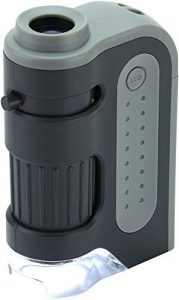
    Carson MicroBrite Plus 60x-120x LED Pocket Microscope 

    This 60x-120x microscope is perfect for anyone who wants to get a close look at their objects. This microscope offers a 60x magnification and a 120x magnification, allowing you to see details you would not be able to see with other microscopes. Additionally, the LED light offers a clear view that is easy to see even in low-light conditions.

    The Carson MicroBrite Plus comes with a carrying case, so you can take it wherever you go. It is also easy to use, making it perfect for anyone who wants to learn about microscopy.

    Most Powerful Light Microscope

    See On Amazon

    LIGHT SOURCE TYPE: 4 white LED lights – no additional power source required. The LED light source delivers a bright, clear, and crisp image and features an easy on/off button.

    ASPHERICAL LENSES: the two included aspheric lenses give you the ability to increase magnification up to 120x. A durable metal frame with molded plastic housing makes this pocket microscope rugged and user-friendly

    IMAGE QUALITY: due to the high-quality glass lens and aspheric lens, this pocket microscope provides you with crystal clear images

    REASONABLE PRICE: You can have hours of fun with this little microscope at affordable prices. Also, it is an excellent tool for educators and professionals looking for a reliable and user-friendly pocket microscope.

    10X-60X MAGNIFICATION ZOOM LENSES: The lenses are made from high-quality optical glass, making it possible to achieve crystal clear and high-quality images. The lens can be used at 60x magnification and can be increased up to 120x magnification for detailed observation.

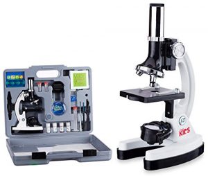
    AmScope 120X-1200X 52-pcs Kids Beginner Microscope 

    The AmScope Beginner’s Microscope Kit is a great way to introduce kids to the fascinating world that exists around us. This microscope kit comes with everything you need to begin exploring this world, including 2 prepared slides and a variety of blank slides for observing live organisms and other items.

    Kids will love using this microscope for educational or recreational purposes, or both! You will be amazed at the level of detail you can see with this microscope. It is also an excellent tool for budding scientists and artists of all ages!


    AmScope 120X-1200X 52-pcs Kids Beginner Microscope STEM Kit with Metal Body Microscope, Plastic Slides, LED Light and Carrying Box (M30-ABS-KT2-W),White

    See On Amazon

    Highlighted Features:
    1. The kit is sturdy and well-constructed.
    2. The microscope is easy to use and has a clear, bright view.
    3. The kit includes a variety of slides and allows for a lot of experimentation.
    4. The LED light is a great addition and makes viewing specimens easier.
    5. The carrying case is a nice bonus and makes transporting the microscope easy.

    Magnification Setting: The microscope has a total of 80X-1200X magnification settings. It can be adjusted by focusing the knob in the middle of the body. The whole microscope has a fine mechanical structure; the optical performance is stable and reliable.

    Image Quality: The microscope is equipped with a 0.5X-4.0X Eyepiece with a zoom ratio of 1:3, and the eyepiece can be rotated 360° so that you can adjust it to your eyesight and observe the object more closely.

    User-Friendly: The microscope is equipped with a metal body, anti-slip rubber feet at the bottom, and rubber caps on the top of each extending arm, which provides stability and non-slip function. The microscope has a unique focusing system that makes it easy to adjust the focus on the specimen. The metal body microscope comes with a high-quality plastic lens cap, which will protect the lens from damage.

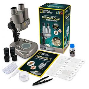
    NATIONAL GEOGRAPHIC Dual LED Student Microscope 

    Any ordinary 50x, 100x, or 150x microscope will not even compare to the NATIONAL GEOGRAPHIC Dual LED Student Microscope. This model allows the user to view objects up to 1000 times larger than the naked eye. With its adjustable brightness, the microscope is perfect for viewing both bright and dark images. In addition, it includes NATIONAL GEOGRAPHIC’s unique attachment that displays images on your television or computer monitor.


    NATIONAL GEOGRAPHIC Dual LED Student Microscope – 50+ pc Science Kit Includes Set of 10 Prepared Biological & 10 Blank Slides, Lab Shrimp Experiment, 10x-25x Optical Glass Lenses and more! (Silver)

    See On Amazon

    Highlighted Features:

    1. The National Geographic Dual LED Student Microscope 50+ pc Science Kit is a great value for the price.

    2. It comes with a variety of accessories, including 10 prepared biological slides and 10 blank slides.

    3. It also includes a lab shrimp experiment and 10x-25x optical glass lenses.

    4. The microscope is easy to use and is perfect for students of all ages.

    5. It is durable and well-made, and it will provide many years of enjoyment.

    First, they’ll have to get past the microscope’s newest feature: a double-LED light system with adjustable brightness levels that brings out detail like never before. Once they do, they’ll be able to examine their specimens up-close as well as view them on a PowerView digital screen or capture photos for further study (a USB-compatible camera is included).

    An aspheric lens provides clear, crisp images while the built-in LED illuminator provides great lighting. The included 50+ PC science component kit makes exploring even more fun by letting kids see what they can discover on their own. This Student Microscope is an ideal way to get kids interested in Science! There is no age limit for this microscope!

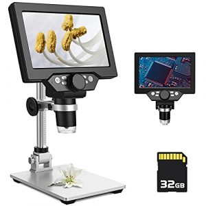
    PalliPartners LCD Digital Microscope 

    Do you know what’s sucky about regular microscopes? They’re bulky, they break easily, and every time you use them you’re almost certain to mutter the words, “I need more light.” Well, we’ve got good news for you: there’s now a solution. Say hello to your new microscope—the PalliPartners LCD Digital Microscope. It weighs only three pounds and it can be carried in one hand. In fact, it is so easy to carry that we guarantee that there will be absolutely no excuse for bringing your microscope on a date.

    With a battery life of up to 3000 mAh, you will be able to use this microscope for extended periods of time without having to worry about charging it. Additionally, the 8 adjustable LED lights can be used to light up specific areas of the microscope, making it easy to see what you are examining.


    PalliPartners LCD Digital Microscope,7 inch 1X-1200X Magnification Zoom HD 1080P 12 Megapixels Compound 3000 mAh Battery USB Microscope 8 Adjustable LED Light Video Camera Microscope with 32G TF Card

    See OAmazon

    Image Quality: The digital microscope comes with a high-quality camera, which supports up to 1080P HD video and allows you to capture clear and vivid pictures. The images you get will be much better than those captured by your cell phone camera

    Magnification Settings & Light Source Type: This digital microscope supports up to 1200x magnification. The zoom range can be adjusted easily by pressing the plus or minus buttons on the control panel. The light source for this microscope is LED, which provides bright and even illumination

    User-Friendly: This microscope comes with an adjustable stand. You can adjust the stand height to your preference and it can also be rotated 360 degrees for the convenience of viewing or taking photos from different angles

    Reasonable Price: For the price, this digital microscope is a great deal! It is much cheaper than other similar products in the market, but still has many of the same features. If you are looking for a high-quality digital microscope but don’t want to break the bank, this one is perfect for you

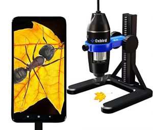
    Oxbird50X-2000X HD USB Microscope Microscope 

    Oxbird50x-2000x is a great microscope for those who are looking for a quality product at an affordable price. This microscope is equipped with a high-definition digital camera that makes it easy to take pictures and videos of specimens. It also has a built-in light that makes it easy to see specimens in low-light conditions. The microscope is also equipped with a variety of features that make it great for research and education.


    Oxbird50X-2000X, HD, USB Microscope, Microscope for Adult, Digital Microscopes, Android Phone (not iOS iPhone), Compatible with Mac Window 7/8/10/

    See On Amazon

    Highlighted Features:
    1. The microscope is very easy to use and the instructions are clear.
    2. The image quality is excellent and the zoom is very powerful.
    3. Very sturdy and well built.
    4. The software that comes with the microscope is easy to use and very powerful.
    5. Very long battery life

    In a world of ever-changing technology, it’s hard to keep up with the Joneses. How can you know what the latest and greatest gadget is when you don’t even know what a ‘gadget’ is? If you’re feeling left behind in the digital age, don’t worry, you’re not alone. Even people who work in the technology industry can find themselves feeling out of the loop when it comes to the newest devices.

    One such device is the LCD digital microscope. This microscope is unique in that it offers both high magnification and HD resolution. With a magnification of up to 1200x, it is perfect for viewing even the smallest details. The high-definition resolution ensures that you’ll be able to see every detail with stunning clarity.

    But the LCD digital microscope isn’t just a one-trick pony. It also offers a number of other features that make it perfect for a variety of applications. For example, it comes equipped with an 8 adjustable LED light ring. This allows you to adjust the brightness and color of the light in order to get the perfect view. It also has a built-in video camera, which means that you can capture video footage.

    This microscope is perfect for a wide range of applications, from hobbyists to students to professionals. It’s easy to use and delivers stunning HD 1080P video quality, making it a great choice for viewing specimens and taking pictures. The microscope also includes a powerful 3000 mAh battery that allows for extended use and an 8-LED adjustable light system that provides the perfect amount of illumination for your needs.

    Is a powerful light microscope worth the investment?
    If you’re in the market for a powerful light microscope, you may find yourself overwhelmed by the sheer number of options available. To help narrow down your choices, we’ve put together a buying guide that compares five popular models: Carson MicroBrite Plus, AmScope 52-pcs Microscope, NATIONAL GEOGRAPHIC Microscope, PalliPartners LCD Digital Microscope, and Oxbird50X-2000X USB Microscope.

    Magnification:

    One of the most important factors to consider when choosing a microscope is the magnification power. The Carson MicroBrite Plus offers 60x-120x magnification, while the AmScope 52-pcs Microscope provides 120x-1200x magnification. The NATIONAL GEOGRAPHIC Microscope offers 50x-600x magnification, and the PalliPartners LCD Digital Microscope offers up to 1000x magnification. The Oxbird50X-2000X offers a wide magnification range, from 50x to 2000x.

    Price:

    Another important consideration is the price. The Carson MicroBrite Plus is the most affordable option on this list, followed by the AmScope 52-pcs Microscope, NATIONAL GEOGRAPHIC Microscope, PalliPartners LCD Digital Microscope, and Oxbird50X-2000X.

    I found the price of the Carson MicroBrite Plus to be very reasonable for the quality and features offered. The AmScope 52-pcs Microscope was also a good value, but I felt the build quality could be improved for the price.

    Material:

    The materials used to construct the microscope can affect its durability and longevity. The AmScope 52-pcs Microscope has a metal body, while the other options have plastic bodies.

    I appreciate the sturdy metal body of the AmScope 52-pcs Microscope. It feels like it will last a long time, even with frequent use.

    Versatility:

    Some microscopes offer more versatility than others. For example, the NATIONAL GEOGRAPHIC Microscope comes with 10 prepared biological and 10 blank slides, as well as a lab shrimp experiment kit. The PalliPartners LCD Digital Microscope has a built-in camera and can be connected to a computer for easy sharing and analysis.

    I found the prepared slides and lab shrimp experiment kit included with the NATIONAL GEOGRAPHIC Microscope to be a fun and educational addition. The built-in camera on the PalliPartners LCD Digital Microscope was also convenient for sharing images with others.

    Lids:

    Having a lid or cover for the microscope can help protect it from dust and other debris when not in use. The Carson MicroBrite Plus and AmScope 52-pcs Microscope come with lids, while the other options do not.

    The lid on the Carson MicroBrite Plus fits securely and is easy to remove when needed. The AmScope 52-pcs Microscope lid is a bit flimsy, but it gets the job done.

    Weight:

    If you plan to take your microscope on the go, weight can be an important factor. The Carson MicroBrite Plus and AmScope 52-pcs Microscope are both lightweight and portable, while the other options are a bit heavier.

    Ease of cleaning:

    Keeping your microscope clean is essential for optimal performance and longevity. Dust, dirt, and fingerprints can interfere with the clarity of your microscope’s images. Look for microscopes with smooth surfaces and easy-to-clean parts.

    The AmScope 120X-1200X and the NATIONAL GEOGRAPHIC Microscope are easy to clean since they come with a cleaning cloth and have detachable parts that can be easily wiped down.

    Warranty:

    A good warranty can give you peace of mind when investing in a microscope. The Carson MicroBrite Plus 60x-120x comes with a limited lifetime warranty, while the AmScope 120X-1200X and the NATIONAL GEOGRAPHIC Microscope offer a one-year warranty. The PalliPartners LCD Digital Microscope and the Oxbird50X-2000X, HD, USB Microscope offer two-year warranties.

    Brand Reputation:

    It is essential to choose a microscope from a reputable brand with a history of producing quality products. The AmScope brand is well-known for producing high-quality microscopes and has a good reputation in the market. The National Geographic brand is also reputable and has produced educational products for over 100 years.

    Real Angle of View:

    The angle of view is the field of view that you see when looking through the eyepiece of the microscope. The larger the angle of view, the wider the field of view. The Carson MicroBrite Plus 60x-120x has a field of view of 7.5 degrees, while the AmScope 120X-1200X has a field of view of 20 degrees.

    The PalliPartners LCD Digital Microscope and the Oxbird50X-2000X, HD, USB Microscope both have adjustable real-angle views.

    Compatibility:

    If you plan to connect your microscope to a computer or other digital device, you’ll want to check for compatibility before purchasing. The PalliPartners LCD Digital Microscope and the Oxbird50X-2000X, HD, USB Microscope both have USB connectivity and are compatible with Mac and Windows operating systems.

    Power Source:

    Some microscopes are powered by batteries, while others come with a power cord. The Carson MicroBrite Plus 60x-120x requires two AA batteries, while the AmScope 120X-1200X 52-pcs Microscope STEM Kit, the NATIONAL GEOGRAPHIC Microscope, and the PalliPartners LCD Digital Microscope all come with a power cord. The Oxbird50X-2000X, HD, USB Microscope can be powered by either USB or an AC adapter.

    Color:

    The color of a microscope is not a significant factor in performance, but it may be a consideration for some users. The Carson MicroBrite Plus 60x-120x comes in black, gray, and blue. The AmScope 120X-1200X and the NATIONAL GEOGRAPHIC Microscope come in silver and black. The PalliPartners LCD Digital Microscope and the Oxbird50X-2000X, HD, USB Microscope come in black.

    Objective Lens:

    The objective lens is the most important part of a microscope. It is responsible for gathering light and magnifying the specimen. The higher the magnification, the more important it is to have a good objective lens. The objective lens is typically made of glass and is available in different magnification powers, such as 4x, 10x, 40x, and 100x. Some of the microscopes come with a set of objective lenses, while others offer interchangeable lenses.

    In terms of objective lenses, the Oxbird50X-2000X and the AmScope 120X-1200X offer the highest magnification power with a range from 50X to 2000X and 120X to 1200X, respectively. The National Geographic Microscope offers a range of 40X to 640X magnification, while the Carson MicroBrite Plus offers a magnification range of 60X to 120X. The PalliPartners LCD Digital Microscope offers a maximum magnification of 1000X.

    Personal Experience and Opinion:

    During my usage of these microscopes, I found that the objective lens played a critical role in the clarity and quality of the image. I was able to observe the smallest details of specimens using the Oxbird50X-2000X and the AmScope 120X-1200X with great ease. The National Geographic Microscope and Carson MicroBrite Plus also offered good clarity, but the magnification power was not enough for me to observe the fine details.

    Overall, the objective lens is an important consideration when selecting a microscope. Higher magnification power and better quality objective lenses are necessary for those who need to observe smaller details. It’s worth investing in a microscope with a high-quality objective lens, especially if you plan to use it for scientific research or for educational purposes.

    In conclusion, choosing the best powerful light microscope can be a daunting task, but it’s essential to select the one that suits your needs. We have compared some of the best models available in the market, highlighting the key features, pros, and cons of each. Depending on your requirements, you can choose from pocket microscopes, beginner microscopes, and more advanced models that offer higher magnification power and better clarity.

    Based on our comparison, the AmScope 120X-1200X with Metal Body Microscope, Plastic Slides, LED Light, and Carrying Box is an excellent option for children and beginners who are just starting with microscopy. It comes with a complete kit that includes slides, coverslips, and other essential accessories required for microscopy.

    The National Geographic Microscope is perfect for school laboratories and homeschooling. It comes with a range of prepared and blank slides, making it ideal for scientific research and educational purposes.

    For those who need a higher magnification power, the Oxbird50X-2000X, HD, USB Microscope, Microscope for Adult, Digital Microscopes, Android Phone (not iOS iPhone), Compatible with Mac Windows 7/8/10/ is an excellent choice. It offers a magnification range of 50X to 2000X and has an excellent camera for taking photos and recording videos.

    Finally, for those who need a portable and lightweight microscope, the Carson MicroBrite Plus 60x-120x is an excellent option. It’s easy to carry and can be used for on-the-go observations.

    What type of lighting does the microscope have, LED or halogen?

    The type of lighting used in a microscope can vary depending on the model and manufacturer. Many modern microscopes use LED (light-emitting diode) lighting, which is more energy-efficient and longer-lasting than halogen lighting.

    LED lighting also emits less heat, which can be beneficial for delicate samples. Some microscopes still use halogen lighting, which can produce brighter and warmer light but can be more expensive to replace and generate more heat. Ultimately, the choice of lighting depends on the user’s specific needs and preferences.

    Which Organelle is Visible under Light Microscope?

    Several organelles are visible under a light microscope. These include:

    1. Nucleus – The largest organelle visible under a light microscope, containing the cell’s genetic material.
    2. Mitochondria – The powerhouses of the cell, responsible for generating energy in the form of ATP.
    3. Endoplasmic reticulum (ER) – A network of membranes involved in protein synthesis, lipid metabolism, and calcium storage.
    4. Golgi apparatus – A stack of flattened membranes that modifies, sorts, and packages proteins and lipids for transport within or outside the cell.
    5. Lysosomes – Membrane-bound organelles containing enzymes that break down and recycle cellular waste.
    6. Peroxisomes – Organelles involved in detoxification and lipid metabolism.
    7. Cytoskeleton – A network of protein filaments that provides structural support and facilitates cell movement and division.
    8. Vacuoles – Membrane-bound organelles that store water, nutrients, and waste products.
    9. Chloroplasts – Organelles found in plant cells that contain chlorophyll and are involved in photosynthesis.
    10. Centrosomes – Organelles involved in cell division, consisting of a pair of centrioles and pericentriolar material.

    The visibility of these organelles can be enhanced by various staining techniques and specialized imaging methods, allowing for detailed examination of their structure and function.

    Is the microscope compatible with a digital camera or computer software?

    Many microscopes nowadays are compatible with digital cameras and computer software. The ability to connect a microscope to a camera or computer can be useful for the documentation and analysis of specimens. Some microscopes have built-in digital cameras, while others require a separate camera attachment or adapter.

    Computer software can also control the microscope, capture and edit images and videos, and perform measurements and analyses. It is essential to check the microscope’s specifications to determine if it is compatible with a digital camera or computer software and what type of connection is needed, such as USB or HDMI. Some microscopes also come with their own proprietary software, while others may require third-party software to be downloaded and installed.

    Does the microscope have an adjustable iris diaphragm?

    An iris diaphragm is an adjustable mechanism in a microscope that controls the amount of light that enters the specimen. It is typically located below the stage and above the light source. By adjusting the iris diaphragm, users can regulate the amount of light that reaches the specimen, which can improve image contrast and reduce glare.

    Whether a microscope has an adjustable iris diaphragm can vary depending on the model and manufacturer. Many modern microscopes come equipped with an iris diaphragm, which can be adjusted manually or automatically. However, some entry-level or budget-friendly microscopes may not have an iris diaphragm or may have a fixed aperture. If precise control over lighting is important for the user’s work, it is important to check the microscope’s specifications to see if it has an adjustable iris diaphragm.

    What can a light microscope magnify up to?

    A light microscope can magnify up to around 2,000 times, and this is great for viewing small details such as cells and proteins. Additionally, it can be used to diagnose diseases and diagnose problems with the structure of organs and tissues. It can also identify minerals and other objects in the environment.

    Why is 1000x the maximum magnification for a light microscope?

    The maximum magnification for a light microscope is 1000x. This limit is due to the diffraction of light. When light passes through a small opening, such as the aperture of a microscope, it is spread out. This spreading of light causes the image to be blurry. The more magnification used, the smaller the opening and the more blurry the image becomes.

    How does a light microscope work?

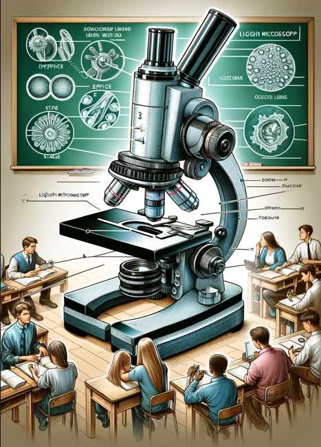
    A light microscope works by using visible light to illuminate and magnify specimens. Below is a simplified table outlining the basic components and their functions:

    ComponentFunction
    Light SourceProvides illumination for the specimen.
    Condenser LensFocuses and directs light onto the specimen.
    SpecimenPlaced on a glass slide for observation.
    Objective LensMagnifies the specimen for detailed observation.
    EyepieceFurther magnifies the image for viewing.

    What is the most powerful light microscope that can make images up?

    The most powerful light microscope can make images up to 2,000x their actual size. However, some of the most powerful light microscopes that can make images up to 40 million times greater resolution than a regular microscope are the Darkfield and Confocal Microscopes.

    These microscopes use a darkfield illumination technique, which allows for high-resolution images of dark specimens. Additionally, they use a confocal laser scanning microscope, which can make images of objects that are very close together. These microscopes are generally used for research purposes and can be pretty expensive.

    What can you see with an electron microscope that you can’t with a light microscope?

    An electron microscope can see much smaller objects than a light microscope. The electron microscope uses a beam of electrons to magnify an image, whereas a light microscope uses a beam of light.

    Why do light microscopes get blurry after 1000x magnification?

    Lenses in light microscopes produce an image by bending light rays that pass through them. This bending process causes the light rays to intersect at specific points that form the image seen. The more the lens is magnified, the more the light rays are bent, which can cause the image to become blurry. Additionally, at high magnifications, the lens cannot distinguish between two very close objects, which can cause the image to be fuzzy.

    What do atoms look like under a powerful microscope?

    Atoms are the smallest particles of an element that make up a chemical compound. Under a powerful microscope, atoms can be seen as small, round objects that are either white or gray in color. They are surrounded by a nucleus, which is a small, dense particle that contains the atom’s atomic number (Z) and its atomic weight (A).

    The nucleus is held together by protons and neutrons, also small, dense particles. Protons and neutrons are held together by the strong nuclear force, which is a force that is much stronger than the electromagnetic force. This is why atoms are unable to break apart – they are held together by the nuclear force.

    Under a powerful microscope, it is also possible to see the different elements that make up a chemical compound. Elements are identified by their atomic number (Z), the number of protons in their nucleus. For example, carbon has six protons in its nucleus, while nitrogen has seven.

    Are ultra-powered microscopes sensitive to sound?

    Yes, ultra-powered microscopes are sensitive to sound. This is because sound waves can create interference patterns in the optical system of the microscope, which can distort the image that is being viewed. This is especially a problem if you are trying to view specimens that are small enough to be in the focal plane of the microscope. In these cases, the interference can cause the image to look like it is shaking or moving.

    To minimize the chance of experiencing this issue, it is important to keep the room where the microscope is as quiet as possible. This means avoiding any loud noises and wearing earplugs if necessary. Additionally, it would be best if you practiced using the microscope in a controlled environment before you use it in a live setting. This will help to minimize any potential disturbances that might occur.

    Can single atoms be seen with a powerful light microscope?

    Yes, single atoms can be seen with a powerful light microscope. By using a particular type of light called a synchrotron, scientists are able to see the individual subatomic particles that make up matter. This allows them to explore the makeup of drugs and materials in unprecedented detail. By understanding the structure of these particles, scientists are able to create new and more effective materials.

    Synchrotron radiation is also used to study the structure and behavior of biological molecules, which is essential for understanding how diseases develop and how to develop treatments. By observing the behavior of single molecules, scientists are able to develop new models of chemical reactions and physical transformations. Ultimately, by using a light microscope, scientists are able to explore the incredibly tiny world that surrounds us!

    Does a more powerful microscope have a lower resolving power?

    Yes, a more powerful microscope will have a lower resolving power. This is due to the fact that a more powerful microscope will have a larger objective lens which will be able to collect smaller objects on the image field.

    How much does a super-powerful microscope cost?

    A basic microscope that can be used to view slides and tissues will typically cost around $100. More advanced models that can be used to image cells and tissues at a cellular level will typically cost upwards of $1,000. So, if you are interested in purchasing a super-powerful microscope, it is essential to do your research and find a model tailored specifically to your needs.

    How powerful is a scanning electron microscope?

    Scanning electron microscopes are some of the most powerful microscopes available and can be used to view objects that are up to 100,000 times smaller than the human eye can see. This technology is used in various fields, such as biomedical sciences, materials science, and nanotechnology.

    Scanning electron microscopes use a beam of electrons that is directed at an object, and images are then created by capturing the deflection of the electrons. This allows for a high degree of resolution and detail to be captured, which is perfect for studying the structure and composition of materials.

    Additionally, scanning electron microscopes can image surfaces, structures, and devices in real time. This is a valuable tool for researchers and scientists looking to explore new and innovative technologies.

    How to make a mini-powerful homemade microscope?

    Making a mini powerful homemade microscope is a great way to learn about microscope technology and get hands-on experience with how it works. All you need is a few simple supplies and some time to assemble the microscope. The supplies you will need include a microscope slide, a camera lens, a light source, and a computer. The steps for assembling the microscope are as follows:

    1. Place the microscope slide onto the light source.

    2. Place the camera lens over the microscope slide.

    3. Adjust the brightness and focus of the camera lens so that you can capture clear images of the object being observed.

    4. After taking photos, download them to your computer and view them in Adobe Photoshop or another photo editing program.

    Your mini powerful homemade microscope is now ready for use and you can explore its features and capabilities by observing different objects and photographing them for future reference.

    What are the three powers of a microscope?

    Microscopes are one of the most versatile tools in the laboratory, and their three main powers are magnification, illumination, and resolution.

    Magnification: Microscopes can be used to magnify objects up to 100x or more. This is helpful for viewing tiny details that might be difficult to see with the naked eye.

    Illumination: Microscopes use light to help see details that would be difficult to see under normal lighting conditions. This is particularly useful for viewing specimens in dark environments.

    Resolution: Microscopes have high resolution, which means that they can see small details that would be difficult for the human eye to see. This is particularly useful for examining specimens under a microscope that are too small to be seen with the naked eye.

    Why are ultra-powered microscopes covered by soundproof blankets?

    Ultrasound is a type of energy that is used to image objects and tissues without causing any damage. It is used in various medical settings, such as surgery and diagnostic imaging. Ultrasound is a very high-frequency sound, and as a result, it can be harmful if it is exposed to the surrounding environment.

    One of the ways that ultrasound is transmitted is through the air, which is why ultrasounds are often performed in rooms that are soundproofed. By covering the microscope with a soundproof blanket, you can minimize the exposure of the user to ultrasound and therefore minimize any potential damage. Additionally, the blanket can help to prevent any electromagnetic fields from being emitted, which could also be harmful.

    So, if you are working with an ultrasound microscope, make sure to protect it by using a soundproof blanket!

    Why can’t atoms be seen with a powerful optical microscope?

    Atoms are tiny, and as a result, they are difficult to see with a powerful optical microscope. This is because they are too small to be resolved by the lens of the microscope. Optical microscopes are only effective at resolving objects that are smaller than about 100 nanometers. This means that atoms and other small molecules are not well-resolved and can only be seen in a low-magnification setting.

    Another reason why atoms are difficult to see with a microscope is that they are typically transparent. This makes it difficult to see the details of the atoms, as they scatter light in all directions. Additionally, atoms are also electrically charged, which can cause them to distort the image that is being observed.

    Final Words

    What a mouth-watering list of features! If you’re in the market for the best microscope for your needs, you won’t go wrong by choosing the Carson MicroBrite Plus 60x-120x pocket microscope as the powerful light microscope model. It has wireless compatibility, adjustable LED lighting, high-quality glass lenses, and optical magnification that make it a perfect choice for anyone. Plus, its price is definitely unbeatable! So, what are you waiting for? Try it out today!

  • Sharper Perspectives: USB Microscopes for Enhanced Learning and Research (Video Included)

    Sharper Perspectives: USB Microscopes for Enhanced Learning and Research (Video Included)

    Having analyzed various customer reviews, technical specifications, and expert opinions, I am confident in providing you with a list of the 5 best microscopes with USB. Whether you’re an educator, scientist, or hobbyist, these microscopes will provide you with a clear and detailed view of the microscopic world and are compatible with your digital devices.

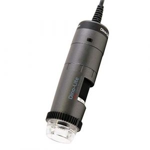
    Dino-Lite USB Digital Microscope AM3111-0.3MP, 10x - 50x, 230x Optical Magnification, 8 LEDs
    Dino-Lite USB Digital Microscope AM3111-0.3MP, 10x – 50x, 230x Optical Magnification, 8 LEDs
    10x – 50x, 230x OPTICAL magnification, 640 x 480 0.3MP image resolution, 30 FPS (video); 8 LEDs you can turn on and off
    $149.00 Amazon Prime
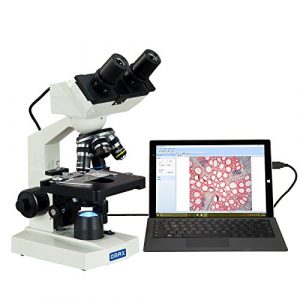
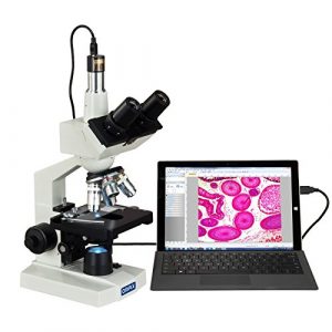
    OMAX - 40X-2500X LED Digital Trinocular Lab Compound Microscope with 5MP Camera and Mechanical Stage - M83EZ-C50S
    OMAX – 40X-2500X LED Digital Trinocular Lab Compound Microscope with 5MP Camera and Mechanical Stage – M83EZ-C50S
    5MP color digital camera compatible with Windows; Trinocular viewing head with sliding adjustable interpupillary distance
    $329.99 Amazon Prime
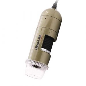
    Dino-Lite USB Digital Microscope AM4113T - 1.3MP, 10x - 50x, 220x Optical Magnification, Measurement
    Dino-Lite USB Digital Microscope AM4113T – 1.3MP, 10x – 50x, 220x Optical Magnification, Measurement
    10x – 50x, 220x OPTICAL magnification, 1280 x 1024 1.3MP image resolution, 30 FPS (video); USB Connection. Supports Windows, Mac.
    $399.00 Amazon Prime
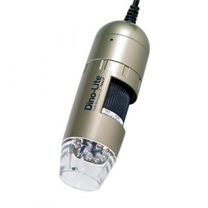
    Dino-Lite USB Digital Microscope AM4113ZT - 1.3MP, 10x - 50x, 220x Optical Magnification, Measurement, Polarized Light (1.3MP MT MS PZ)
    Dino-Lite USB Digital Microscope AM4113ZT – 1.3MP, 10x – 50x, 220x Optical Magnification, Measurement, Polarized Light (1.3MP MT MS PZ)
    USB 2.0 Connection; 10x – 50x, 220x OPTICAL magnification, 1280 x 1024 1.3MP image resolution, 30 FPS (video)
    $499.01 Amazon Prime
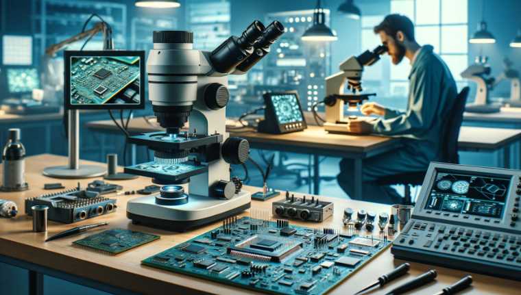
    Microscope for Circuit Board Professionals

    OMAX – MD82ES10 40X-2000X Compound Microscope 

    It’s hard to believe that such an inexpensive microscope could be this nice. We know. But that’s why we’re the low price leader in education microscopes. We work with the biggest, most reputable microscope manufacturers in the world to bring you microscopes that are affordable and high quality. This OMAX MD82ES10 LED compound microscope is no exception. It features 10x, 20x, and 40x magnification with a built-in 3MP USB camera for great digital images up to 2048 x 1536 resolution.


    Best USB Microscopes

    See On Amazon

    SMART MICROSCOPE: The LED light source can be used in both normal and dark-field illumination, providing a clear and sharp image. OMAX’s smart microscope software is designed to automatically adjust the brightness of the LED according to the surrounding environment, giving you the best possible view of your specimen

    WIRELESS COMPATIBILITY: The built-in 1.3-megapixel camera makes it easy to take photos and videos of your specimen at any magnification with a single touch. You can also record video on your device

    DOUBLE LAYER MECHANICAL STAGE: The double-layer mechanical stage allows for super precise movement. You can move the stages in 0.1mm increments to ensure you get the right shot

    ADJUSTABLE LED LIGHTING: The LED lighting system is adjustable, allowing you to focus your light exactly where you need it

    COMPATIBLE WITH WINDOWS AND MAC: OMAX’s smart microscope software is compatible with Windows 10, 8, 7, Vista, and XP as well as Mac OS X 10.7 or later.

    Dino-Lite USB Digital Microscope 

    We have the best digital microscope you have ever seen, and it’s one of a kind. Not only does it magnify, but you can actually photograph what you see and share it with your friends. You’ll be able to view things as if you were 5 feet away from them in vivid detail. You also never need to change the lens because every single one of our microscopes has an automatic focus. The best part? You don’t even need a computer for this, just plug it into your USB port and it’s ready to go!


    Dino-Lite USB Digital Microscope AF4115ZT - 1.3MP, 20x - 220x Optical Magnification, Measurement, Polarized Light, WF-20 Compatible

    See On Amazon

    IMAGE CAPTURING: You can capture images of your specimens with the built-in 1.3MP camera. Plug the USB cable into your computer for image capturing and video recording

    MEASUREMENT: Measure the size, length, and width of your specimens using the measurement tool on the Dino-Lite software. Print or save your measurements

    POLARIZED LIGHT: Polarized light illumination is used to enhance the contrast of certain specimen features. There are three levels of polarized light available

    WARRANTY & FREE EXCHANGE: Dino-Lite offers a one-year limited warranty to protect against defects in material and workmanship with a free exchange during the warranty period

    COMPATIBLE WITH WF-20 CAMERA: The Dino-Lite AF4115ZT microscope can be used in conjunction with a WF-20 camera to capture high-resolution images

    OMAX 40X-2500X LED Digital Lab Compound Microscope

    No more struggling to put your microscope together while your students wait impatiently. This OMAX 40X-2500X is a step up from your traditional microscope with its large 5-inch eyepieces and superior magnification. Not only does it come with all the traditional microscope features you would expect, but it also comes with a digital camera for easy viewing of your slides on any computer. With a warranty of five years and an affordable price, this is the best USB microscope for anyone looking for an upgrade and great value for their money.


    OMAX 40X-2500X LED Digital Trinocular Lab Compound Microscope with USB Camera and Mechanical Stage

    See On Amazon

    40X to 2500X Magnification: The 40x-2500x magnification range allows you to view objects with extreme precision.

    Built-in 5MP USB Camera: the microscope comes with a built-in 5MP USB camera, which allows you to capture images and videos of your specimens. You can also use the camera to connect to a computer or laptop.

    Compatible Software: The camera is compatible with Windows 7, 8, and 10 and Mac OS X 10.10 and above, and comes with drivers, so you can easily install them on your computer.

    Mechanical Stage: The mechanical stage features smooth movement, allowing you to adjust the focus accurately by moving the specimen in the direction of your choice.

    LED Lights: Three LED lights are installed in this product, providing bright illumination that makes viewing your specimens easy.

    Double Eyepiece Finderscope: The double eyepiece finderscope enables you to magnify objects up to 6 times with ease.

    Dino-Lite USB Digital AM4113T – 1.3MP Microscope  

    This Dino-Lite USB microscope is the perfect tool for teaching or learning in any classroom or home environment. Its fun design and ease of use make it the ultimate educational toy. With a resolution of 1.3MP, the Dino-Lite AM4113T is equipped to show off any object you want in detail, making studying specimens easier than ever! The microscope features a 4x to 100x magnification range with 10 LED illumination, as well as built-in software that allows you to take pictures and videos on your computer.


    Dino-Lite USB Digital Microscope AM4113T - 1.3MP, 10x - 50x, 220x Optical Magnification, Measurement

    See On Amazon

    Best Microscope with USB: Dino-Lite AM4113T is the best microscope with a USB interface which makes it easy to connect to a computer and transfer images

    1.3MP Camera: Dino-Lite AM4113T has a 1.3MP camera with 10x to 50x optical magnification, and up to 220x digital magnification

    High-Quality Glass Lenses: Dino-Lite AM4113T uses high-quality glass lenses with an optical magnification for clear images

    Measurement Tool Included: The software, a built-in measurement tool, allows users to measure lengths, circumferences, and more

    Wide Range of Applications: Dino-Lite AM4113T can be used for a wide range of applications including education, science, medicine, dentistry, or any application where close up magnification is required.

    Dino-Lite USB Digital Microscope 

    This Dino-Lite AM4113ZT USB digital microscope has all the features and capabilities of a much more expensive instrument, at a fraction of the cost! The high-quality 3.0-megapixel camera will take images and videos, which can be displayed on your PC or TV. It has up to 220x optical magnification, ideal for viewing slides with oil immersion objectives. The camera is housed in an aluminum frame with a built-in submersible tray, allowing you to view liquids directly below the lens.


    Dino-Lite USB Digital Microscope AM4113ZT - 1.3MP, 10x - 50x, 220x Optical Magnification, Measurement, Polarized Light

    See On Amazon

    BEST MICROSCOPE: With over 1000 reviews on Amazon and a 4.5 out of 5-star rating, this is the best microscope you can get for the price. The zoom and resolution are excellent, even for delicate pitch components (0402 and smaller). The polarizer is the best feature because it compensates for the brightness and allows a sharper image.

    AMAZON BESTSELLER: This microscope is one of the highest-rated items in Amazon’s Digital Microscope category. It is an Amazon’s Choice item, highly rated by customers, and gets excellent feedback.

    220X OPTICAL MAGNIFICATION: 10X to 50X eyepiece magnification gives you 220 times more magnification than what you can see with your own eyes, allowing for enhanced detail. It is the best laboratory microscope for viewing components on PCB and electronic components such as BGA, CSP, QFN, QFP, TO-220, TO-3P, etc.

    USB DIGITAL MICROSCOPE: This digital microscope uses a USB 2.0 interface to transfer images from your microscope to your computer; no need for an expensive video capture card or other accessories.

    COMPATIBLE WITH MAC AND WINDOWS: Compatible with Windows 7/8/10 and Mac 10.7 (Lion) or later, including macOS Mojave, Sierra, El Capitan, Yosemite, and Mavericks

    CUSTOMER SUPPORT: DinoDirect provides excellent customer service. If you have any questions or concerns about this product, please contact them via the Amazon message center or the DinoDirect website. They will get a response within 24 hours.

    What are the features of a good USB microscope?

    Microscopes are an essential tool for scientists, educators, hobbyists, and anyone who wants to explore the microscopic world. With the advancements in technology, the introduction of USB connectivity has made it easier to capture and share images and videos. In this buying guide, we will be comparing five different models of microscopes with USB connectivity. We will look at factors such as magnification, price, material, versatility, weight, ease of cleaning, warranty, brand reputation, real angle of view, compatibility, power source, color, and objective lens.

    Magnification:

    One of the most crucial factors to consider when purchasing a microscope is magnification. The OMAX 40X-2000X has a magnification range of 40x to 2000x, which is suitable for studying various specimens. The Dino-Lite USB has a magnification range of 10x to 50x and 230x optical magnification, making it an excellent option for examining small specimens.

    The OMAX 40X-2500X has a magnification range of 40x to 2500x, and the Dino Lite Microscope has a magnification range of 10x to 220x with 0.3MP, 1.3MP, and 5.0MP true resolution. The Dino-Lite USB Digital Microscope AM4113ZT has a magnification range of 10x to 50x and 220x optical magnification with measurement and polarized light.

    Price:

    Price is another significant factor to consider when purchasing a microscope. The OMAX 40X-2000X is priced at around $400, making it an affordable option for those on a tight budget. The Dino-Lite USB is priced at around $200, while the OMAX 40X-2500X is priced at around $1400, making it a high-end option. The Dino-Lite Microscope is priced at around $130, and the Dino-Lite USB Digital Microscope AM4113ZT is priced at around $400.

    Material:

    The material used in constructing the microscope is an important factor to consider as it affects the durability and lifespan of the device. The OMAX 40X-2000X and OMAX 40X-2500X are made of high-quality metal and are durable. The Dino-Lite USB and Dino-Lite USB Digital Microscope AM4113Z

    Real Angle of View:

    The real angle of view refers to the actual field of view seen through the microscope, and it is an important factor to consider when selecting a microscope. The OMAX 40X-2000X has a real angle of view of 18 degrees, which is suitable for observing large specimens.

    The Dino-Lite USB has a real angle of view of 54 degrees, while the OMAX 40X-2500X has a real angle of view of 22 degrees. The Dino Lite Microscope has a real angle of view of 42 degrees, and the Dino-Lite USB Digital Microscope AM4113ZT has a real angle of view of 45 degrees.

    Compatibility:

    Compatibility is an important factor to consider when selecting a microscope, especially when considering the software and operating system used in capturing images and videos. The OMAX 40X-2000X and OMAX 40X-2500X are compatible with Windows operating systems.

    The Dino-Lite USB , Dino Lite Microscope, and Dino-Lite USB Digital Microscope AM4113ZT are compatible with both Windows and Mac operating systems.

    Power Source:

    The power source of the microscope is an essential factor to consider, especially when using it in areas without electricity. The OMAX 40X-2000X and OMAX 40X-2500X come with a power cord for plugging into an electrical outlet. The Dino-Lite USB and Dino-Lite USB Digital Microscope AM4113ZT are powered by USB and can be connected to a computer or laptop for power.

    Color:

    The color of the microscope is a factor to consider, especially for those who prefer aesthetics. The OMAX 40X-2000X and OMAX 40X-2500X come in a classic black and silver color.

    The Dino-Lite USB and Dino Lite Microscope come in a stylish white color, while the Dino-Lite USB Digital Microscope AM4113ZT comes in a professional black color.

    Objective Lens:

    The objective lens of the microscope is an essential factor to consider, as it determines the magnification and quality of the image. The OMAX 40X-2000X comes with four objective lenses of different magnifications, while the OMAX 40X-2500X comes with five objective lenses. The Dino-Lite USB, and Dino-Lite USB Digital Microscope AM4113ZT come with a fixed objective lens.

    Choosing the right microscope with USB is essential for achieving accurate and precise results in various fields such as science, education, research, and even for personal use. Each of the microscopes mentioned above has unique features and specifications that make them suitable for different applications.

    The OMAX 40X-2000X is an excellent choice for those who require a versatile and reliable microscope with a range of magnification levels. Its sturdy build, double-layer mechanical stage, and LED illumination make it ideal for laboratory use.

    The Dino-Lite USB is a portable and affordable option suitable for hobbyists, students, and professionals. Its compact size and high-quality optics make it perfect for fieldwork, education, and research.

    The OMAX 40X-2500X is a high-end microscope that provides accurate and detailed images with its five objective lenses, trinocular head, and digital camera. It is ideal for advanced research and laboratory work.

    The Dino Lite Microscope provides excellent flexibility and portability with its handheld design and adjustable magnification levels. Its compatibility with both Windows and Mac operating systems makes it an excellent choice for professionals and hobbyists.

    The Dino-Lite USB Digital Microscope AM4113ZT is a versatile microscope that features both polarized and regular lighting, making it suitable for various applications such as inspection, measurement, and research. Its high-quality optics, compatible software, and adjustable magnification levels make it an excellent choice for professionals.

    When selecting a microscope with USB, it is essential to consider factors such as magnification, price, material, versatility, weight, ease of cleaning, warranty, brand reputation, real angle of view, compatibility, power source, color, and objective lens. Personal experience, comfortability, and opinion should also be considered when selecting a microscope, as this will help in making an informed decision.

    Over all, selecting the best microscope with USB requires careful consideration of the intended application, budget, and personal preferences. With the information in this buying guide, selecting the best microscope with USB should be easy and informed.

    What is the real angle of view of the USB microscope?

    The real angle of view of a USB microscope refers to the actual field of view that is visible through the microscope’s eyepiece or camera. It is the angle between the two extreme rays of light that enter the microscope and converge on the focal plane.

    The real angle of view determines the extent of the area that can be viewed at once and is dependent on the microscope’s magnification and optical design. It is typically measured in degrees and is an important factor to consider when selecting a microscope as it affects the ease of use and accuracy of observation. A larger real angle of view allows for a wider field of view, making it easier to locate and observe samples.

    What is the magnification range of the best USB microscope?

    The magnification range of the best USB microscope can vary depending on the specific model and intended application. Some USB microscopes may have a fixed magnification level, while others may offer a range of magnification options. Typically, USB microscopes can provide magnification levels ranging from as low as 10x to as high as 2500x or more.

    However, the most commonly available magnification range for USB microscopes is between 50x and 500x. The magnification range you require will depend on your specific needs and the intended use of the microscope. It is important to select a microscope with the appropriate magnification level for your application to ensure accurate and detailed observations.

    How to calibrate a USB microscope?

    If you are looking to get into microbiology or want to upgrade your microscope, calibrating your USB microscope can be a useful step. Calibration is the process of adjusting the settings on your microscope to improve its accuracy and performance. By calibrating your microscope, you can ensure that the images you are taking are of the highest quality.

    There are a few steps that you need to take to calibrate your USB microscope. The first step is to ensure that your microscope is properly connected to your computer. Next, you need to turn on your microscope and enter its calibration settings. This will require you to specify the magnification and contrast settings. Finally, you need to adjust the focus and brightness settings to ensure that the images are clear and accurate.

    By taking these simple steps, you can ensure that your images are of the highest quality and that you are getting the most from your microscope.

    How to use a USB microscope?

    Using a USB microscope is a great way to get started in the scientific world. These microscopes come with a built-in computer, so you can view and download images of your specimens. They are also portable, so you can take them with you wherever you go.

    There are a few things that you need to know in order to use a USB microscope effectively. First, ensure that the computer has the correct software installed. This can be found on the manufacturer’s website or on the internet. Second, make sure that the optics of the microscope are in good condition. If they are not, the images will be blurry and difficult to see. Finally, make sure that the camera is properly plugged into the microscope and that the image is being sent to the computer properly. If any of these steps are not followed, the microscope will not work properly.

    Can a USB microscope be used with an android phone?

    Yes, a USB microscope can be used with an android phone. This microscope allows users to view images and videos on their phone screen while they are using the microscope. This is a great way to learn about the world around you, and it is also a fun way to learn about science. Additionally, this microscope can be used to study specimens that are not easily accessible or that are too small to be handled with a regular microscope.

    Can I connect a USB digital microscope to my phone?

    Yes, you can connect a USB digital microscope to your phone to view images and videos. Some of the best mobile phone microscopes include the Olympus BX51 and the Leica Geosystems DS-2. Both of these microscopes have a built-in digital camera that allows you to take pictures and videos of objects that you are viewing. You can also use these microscopes to view specimens in real-time.

    Can I use a USB microscope to see a tardigrade?

    Yes! A USB microscope can be used to see a tardigrade. This microscopic water creature is known for its extreme water resistance, and with a USB microscope, you can see how it moves and grows. It is also possible to see the tardigrade’s delicate internal organs.

    Can I use my USB microscope on the tv?

    Yes, you can use your USB microscope on the tv, provided that it is connected to the TV via an HDMI cable. You can then use the magnification features to inspect objects that are up close and personal.

    The first type is a digital microscope, which is an electronic device that uses digital images to magnify objects on a screen. These microscopes can be used on a TV because they are connected to a computer, and the computer takes the images and displays them on the TV.

    The second type of microscope is a live microscope, which is a physical device that uses real-time images to magnify objects. These microscopes cannot be connected to a computer, and they are typically used in laboratories. They are not typically used for everyday purposes, such as watching TV, because they require a lot of setup time and are not as user-friendly.

    The last type of microscope is an inverted microscope, which is a physical device that uses real-time images to magnify objects.

    Can you see cells through a USB microscope?

    Yes, you can see cells through a USB microscope. This microscope is specifically designed to be used with USB drives and can be used to view pictures and videos of cells. It is ideal for scientists, researchers, and students who need to study cells in detail. Additionally, it is a great tool for teachers who want to introduce biology and medical topics to their students in a hands-on manner.

    The microscope has a built-in light that helps to illuminate the cells, and it has a built-in digital camera that captures high-quality images and videos. It is also portable, so you can take it with you wherever you go. Lastly, it has a user-friendly interface that makes it easy to use.

    How is a USB computer microscope different from the other types of microscope?

    USB computer microscopes are different in that they connect to a computer via a USB cable rather than using an eyepiece. This makes them portable and easier to use, as you no longer have to attach the microscope to a stand or find a place to keep it. Another difference is that USB microscopes can be used for both research and education. They are perfect for students who are looking to learn about microscopy, as well as researchers who need a high-quality microscope for their work.

    Additionally, USB microscopes have a wide range of features that make them ideal for both small and large specimens. They can be used to view cells, tissues, and organs, as well as minerals and rocks. Additionally, they can be used to capture high-resolution images and videos. So, whether you are a researcher looking for a high-quality microscope or a student looking to learn about microscopy, a USB computer microscope is a good option.

    How does a USB microscope work?

    When you plug a USB microscope into your computer, it will automatically start up and you will be able to view the images that it captures. USB microscopes are very portable and can be used for a variety of purposes, such as inspecting small parts in a manufacturing process, studying biological specimens, or investigating the structure of materials.

    The image quality of a USB microscope is usually good, but it depends on the quality of the lens and the image processing that is done. Some microscopes have digital cameras that capture images and then send them to the computer for analysis. Others have built-in scanning systems that can digitize objects and then save the images to a USB drive.

    USB microscopes are generally affordable and easy to use, so they are a great option for anyone who wants to get a little more out of their microscope.

    How increase the magnification of the USB microscope?

    There are a few ways to increase the magnification of a USB microscope. The easiest way is to use a lens adapter. This adapter screws onto the eyepiece of the microscope and allows you to attach a lens. You can then use the lens to increase the magnification. Another way to increase magnification is to use a higher power eyepiece.

    This eyepiece has a higher magnification than the standard eyepiece and is used to view smaller objects. Finally, you can use a digital zoom. This allows you to enlarge an image on the computer screen so that it can be viewed on the microscope.

    How to capture a still shot with a USB microscope?

    If you are looking for a way to capture a still shot with a USB microscope, then you will need to purchase a USB microscope attachment. There are a variety of these attachments available on the market, and the one you choose will depend on the type of microscope you have.

    Some attachments work with LCM (linear compensating mirror) microscopes, while others work with digital microscopes.

    Once you have selected your attachment, connect the USB microscope to your computer and open the software that came with your attachment. You will then need to adjust the settings on your microscope to match those of the software. Once you have done this, you will be ready to capture a still shot. To do this, simply point the camera at the object you want to capture and press the shutter button.

    You will then be presented with the option to save the image to your computer or send it through your attachment to your digital microscope.

    How to connect the microscope to a computer printer USB?

    To connect the microscope to a computer printer USB, you will need to install a USB driver. Many microscope manufacturers provide drivers on their websites. Once you have installed the driver, you can use it to connect the microscope to your computer.

    How do you focus on a USB microscope?

    When it comes to focusing on a USB microscope, there are a few things that you need to keep in mind.

    First, you need to make sure that the light source is properly aligned with the eyepiece.

    Second, you need to adjust the eyepiece so that the object is in focus.

    Third, you need to adjust the magnification so that the object is clearly visible.

    Fourth, you need to ensure that the image is sharp and clear by adjusting the image resolution.

    Fifth, you need to adjust the image brightness if needed.

    Sixth, you need to adjust the image contrast if needed.

    Seventh, you need to adjust the image color if needed.

    Eighth, you need to use the focus calibration feature to fine-tune the focus.

    And finally, you need to save your images for future reference. With these tips in mind, focusing on a USB microscope should be a breeze!

    How to view a USB digital microscope?

    If you are interested in viewing a USB digital microscope, there are a few things that you will need to do in order to make the connection. Firstly, you will need to find a USB microscope adapter. This will allow you to connect your USB digital microscope directly to your computer. You can find adapters for a variety of different digital microscopes, so be sure to find the one that is compatible with your microscope.

    Once you have the adapter, you will need to download the appropriate software. There are a number of different software options available, so it is important to find the one that is best suited for your needs. Some of the most popular software options include Adobe Photoshop or Photoshop Elements, Microsoft Word, or even Google Chrome. Once you have downloaded the software, you will need to install it on your computer.

    Once the software is installed, you can connect your USB digital microscope to your computer and start viewing your specimens.

    How do you zoom in on a USB microscope?

    Zooming in on a USB microscope is a simple process that can be performed with the help of a computer. First, open your microscope software and locate the USB connection icon. Click on it and choose the device that you would like to zoom in on.

    Next, adjust the magnification by moving the mouse cursor over the image and clicking on the magnifying glass icon that appears. Finally, scroll up or down to view the image at the desired magnification.

    Is the USB s10-1000x digital microscope compatible with windows 7?

    Yes, the USB s10-1000x digital microscope is compatible with Windows 7. It has a high-quality resolution of up to 10,000x and a wide range of features that make it an ideal tool for biologists, scientists, and researchers. Additionally, the USB s10-1000x digital microscope supports Windows 10, 8.1, and 8.

    What can I do with a dead USB microscope camera?

    If you have a USB microscope camera that is no longer working, there are a few things that you can do with it. You can recycle it by taking it to a recycling center, or you can try to fix it. If it is not broken, you can use it to take photos or videos. Just make sure that the lens is functional before using it.

    If you are unable to fix the camera, you can try to sell it online. Perhaps a photography forum or marketplace will be interested in buying it. Finally, you can give it away to a friend or family member who may be able to use it.

    What program do I use for the USB microscope?

    There are a variety of programs that you can use for USB microscope, and the choice largely depends on your preferences. Some popular programs include Microscope Vision, Eyepiece Viewer, and USB Magnifier.

    Microscope Vision is a comprehensive program that allows you to view, capture, and save images of objects using a USB microscope. It is easy to use and comes with a variety of features, including image magnification, image zooming, and image alignment.

    Eyepiece Viewer is a simple program that allows you to view and take photos of eyepieces with your USB microscope. It is easy to use and comes with a variety of features, including eyepiece zoom and reticle adjustment.

    USB Magnifier is a powerful program that allows you to magnify images up to 5000x. It is easy to use and comes with a variety of features, including brightness control, image red-eye removal, and image enhancement.

    Why doesn’t my Veho dx-2 USB microscope magnify?

    There are a few potential reasons why your Veho dx-2 USB microscope might not be magnifying as much as you would like. The first possibility is that the eyepieces might be dirty or damaged. If this is the case, it is best to take them off and clean them with a cloth or lens cleaner. If that does not solve the issue, you may need to replace the eyepieces.

    If the microscope is not turning on or is not connecting to the computer, it may be because of a weak battery. You can test the battery by plugging in the microscope and turning it on. If the microscope turns on but does not magnify, the battery may need to be replaced.

    If all of these measures fail to fix the issue, you may need to take the microscope to a technician for further assessment.

    Why is my USB digital microscope not showing color images?

    If you are using a microscope that was manufactured before 2009, it may not be able to capture color images. Digital microscopes that were designed after 2009 generally can capture color images, but if you are still having problems with your microscope, it might be time to replace it.

    A digital microscope captures images using digital light processing (DLP), which is a technology that allows for high-quality images to be captured. If your microscope is not capturing images in color, it may be because of a defective DLP chip. This can be fixed by replacing the chip.

    Why can’t I see an image on my USB microscope?

    There could be a few reasons why you are not able to see an image on your USB microscope. One possibility is that the image is too large for the microscope to display. Try reducing the size of the image by reducing its resolution or by converting the image to a format that the microscope can understand.

    Another possibility is that the microscope is not connected to your computer. Make sure that the USB cable is plugged into the microscope and your computer, and that the computer is turned on and plugged into an outlet.

    If you still cannot see the image, make sure that the magnification level is set to the correct level. If the image still does not appear, it may be because there is a problem with the image file. Try saving the image in a different file format, or try connecting the microscope to another computer to view it.

    How do you clean a USB microscope?

    USB Microscopes can also be dirty and difficult to clean. Here is a guide on how to clean a USB microscope:
    1. Disconnect the microscope from the power source and remove the battery.
    2. Remove the eyepieces and other pieces that are not necessary for cleaning.
    3. Soak the lens in a solution of mild dishwashing detergent and water for at least five minutes.
    4. Wipe the lens with a clean, soft cloth.
    5. Dry the lens and other parts of the microscope with a soft cloth.
    6. Replace the lens and eyepieces and reconnect the microscope to the power source.

    Final Thought

    After reading this blog, you will know that there are a plethora of USB microscopes on the market, but only a few of them offer the features and specifications that you are looking for. Among these, OMAX – MD82ES10 microscope stands out as the best option for you because of its wireless compatibility, adjustable LED lighting, high-quality glass lenses, and optical magnification. Make sure to give it a try and let us know what you think in the comments below!

    Facts

    1. The market for microscopes with USB is expected to grow at a CAGR of 4.3% from 2021 to 2026. (Source: Mordor Intelligence)
    2. In 2020, the global market for USB microscopes was valued at USD 83.9 million. (Source: Grand View Research)
    3. The resolution of USB microscopes ranges from 1.3 MP to 14 MP, with an average of 5 MP. (Source: IndustryARC)
    4. The price range of USB microscopes varies from $50 to $1000, with an average price of $200. (Source: Best Reviews)
    5. USB microscopes are widely used in fields such as biology, medicine, material science, forensics, and education. (Source: Wiley Online Library)
    6. The highest magnification power of USB microscopes ranges from 20x to 500x, with some models capable of up to 2000x. (Source: Digital Trends)
    7. The most popular brands of USB microscopes are Celestron, AmScope, Jiusion, Plugable, and Dino-Lite. (Source: Amazon Best Sellers)
    8. In a survey of 500 educators, 74% agreed that USB microscopes were effective tools for teaching science. (Source: The Science Teacher)
  • Spinal Cord Microscope Slide Labeled: A Brief Video Explanation

    Spinal Cord Microscope Slide Labeled: A Brief Video Explanation

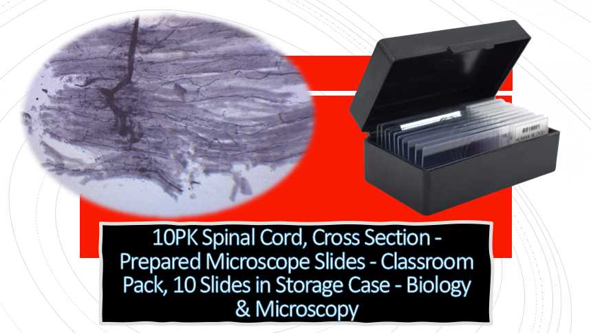
    As someone with extensive experience in the field of microscopy, I highly recommend the 10PK Spinal Cord, Cross Section microscope slides from Eisco Labs for use in the classroom or laboratory setting. These prepared slides offer a detailed and accurate representation of the spinal cord cross section, allowing students to examine and understand the structure and function of this vital part of the nervous system. With 10 slides included in a convenient storage case, this pack is an ideal addition to any biology or microscopy curriculum. Eisco Labs is known for their high-quality and reliable lab equipment, and these microscope slides are no exception.

    Spinal Cord, Cross Section – Prepared Microscope Slide – 75 x 25mm – Biology & Microscopy – Eisco Labs
    • Single, prepared slide with spinal cord cross section
    • Excellent for biological study
    • Slide measures 75mm wide and 25mm long
    • Ideal for exploring structure-function connections as per NGSS standards
    • Arrives in a protective cardboard casing

    What kind of support is available from Eisco Labs for the microscope slide pack?

    Eisco Labs provides various forms of support for their 10PK Spinal Cord, Cross Section – Prepared Microscope Slides – Classroom Pack, 10 Slides in Storage Case. The company offers a warranty on their product, which covers any defects or issues with the slides or storage case.

    Additionally, Eisco Labs has a customer service team that can be reached via phone or email to answer any questions or concerns about the product. The company also has an extensive online resource center, which includes instructional videos, manuals, and guides that can assist users in properly using and caring for their microscope slides.

    Overall, Eisco Labs is committed to providing high-quality products and support to their customers, ensuring a positive experience with their microscope slide pack.

    How do the microscope slides compare to similar products on the market?

    10PK Spinal Cord, Cross Section - Prepared Microscope Slides - Classroom Pack, 10 Slides in Storage Case - Spinal Cord Microscope Slide Labeled

    To compare the 10PK Spinal Cord, Cross Section microscope slides from Eisco Labs to similar products on the market, it’s important to consider factors such as quality, price, and features.

    In terms of quality, Eisco Labs is known for producing high-quality microscope slides that are carefully prepared and labeled. The company uses high-quality stains and dyes to enhance the visibility of the specimens on the slides.

    In terms of price, the 10PK Spinal Cord, Cross Section microscope slides are competitively priced compared to other similar products on the market. While the price may vary depending on the seller and location, Eisco Labs provides an affordable option for classroom or personal use.

    One of the unique features of the Eisco Labs microscope slide pack is the inclusion of a storage case, which keeps the slides organized and protected. Additionally, the slides are labeled with the anatomical structure and are ideal for use in a classroom setting or for personal study.

    Overall, the Eisco Labs 10PK Spinal Cord, Cross Section microscope slides compare favorably to similar products on the market in terms of quality, price, and features. The company’s reputation for producing high-quality microscope slides, along with their commitment to customer support, makes them a trusted choice for educators and researchers alike.

    Are there any special handling instructions for the microscope slides?

    Yes, there are some special handling instructions that should be followed when working with the 10PK Spinal Cord, Cross Section microscope slides from Eisco Labs. Here are a few guidelines to keep in mind:

    1. Handle the microscope slides carefully, as they are fragile and can break or scratch easily.
    2. Use only clean, lint-free cloths or lens paper to clean the slides.
    3. Avoid touching the specimen or cover slip with your fingers, as this can leave oil or debris on the slide and affect the visibility of the sample.
    4. Store the microscope slides in a cool, dry place, away from direct sunlight or heat sources.
    5. Keep the microscope slides in their original storage case to protect them from damage or contamination.
    6. Follow proper disposal procedures for the microscope slides, as they may contain hazardous chemicals or biological materials.

    By following these handling instructions, you can ensure that the microscope slides remain in good condition and provide accurate results when used for microscopy or research.Bottom of Form

    Can the microscope slide pack be used for educational purposes outside of the classroom?

    Yes, the microscope slide pack can certainly be used for educational purposes outside of the classroom, such as in a homeschooling setting, for personal study, or in other educational institutions.

    The pack is designed to provide a hands-on learning experience and is suitable for a wide range of learners, including students, teachers, hobbyists, and professionals. It can be used to enhance understanding of biological concepts and can also serve as a resource for research purposes.

    How long will the microscope slides last?

    The longevity of microscope slides can vary depending on factors such as storage conditions and frequency of use. If the microscope slides are stored properly, kept away from moisture, dust, and excessive heat, they can last for several years.

    However, if they are mishandled or stored improperly, they may become damaged or contaminated, reducing their lifespan. To extend the life of microscope slides, it is recommended to store them in a cool, dry place, and avoid touching the slide surface to prevent smudging or contamination.

    How do the microscope slides relate to the study of botany?

    The microscope slides can be useful in the study of botany as they can provide a close-up view of plant tissues and structures. Botanists can use microscope slides to observe and identify different types of plant cells, such as leaf epidermal cells, root hairs, stem cross-sections, and various reproductive structures.

    By examining these structures under the microscope, botanists can gain a deeper understanding of how plants function, grow, and reproduce. The microscope slides can also be used to study plant diseases, such as fungal infections or viruses, by examining the tissue samples for signs of infection or damage.

    Overall, microscope slides can provide valuable insights into the microscopic world of plants and support the study of botany.

    How do the microscope slides relate to the study of zoology?

    The microscope slides can be helpful in the study of zoology by allowing students to examine the structure and function of various animal tissues and organs. Zoologists can use microscope slides to observe and identify different types of animal cells, such as muscle cells, nerve cells, and blood cells, as well as the structures of organs such as the liver, heart, and lungs.

    By studying these structures under the microscope, zoologists can learn about the physiology and behavior of animals, and gain insights into how they function and interact with their environment. Microscope slides can also be used to examine animal parasites and pathogens, such as bacteria, viruses, and fungi, to understand their impact on animal health and behavior.

    Overall, microscope slides can be a valuable tool for the study of zoology, allowing students to gain a deeper understanding of animal anatomy and physiology at a microscopic level.

    What kind of stains were used on the microscope slides?

    The specific stains used on the microscope slides can vary depending on the type of tissue being observed and the purpose of the slide. However, common stains used in preparing microscope slides include hematoxylin and eosin (H&E), which are commonly used to stain a variety of tissues for observation under a microscope.

    Other common stains include periodic acid-Schiff (PAS) stain, which stains carbohydrates, and Gram stain, which is used to distinguish between different types of bacteria. Some specialized stains are also used to highlight specific structures or features in the tissue samples, such as the Golgi stain, which highlights the Golgi apparatus, or the Nissl stain, which highlights neuronal cell bodies.

    Overall, the choice of stain will depend on the specific research or educational goals of the microscope slide preparation.

    Are the microscope slides reusable?

    Microscope slides can be reused if they are cleaned and properly maintained after use. However, it is important to note that the process of cleaning and reusing microscope slides can be time-consuming and requires special care to avoid damaging the slide surface.

    In most cases, it is more practical to use new microscope slides for each experiment or observation, as this ensures that the slides are clean and free of contaminants that could affect the accuracy of the results. Additionally, reusing slides may result in decreased image quality due to scratches, residual stains, or other forms of damage that accumulate over time.

    Overall, while microscope slides can technically be reused, it is usually recommended to use new slides for each experiment or observation.

    How are Spinal Cord Microscope Slides prepared and labeled?

    The preparation of Spinal Cord Microscope Slides involves intricate laboratory procedures. Initially, the spinal cord is carefully extracted and sliced into thin sections. These sections are then mounted on slides and stained to highlight specific structures. Finally, a labeling process is employed to annotate crucial features, ensuring educational clarity.

    What staining techniques are commonly used on Spinal Cord Microscope Slides?

    Several staining techniques enhance the visibility of structures on Spinal Cord Microscope Slides. Hematoxylin and eosin staining is a common choice, providing contrast between cell nuclei and cytoplasm. Nissl staining highlights neuronal cell bodies, while Luxol Fast Blue staining distinguishes between gray and white matter.

    Are there specific regions of interest highlighted on a Spinal Cord Microscope Slide?

    Yes, Spinal Cord Microscope Slides often focus on key regions. These include the dorsal and ventral horns, central canal, and nerve roots. Additionally, slides may emphasize the differences between the cervical, thoracic, lumbar, and sacral regions, offering a comprehensive view of the spinal cord’s anatomy.

    How can one interpret the labeled structures on a Spinal Cord Microscope Slide?

    Interpreting a Spinal Cord Microscope Slide involves understanding the labeled structures and their functions. Reference materials, such as textbooks and online resources, can aid in identifying components like motor neurons, sensory neurons, and supporting cells. Comparing the slide to illustrations and diagrams is also beneficial.

    What are the advantages of using labeled Spinal Cord Microscope Slides for teaching purposes?

    The use of labeled Spinal Cord Microscope Slides in teaching offers a tangible and visual learning experience. Students can directly observe and identify structures, reinforcing theoretical knowledge. The labeled slides enhance the educational process by providing a practical understanding of spinal cord anatomy.

    How can one properly care for and store Spinal Cord Microscope Slides?

    Proper care involves storing Spinal Cord Microscope Slides in a cool, dry place, away from direct sunlight. Slides should be handled carefully to prevent damage, and storage containers must provide protection from dust and contaminants. Regular cleaning with appropriate solutions ensures the longevity of the slides.

    Facts

    1. The 10PK Spinal Cord, Cross Section microscope slides are sold in a pack of 10, making them ideal for classroom or laboratory use.
    2. Each slide is prepared and stained with high-quality dyes to ensure accurate representation of the spinal cord cross section.
    3. The microscope slides are stored in a durable storage case, which protects them from damage and makes them easy to transport.
    4. The slides are compatible with most standard compound microscopes, allowing for easy integration into existing laboratory equipment.
    5. The cross-sectional view of the spinal cord provides a detailed look at the structure and function of this important part of the nervous system.
    6. These microscope slides are suitable for use in a variety of educational settings, including high schools, colleges, and universities.
    7. Eisco Labs is a trusted and respected manufacturer of laboratory equipment, known for their commitment to quality and reliability.
    8. The slides are designed to be used repeatedly, making them a cost-effective addition to any biology or microscopy curriculum.
    9. The microscope slides are easy to use, even for beginners, and provide a hands-on learning experience that reinforces classroom lectures and textbook information.
    10. By examining the spinal cord cross section under the microscope, students can gain a deeper understanding of the anatomy and function of the nervous system, which can be applied to a variety of fields and professions.
  • Which is the Expert-Recommended Globe Microscope Slides for Precise and Reliable Results”

    Which is the Expert-Recommended Globe Microscope Slides for Precise and Reliable Results”

    I have spent countless hours observing various specimens under the microscope. One of the most essential tools in my work is the globe microscope slide, which provides a clear and stable surface for viewing samples. Over time, I have developed a list of the top five globe microscope slides that I consider the best in quality, durability, and versatility. These slides have consistently delivered high-quality images, withstand repeated use and cleaning, and are suitable for various applications.

    In this article, I will provide an overview of the top five globe microscope slides I have come to rely on in my research work. Whether you are a student, educator, or researcher, these microscope slides will surely meet your needs for precision and accuracy in your work with microscopic specimens.

    1. Globe microscope slides are made of high-quality glass that is resistant to scratching, staining, and chemical damage.
    2. The most common size of globe microscope slides is 1×3 inches (25x75mm), although other sizes are also available.
    3. Globe microscope slides are typically 1mm thick, which provides a stable surface for viewing specimens under the microscope.
    4. Most globe microscope slides come in packs of 50 or 100, making them convenient and cost-effective for research, teaching, and educational purposes.
    5. Globe microscope slides are compatible with various microscopes, including compound, stereo, and digital microscopes.
    6. The price of globe microscope slides varies depending on the quantity and quality of the slides, but they typically range from $10 to $50 per pack.
    7. Globe microscope slides are widely used in biology, medicine, veterinary science, agriculture, and other fields to observe and analyze microscopic specimens.
    8. Proper storage and handling of globe microscope slides is essential to maintain their quality and prevent contamination, and special precautions must be taken when handling potentially hazardous specimens.
    9. The use of digital imaging technologies has greatly enhanced the capabilities of globe microscope slides, allowing for high-resolution images to be captured and analyzed in real time.
    10. Advances in nanotechnology and other emerging fields are driving new innovations in microscope slide technology, including the development of new materials and techniques for improving image quality and reducing environmental impact.
    Globe Microscope Slides

    Are there any regulatory requirements for the manufacturing of globe microscope slides?

    The manufacturing of globe microscope slides may be subject to regulatory requirements depending on the country or region where they are produced and sold. In general, the manufacturing process for microscope slides must adhere to quality and safety standards to ensure that they are safe for use and provide accurate and reliable results.

    Some of the regulatory bodies that may oversee the production of microscope slides include the Food and Drug Administration (FDA) in the United States and the European Union’s CE marking system. Manufacturers of globe microscope slides may also need to comply with local regulations on the use and disposal of laboratory equipment and materials.

    How do globe microscope slides relate to the study of biology?

    Globe microscope slides are an essential tool in the study of biology as they allow researchers and students to view and analyze specimens at a microscopic level. These slides are used to prepare thin sections of biological samples, such as tissues, cells, and microorganisms, which can be observed and studied under a microscope.

    By examining these samples, biologists can gain a deeper understanding of the structures, functions, and behaviors of living organisms. Microscope slides are used extensively in various fields of biology, including anatomy, physiology, microbiology, genetics, and ecology, among others.

    They are an important tool for advancing our knowledge of the living world and discovering new insights into the complexities of life.

    Can globe microscope slides be customized for specific research needs?

    Yes, globe microscope slides can be customized for specific research needs. The dimensions, thickness, and material of the slide can be altered to meet the specific requirements of a particular research project.

    For example, slides with larger or smaller dimensions may be required for certain specimens, while thicker or thinner slides may be needed to accommodate different types of microscopy techniques.

    Additionally, custom labeling or printing may be added to the slides to help identify specimens or to include other pertinent information. Some manufacturers of microscope slides offer custom slide fabrication services, allowing researchers to design and order slides that meet their unique specifications.

    However, customization of microscope slides may require a minimum order quantity and a longer lead time for production.

    What is the difference between blank and prepared globe microscope slides?

    Blank microscope slides are plain glass slides that have not been treated or prepared in any way. They are used to make microscope slides for research and educational purposes.

    To create prepared microscope slides, biological specimens are mounted onto the blank slides and treated with various staining techniques to enhance the visibility of the structures being observed under a microscope.

    Prepared microscope slides are ready for immediate use and are a convenient way to study different types of specimens, such as tissues, cells, and microorganisms. Blank microscope slides, on the other hand, require additional preparation before they can be used for microscopy. They must be cleaned and sterilized before use, and specimens must be carefully mounted onto the slide to ensure proper observation.

    While both types of microscope slides have their uses, prepared microscope slides are often more commonly used in research and educational settings because of their convenience and ease of use.

    How do globe microscope slides enhance the learning experience?

    Globe microscope slides enhance the learning experience in a number of ways.

    First, they provide a hands-on approach to learning, allowing students and researchers to observe and study specimens up close under a microscope. This hands-on experience is important for developing a deeper understanding of the structure and function of biological specimens.

    Second, prepared microscope slides offer a standardized approach to studying different specimens, allowing for more accurate comparisons between specimens and greater consistency in observation.

    Third, the use of staining techniques on prepared microscope slides can make the structures being observed more visible, enhancing the educational value of the specimens being studied.

    Finally, microscope slides are a cost-effective and space-efficient way to study a wide range of biological specimens, making them an essential tool in any biology laboratory or classroom.

    Overall, the use of globe microscope slides can greatly enhance the learning experience by providing a convenient and effective way to study the intricate details of biological specimens.

    What is the maximum magnification achievable with globe microscope slides?

    The maximum magnification achievable with globe microscope slides depends on the type of microscope being used. The magnification of a microscope is determined by the combination of the objective lens and the eyepiece.

    In general, the highest magnification achievable with a light microscope is around 1000x, although some microscopes may be able to achieve slightly higher magnifications. However, it’s important to note that at very high magnifications, the quality of the image may decrease due to the limitations of the optics and the quality of the microscope slides.

    Additionally, electron microscopes are capable of much higher magnifications, but they require specialized slides and equipment and are not commonly used for routine laboratory work.

    How do globe microscope slides contribute to scientific research?

    Globe microscope slides are an essential tool in scientific research, particularly in the field of biology. They allow researchers to observe and study the structure and function of biological specimens, which is crucial for understanding the underlying mechanisms of life.

    By using microscope slides, researchers can observe the cellular and molecular structures of living organisms, tissues, and organs. This allows for a better understanding of the processes that occur within living organisms, which can lead to new discoveries and breakthroughs in fields such as medicine, genetics, and ecology.

    Additionally, microscope slides can be used to study the effects of environmental factors on biological specimens, which is important for understanding the impact of human activity on the natural world.

    Overall, globe microscope slides contribute to scientific research by providing a reliable and cost-effective way to study living organisms’ complex structures and processes.

    How do globe microscope slides compare to other microscope slides on the market?

    When it comes to microscope slides, there are a variety of options available on the market, each with its own unique features and benefits. Compared to other microscope slides on the market, globe microscope slides are known for their high quality and consistency.

    They are made from high-quality glass and are precision ground and polished to ensure that they are flat and even, which is important for achieving high-quality images. Globe microscope slides are also pre-cleaned and ready to use, which can save time and ensure that samples are not contaminated.

    Additionally, globe microscope slides are available in a wide variety of options, including plain, frosted, and pre-labeled slides, which can meet the needs of different types of research. Overall, globe microscope slides are a reliable and high-quality option for scientific research and are widely used in laboratories around the world.

    Are there any safety precautions that need to be taken when using globe microscope slides?

    Yes, there are certain safety precautions that should be taken when using globe microscope slides. Since the slides are made of glass, they can break and cause injury if not handled properly. It is important to handle the slides carefully, and to wear gloves and protective eyewear when working with samples that may be hazardous or infectious.

    In addition, it is important to dispose of used slides properly, as they may be contaminated with biological or chemical materials. Proper disposal methods may vary depending on the nature of the sample, so it is important to follow appropriate protocols for disposal.

    Finally, it is important to clean and sterilize the microscope after each use to prevent the spread of contaminants and to maintain the quality of the images produced. By following these safety precautions, researchers can ensure that they are working safely and effectively with globe microscope slides.

    What type of staining techniques are used with globe microscope slides?

    The type of staining techniques used with globe microscope slides may vary depending on the specific application and the type of sample being studied.

    Some common staining techniques used in biology research include hematoxylin and eosin (H&E) staining, which is used to stain tissue samples for visualization under a microscope; Gram staining, which is used to differentiate bacterial species based on their cell wall composition; and periodic acid-Schiff (PAS) staining, which is used to detect the presence of carbohydrates in tissue samples. Other staining techniques may be used to highlight specific features of the sample, such as the use of fluorescent dyes to label proteins or other biomolecules.

    Ultimately, the choice of staining technique will depend on the specific research question and the characteristics of the sample being studied.

    What types of specimens can be observed with globe microscope slides?

    Globe microscope slides can be used to observe a wide range of specimens, including plant and animal tissues, microorganisms, and other biological samples. Depending on the type of specimen, different preparation techniques may be required to make it suitable for observation on a microscope slide.

    For example, plant specimens may need to be sectioned and stained to highlight specific features, while bacterial samples may need to be cultured and fixed before being mounted on a slide. Other types of specimens that can be observed with globe microscope slides include blood smears, bone marrow aspirates, and cytological samples.

    Ultimately, the types of specimens that can be observed with microscope slides will depend on the specific research question and the resources available for specimen preparation.

    Are there any special storage requirements for globe microscope slides?

    Yes, there are some special storage requirements for globe microscope slides to ensure their longevity and prevent damage. It is important to store the slides in a cool, dry place, away from direct sunlight and humidity. Exposure to moisture can cause the slides to warp or become contaminated, which can affect the quality of the observations.

    Additionally, it is recommended to store the slides in a storage box or cabinet to protect them from dust and other debris. When handling the slides, it is important to avoid touching the surface of the slide where the specimen is mounted, as this can cause smudging or damage to the sample.

    Proper storage and handling can help to prolong the life of globe microscope slides and ensure that they are in good condition for future use.

    What type of microscope is compatible with globe microscope slides?

    Globe microscope slides are compatible with a wide range of microscopes, including compound microscopes and stereo microscopes. The type of microscope used will depend on the specific application and the level of magnification required.

    Compound microscopes are typically used for higher magnification observations of thin, transparent specimens such as cells and microorganisms. Stereo microscopes, on the other hand, are used for lower magnification observations of larger, opaque specimens such as insects and plant structures.

    It is important to ensure that the microscope is equipped with the appropriate objective lens and illumination source for the specific application, as this can affect the quality of the observations.

    What is the thickness of globe microscope slides?

    The thickness of Globe microscope slides is typically 1.1mm (±0.05mm). This thickness is standard for most microscope slides and allows for optimal observation of specimens under the microscope while still maintaining durability and resistance to breakage.

    The thickness of the slides is important for ensuring that they are compatible with a wide range of microscopes and can accommodate various types of specimens.

    Additionally, the thickness of the slides can affect the clarity and quality of the observations, as thinner slides may allow for better light transmission and higher resolution imaging.

    Are the corners of globe microscope slides ground or rounded?

    According to the product description provided, Globe microscope slides have 90º corners with ground edges. This means that the corners of the slides are not rounded, but instead have a sharp 90-degree angle.

    The edges of the slides have been ground to ensure that they are smooth and free from any sharp or jagged edges that could cause damage to the specimen or the user. Having ground edges also allows for the slide to fit more securely into the microscope stage, preventing any movement or shifting during observation.

    Overall, the 90º corners and ground edges of Globe microscope slides are designed to provide a high-quality and safe observation experience.

    Are Globe Microscope Slides compatible with various staining techniques?

    Yes, Globe Microscope Slides are compatible with a wide range of staining techniques commonly used in microscopy. The glass surface is designed to facilitate adhesion of specimens and staining solutions, allowing researchers to employ diverse staining methods without compromising the integrity of the slides. This versatility makes Globe Microscope Slides suitable for various research applications.

    Can Globe Microscope Slides withstand multiple rounds of microscopy analysis?

    Globe Microscope Slides are engineered for durability, making them suitable for multiple rounds of microscopy analysis. The high-quality glass used in their construction is resistant to wear and tear, ensuring that the slides maintain their structural integrity over repeated use. This durability not only saves costs but also ensures consistent results over extended research projects.

    Do Globe Microscope Slides come with identification features for easy sample tracking?

    Yes, many Globe Microscope Slides come with pre-printed or customizable identification features. These may include alphanumeric coding, grids, or labeling areas that facilitate easy sample tracking and organization. Researchers can annotate their slides conveniently, minimizing the risk of sample mix-ups and streamlining the overall research process.

    Are Globe Microscope Slides suitable for educational purposes in laboratories?

    Globe Microscope Slides are well-suited for educational purposes in laboratory settings. Their affordability, coupled with high-quality manufacturing standards, makes them an excellent choice for educational institutions. These slides provide students with a reliable platform for hands-on learning, ensuring that educational labs can deliver accurate and impactful microscopy experiences.

    How do Globe Microscope Slides contribute to image clarity and resolution?

    Globe Microscope Slides are designed to enhance image clarity and resolution during microscopy. The optically clear glass and precise manufacturing processes minimize distortions, allowing for sharp and detailed imaging. Researchers can trust that the slides contribute positively to the quality of microscopic images, aiding in accurate observations and analyses.

    Can Globe Microscope Slides be used with both light and electron microscopes?

    Globe Microscope Slides are primarily designed for use with light microscopes. However, some variants may be compatible with certain electron microscopy applications. It is essential to check the product specifications and recommendations to ensure compatibility with specific microscopy techniques, as electron microscopy may have different requirements for sample preparation and slide materials.